ISOLATION, IDENTIFICATION & ANTIBIOGRAM OF BACTERIA ASSOCIATED WITH CAT ORAL CAVITY: A NEGLECTED ISSUE
SHAMIMA AHMED
DEPARTMENT OF MICROBIOLOGY AND PARASITOLOGY SHER-E-BANGLA AGRICULTURAL UNIVERSITY
SHER-E-BANGLA NAGAR, DHAKA-1207
JUNE, 2020
ISOLATION, IDENTIFICATION & ANTIBIOGRAM OF BACTERIA ASSOCIATED WITH CAT ORAL CAVITY: A NEGLECTED ISSUE
BY SHAMIMA AHMED
REGISTRATION NUMBER: 13-05410
A Thesis Submitted to the Faculty of Animal Science and Veterinary Medicine Sher-e-Bangla Agricultural University, Dhaka-1207,
In Partial Fulfillment of the Requirements for the degree of MASTER OF SCIENCE (MS) IN MICROBIOLOGY DEPARTMENT OF MICROBIOLOGY AND PARASITOLOGY
SEMESTER: JANUARY-JUNE/2020 APPROVED BY
Dr. Uday Kumar Mohanta Chairman of Examination Committee Department of Microbiology and Parasitology
Sher-e-Bangla Agricultural University Sher-e-Bangla Nagar, Dhaka-1207 Dr. Uday Kumar Mohanta
Supervisor
Chairman and Associate Professor Department of Microbiology and Parasitology
Sher-e-Bangla Agricultural University Sher-e-Bangla Nagar, Dhaka-1207
Dr. Jahangir Alam Co-Supervisor Chief Scientific Officer (Animal Biotechnology)
National Institute of Biotechnology (NIB) Ganakbari, Ashulia, Savar, Dhaka-1349
DEPARTMENT OF MICROBIOLOGY AND PARASITOLOGY
Sher-e-Bangla Agricultural University Sher-e-Bangla Nagar, Dhaka-1207
Memo No: SAU/MIPA
CERTIFICATE
This is to certify that the thesis entitled “Isolation, Identification & Antibiogram of Bacteria Associated with Cat Oral Cavity: A Neglected Issue” submitted to the department of Microbiology and Parasitology, faculty of Animal Science &
Veterinary Medicine, Sher - e - Bangla Agricultural University, Sher-e-Bangla Nagar, Dhaka - 1207, in partial fulfillment of the requirements for the degree of Master of Science (MS) in Microbiology, embodies the result of a piece of bona fide research work carried out by Shamima Ahmed, registration no. : 13-05410, under my supervision and guidance. No part of the thesis has been submitted for any other degree or diploma.
I further certify that any help or source of information, received during the course of this investigation has been duly acknowledged.
Date: June, 2020
Place: Dhaka, Bangladesh
Dr. Uday Kumar Mohanta Supervisor
Chairman & Associate Professor Department of Microbiology and Parasitology
Sher-e-Bangla Agricultural University, Dhaka - 1207
Dedicated to
My Beloved Parents
i
ACKNOWLEDGEMENT
At the beginning, the author bows the grace and mercy of the “Almighty Allah”, who enabled her to complete this research. The author must express her grateful indebtedness to her beloved father, Rashid Ahmed and mother, Ful Nahar Begum for their ending prayer, encouragement, sacrifice and dedicated supports to educate her this level.
The author with a sense of respect, expresses heart gratitude to her Supervisor Dr. Uday Kumar Mohanta, Chairman & Associate Professor, Department of Microbiology and Parasitology, Sher- e-Bangla Agricultural University, Dhaka-1207, Bangladesh, for his supervision, untiring guidance, invaluable suggestions, timely instructions, inspirations and constructive criticism throughout the tenure of research work.
The author with a sense of respect expresses her gratitude to her Co-Supervisor Dr. Jahangir Alam, Chief Scientific Officer (CSO), Animal Biotechnology Division, National Institute of Biotechnology (NIB), Ganakbari, Ashulia, Savar, Dhaka-1349, Bangladesh, for his untiring guidance, relentless co-operation, invaluable suggestions, and constructive criticism throughout the study. The study was supported by a KrishiGobeshona Foundation (KGF) financed project entitled “Study on zoonotic diseases of pets and assessment of risk factors for human for commonly occurred zoonoses from pet: Implication for better management” awarded to Dr.
Jahangir Alam.
The author is also grateful to Mirza Synthia Sabrin, Assistant Professor, Department of Microbiology and Parasitology, Sher-e-Bangla Agricultural University, Dhaka- 1207, for her kind advice and co-operation in the completion of the study.
The author is grateful to Dr. Forhad Hossain, former Chief Veterinary Officer, CVH, Dhaka- 1200, for his kind support and co-operation during sample collection.
The author is ever grateful to Ministry of Science and Technology, Government of the People’s Republic of Bangladesh for providing the NST fellowship.
The author is also boundless grateful to the staffs of the Animal Biotechnology Division and Environmental Biotechnology Division, NIB; the staffs of the Central Veterinary Hospital, Dhaka and the staffs of the Department of Microbiology and Parasitology, SAU for their cooperation.
The author expresses her boundless gratefulness to her brothers, relatives, friends and senior fellows for their moral support and encouragement a lot in the process of building the academic career which can never repaid.
The Author
ii
ISOLATION, IDENTIFICATION & ANTIBIOGRAM OF BACTERIA ASSOCIATED WITH CAT ORAL CAVITY: A NEGLECTED ISSUE
ABSTRACT
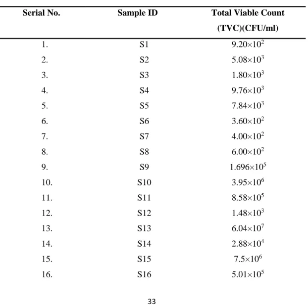
This research work was conducted to isolate, identify and antibiotic sensitivity profiling of bacteria found in oral cavity of pet cat in Dhaka city. A total number of 40 samples were collected aseptically from Central Veterinary Hospital (CVH), Dhaka and transported to National Institute of Biotechnology laboratory, Savar. Total viable count (TVC) of bacteria from all samples (n = 40) was determined. Hundred percent prevalence of organisms was noted with the highest TVC, 1.199×10¹³ and the lowest TVC, 3.60×10². The isolation and identification of bacterial genera/species were performed by cultural characteristics, Gram’s staining, biochemical tests and molecular identification to some extent. The prevalence of Escherichia coli, Salmonella spp., Staphylococcus aureus and Staphylococcus epidermidis were 100%, 5 %, 57.5% and 7.5 % respectively. Both pathogenic and non-pathogenic E. coli were confirmed by PCR and DNA sequencing. Antibiotic sensitivity test by disc diffusion method was performed against seven different antibiotics. E. coli isolates showed the highest sensitive to gentamycin (80%) followed by azithromycin (70%) and the highest resistant to ampicillin. Isolates of Salmonella isolates were found to be highest sensitive against gentamycin (100%) followed by azithromycin (50%). Highest resistant pattern of Salmonella spp. was showed against ampicillin (100%).
Salmonella spp. showed 50% resistant to erythromycin, streptomycin and tetracycline.
S. aureus showed the highest sensitivity to gentamycin (80%), followed by co- trimoxazole (60%) and the highest resistance pattern was shown against ampicillin (100%), followed by erythromycin (80%) and tetracycline (60%). S. epidermidis showed the highest (100%) resistant against ampicillin and the highest (100%) sensitive to gentamycin. The findings from current study recommend that pet cats in Dhaka city contain multi-drug resistant E.coli, Salmonella spp., S. aureus. Only S.
epidermidis was not found as multidrug resistant. This multi-drug resistant phenomenon can cause a potential public health hazard through transmission to humans by direct contact or the food chain or the evolved way of life.
Keywords: TVC, prevalence, antibiogram, multi-drug resistant, public health hazard
iii
LIST OF CONTENTS
CHAPTER TITLE PAGE NO.
ACKNOWLEDGEMENT i
ABSTRACT ii
LIST OF CONTENTS iii-v
LIST OF TABLES vi
LIST OF FIGURES vii-viii
LIST OF APPENDICES ix
LIST OF ABBREVIATION AND SYMBOLS x-xi
CHAPTER 1 INTRODUCTION 1-3
CHAPTER 2 REVIEW OF LITERATURE 4-13
2.1 Cat and public health relation 4-5
2.2 Cat diseases prevalence in Bangladesh 5-6
2.3 Bacterial prevalence in cat 6-9
2.4 Features of Escherichia coli 9
2.5 Features of Salmonella spp. 9-10
2.6 Features of Staphylococcus spp. 10
2.7 Mechanism and origin of antibiotic resistance 10-11 2.8 Antimicrobial sensitivity pattern of E. coli,
Salmonella spp. and Staphylococcus spp.
11-13
CHAPTER 3 MATERIALS AND METHODS 14-32
3.1 Materials 14
3.1.1 Samples 14-15
3.1.2 Bacteriological media 15
3.1.2.1 Agar media 15
3.1.2.2 Liquid media 16
3.1.3 Chemicals and reagents 16
3.1.4 Glass wares and other appliances 16
3.1.5 Antimicrobial discs 16-17
3.2 Methods 17
3.2.1 Brief description of the experimental design 17-18 3.2.2 Collection and transportation of sample 19 3.2.3 Preparation of various bacteriological culture media 19
iv
CHAPTER TITLE PAGE NO.
3.2.3.1 Nutrient broth 19
3.2.3.2 Nutrient agar 19
3.2.3.3 MacConkey’s agar 20
3.2.3.4 Eosine Methylene Blue (EMB) agar 20
3.2.3.5 Salmonella-Shigella agar 20
3.2.3.6 Mannitol salt (MS) agar 20
3.2.3.7 Mueller Hinton Agar 21
3.2.3.8 Phosphate Buffered Saline (PBS) 21
3.2.3.9 Simmons Citrate (SC) agar 21
3.2.3.10 SIM (Sulfide, Indole, Motility) medium 21 3.2.3.11 Methyl Red and Voges–Proskauer (MR-VP) broth 22
3.2.3.12 Sugar solutions 22
3.2.4 Isolation of bacteria 22
3.2.4.1 Preparation of sample 22
3.2.4.2 Serial dilution for bacterial culture (10 fold dilution method)
22-23
3.2.4.3 Primary culture of microorganism 23
3.2.4.4 Isolation in culture media 23
3.2.5 Microscopic identification for the suspected colonies by Gram’s staining method
23 3.2.6 Identification of isolates by different biochemical tests 23
3.2.6.1 Carbohydrate fermentation test 24
3.2.6.2 Methyl Red test 24
3.2.6.3 Voges-Proskauer (V-P) test 24
3.2.6.4 Oxidase test 24
3.2.6.5 Catalase test 24-25
3.2.6.6 SIM (Sulfer, Indole, Motility) test 25
3.2.6.7 Simmons Citrate (SC) agar test 25
3.2.7 Molecular identification of the isolates (only E.coli) 25
3.2.7.1 Bacterial DNA isolation 25-27
3.2.7.2 Polymerase Chain Reaction (PCR) 27-29
3.2.7.3 Sequencing and phylogenetic analysis 30
v
CHAPTER TITLE PAGE NO.
3.2.7.4 Gene Bank accession number 30
3.2.8 Maintenance of stock culture 30
3.2.9 Antimicrobial sensitivity pattern of E. coli, Salmonella spp., S. aureus, S. epidermidis isolated from oral swab of cat
30
3.2.9.1 Disc diffusion method 31-32
CHAPTER 4 RESULTS AND DISCUSSION 33-59
4.1 Results 33-56
4.1.1 Total viable count 33-34
4.1.2 Prevalence of microorganisms in cat oral cavity 35 4.1.3 Results of isolation and identification of E. coli,
Salmonella spp., S. aureus and S. epidermidis
35
4.1.3.1 Results of cultural examination 35
4.1.3.1.1 Culture in nutrient broth 35
4.1.3.1.2 Culture in different selective media 35-38 4.1.3.2 Results of Gram’s staining technique 38-39
4.1.3.3 Results of Biochemical test 39
4.1.3.3.1 Sugar fermentation test 39-42
4.1.3.3.2 MR test, V-P test, Oxidase test, Catalase test, SIM test and Simmons citrate agar test
43-47
4.1.3.4 Molecular identification of E. coli 47
4.1.3.4.1 Result of PCR 47
4.1.3.4.2 Phylogenetic analysis 48-49
4.1.4 Prevalence of specific bacteria 50
4.1.5 Antibiotic sensitivity and resistance pattern of E. coli, Salmonella spp., S. aureus and S. epidermidis isolated from cat oral cavity
50-55
4.2 Discussion 56-59
CHAPTER 5 SUMMARY AND CONCLUSION 60-61
REFERENCES 62-69
vi
LIST OF TABLES
TABLE NO. TITLE PAGE NO.
Table 1 Oral swab from pet cat 14-15
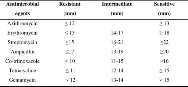
Table 2 Antibiotics with disc concentrations 17 Table 3 Primer sequence with PCR product size and reference 28 Table 4 The zone-size (mm) for Enterobacteriaceae
familyinterpretative table
31 Table 5 The zone-size (mm) for Staphylococcus spp.
interpretative table
32
Table 6 Total viable count of bacteria 33-34
Table 7 Cultural characteristics of E. coli in selective agar 35 Table 8 Cultural characteristics of Salmonella spp. in selective
agar
36
Table 9 Cultural characteristics of S. aureus in selective agar 37 Table 10 Cultural characteristics of S. epidermidis in selective
agar
37 Table 11 Morphology and staining characteristics of different
isolates
38 Table 12 Results of carbohydrate fermentation test 40 Table 13 Results of MR test, V-P test, Oxidase test, Catalase
test, SIM test and Simmons Citrate agar test
47 Table 14 The estimates of evolutionary divergence between
sequences
49 Table 15 Prevalence of E.coli, Salmonella spp., S. aureus and S.
epidermidis
50 Table 16 Sensitivity and resistance pattern of different E. coli
isolates
51
Table 17 Sensitivity and resistance pattern of different Salmonella spp. isolates
52
Table 18 Sensitivity and resistance pattern of different S. aureus isolates
53
Table 19 Sensitivity and resistance pattern of different S.
epidermidis isolates
54
vii
LIST OF FIGURES FIGURE
NO. TITLE PAGE NO.
Figure 1 Layout of the experiment 18
Figure 2 Sample collection from Central Veterinary Hospital (CVH)
19
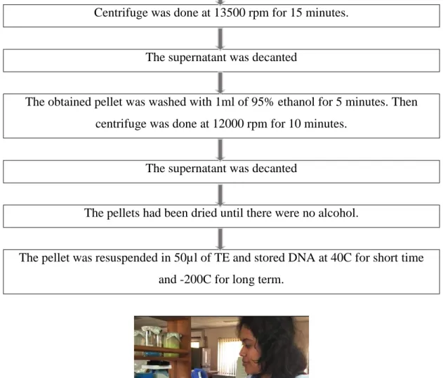
Figure 3 DNA isolation in the laboratory 27
Figure 4 Total Viable count by 10 fold dilution method 34
Figure 5 E.coli in MC agar media 35
Figure 6 E.coli in EMB agar media 35
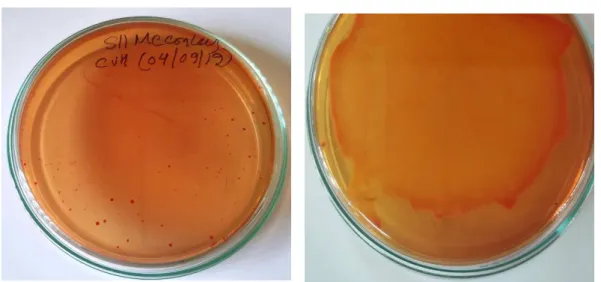
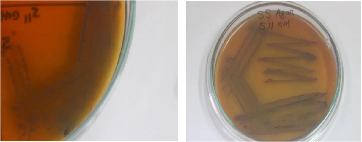
Figure 7 Salmonella spp. in MC agar 36
Figure 8 Salmonella spp. in SS agar 36
Figure 9 S. aureus in MS agar 37
Figure 10 S. epidermidis in MS Agar 37
Figure 11 Gram negative rod shape organism (E.coli) at 100X magnification
38 Figure 12 Gram negative rod shape organism (Salmonella spp.)
at 100X magnification
38 Figure 13 Gram positive circular and cluster organism (S.
aureus)at 100X magnification
39
Figure 14 Gram positive circular clustered organism (S.
epidermidis) at 100X magnification
39
Figure 15 Result of Sugar (Glucose) fermentation test 40 Figure 16 Result of Sugar (Lactose) fermentation test 41 Figure 17 Result of Sugar (Dextrose) fermentation test 41 Figure 18 Result of Sugar (Maltose) fermentation test 42 Figure 19 Result of Sugar (Mannitol) fermentation test 42
Figure 20 Result of MR test 43
Figure 21 Result of V-P test 44
Figure 22 Result of Oxidase test 44
Figure 23 Result of Catalase test 45
Figure 24 Result of SIM test 46
Figure 25 Reaction on SC agar 46
viii FIGURE
NO. TITLE PAGE NO.
Figure 26 Amplification of 584bp DNA from 16S rRNA gene of E. coli
48 Figure 27 Phylogenetic analysis of E. coli 49 Figure 28 Diagram showing the antibiotic sensitivity and
resistance pattern of E.coli
51
Figure 29 Diagram showing the antibiotic sensitivity and resistance pattern of Salmonella spp.
53 Figure 30 Diagram showing the antibiotic sensitivity and
resistance pattern of S. aureus
54 Figure 31 Diagram showing the antibiotic sensitivity and
resistance pattern of S. epidermidis
55
ix
LIST OF APPENDIX NO. OF
APPENDIX TITLE NAME PAGE NO.
APPENDIX 1 Composition of different media 70-71
x
LIST OF ABBREVIATIONS
ABBREVIATION FULL WORD
µg Microgram
µl Microliter
AMP Ampicillin
Approx. Approximately
AZM Azithromycin
CFU Colony Forming Unit
COT Co-trimoxazole
DNA Deoxyribonucleic acid
DX Dextrose
E Erythromycin
E. coli Escherichia coli
EMB Eosin Methylene Blue
ESBL Extended-Spectrum Beta-Lactamase
et al. and others
etc. Etcetra
G Glucose
GEN Gentamicin
H2O2 Hydrogen peroxide
H2S Hydrogen Sulphide
hrs. Hours
IN Intermediate
L Lactose
Lbs Pound
Ltd. Limited
MC MacConkey
MH Muller Hinton
Min. Minute
ML Maltose
Ml Milliliter
MN Mannitol
MS Mannitol Salt
xi
ABBREVIATION FULL WORD
NIB National Institute of Biotechnology
NO. Number
PBS Phosphate Buffered Saline
PCR Polymerase Chain Reaction
R Resistant
S Sensitive/ Streptomycin
S. aureus Staphylococcus aureus
S. epidermidis Staphylococcus epidermidis
SAU Sher-e-Bangla Agricultural University
SC Simmons Citrate
Spp. Species
SS Salmonella-Shigella
TE Tetracycline
TVC Total Viable count
V-P Voges-Proskauer
1
CHAPTER 1 INTRODUCTION
Rearing pet animal has the significance in the history of human civilization. In Bangladesh, people have been keeping cats and dogs as pet animals since early days.
But most of them were not fully indoor animals rather they were more outdoors/stray.
Currently, the tendency of keeping pet as indoor animal is increasing significantly day by day. But local breeds or stray pets are also very common both in urban and rural areas. At present, almost all classes of people have pet. Among all pet animals, cat is the most popular pet in Bangladesh. Now-a-days, people live in nuclear family.
Therefore, the senior citizens and young people mostly pass their time alone. They certainly need continuous and safe company. Hence, in upcoming days the number of cat in home will be increased gradually, and the scenario matches with other countries as well. Owners treat their cats like other family members. Cats supports the owners emotionally, and relives their stress. Owners play with their cats and look after them happily which is indirectly a pleasant source of physical and mental exercise. The owners often kiss their cats, share food and bed with cats in day to day life. People are often aware of fecal and urine contamination from cat, but most of the time the owners are not much conscious of hygiene in case of cat saliva infection. Previous study showed that, cat oral cavity contained a huge number of bacteria, more than 20 types of bacteria. The bacteria did not affect human severely, that means zoonotic importance was comparatively less. On the other hand, only from cat scratch disease approximately 25000 people affected annually in the United States and almost 12000 people die (Jackson et al., 1993). Hence, Cat’s oral swab might be a potential source of infections.
Again, sometimes cat bite may cause mild or serious wound developed on surface of skin. However, delayed presentation may lead to significant morbidity or even mortality. It is recommended that a deep cleaning of the wound and the application of a broad spectrum antibiotic therapy is essential after a cat bite, as animal bites potentially deliver a polymicrobial infection (Talan et al., 1999; Weber et al., 1984;
Wilson and Ho, 2013). The importance of a prompt antibiotic administration must be considered seriously with bacterial contamination. So, identification of the antibiotic sensitivity is essential for choosing the correct antibiotic for effective treatment.
2
Antimicrobial resistance is one of the main causes of failure in antimicrobial therapy.
This mechanism of survival presented by the microorganisms occur naturally or can be acquired. However, acquired resistance is more important due to the fact that it limits viable options of drugs. In recent years, interest in antimicrobial resistance in companion animals has also increased. This is due, in part, to an increasing number of reports of companion animals infected or colonized with clinically and epidemiologically important multiple-drug resistant organisms, such as methicillin- resistant Staphylococcus aureus (MRSA) (Vengust et al., 2006; Moodley et al., 2006).
A comprehensive epidemiological research of antimicrobial resistance in animals should involve the investigation of 3 areas: veterinary pathogens, zoonotic pathogens, and indicator bacteria (Caprioli, 2000). The study of zoonotic pathogens, such as Salmonella spp. and MRSA, in companion animals provides information of special relevance to public health. Companion animals have been a reservoir of Salmonella spp.
for humans through direct contact with pets, contact with feces from pets, preparation of raw meat and bones for pet consumption, and the handling of commercial pet treats (Cherry et al., 2004). Similarly, pets have been colonized and infected with MRSA and have acted as reservoirs of infection for their human contacts (Manian, 2003). The number of multi-drug resistant E. coli are continuously increasing although various antimicrobial agents are being used (Hussain et al., 1982). Uncontrolled use of antibiotics in medicine and animal husbandry for both treatment and prevention of bacterial diseases over the course of decades has fostered the selection of resistant bacteria (Tomasz, 1994 and Singer et al., 2003).
The popularity of cat as pet has already been increased all over the Bangladesh and it will be more popular in upcoming days. Therefore, the chances of spreading infectious diseases from cat will be increased as well. Again, the number of outdoor pet cat is more common than indoor cat in Bangladesh. If the cat is completely indoor since birth, the chances of infection might be less but it is not certain. Therefore, like feces, urine, fur, cat saliva etc. may be a direct or indirect source of human infections. A wider awareness of this problem may be useful in order to prevent life-threatening conditions after cat bite. A study found that, approximately 28% to 80% of cat bites are source of infections and most clinically infected cat bite wounds are mixed infections of aerobic and anaerobic bacteria (John, 2006).
3
Unfortunately, there has been only three studies (two survey and one parasitological) but no microbiological research on cat yet in Bangladesh. Therefore, it is essential to study the bacteria found in cat in Bangladesh. Hence, bacteria identification from oral cavity of pet cat has been chosen for the current study.
Considering the above facts, this study was carried out with the following objectives:
I. To isolate and identify the prevalence of bacterial flora in cat's saliva.
II. To investigate the antibiotic sensitivity patterns of the isolated bacteria.
4 CHAPTER 2
REVIEW OF LITERATURE
Isolation, characterization and antibiotic sensitivity determinati on of the bacteria observed in oral swabs of companion cat was performed using the knowledge gathered from the subsequent related review of literature.
2.1 Cat and Public health relation
Domestic cats (Felis catus) are a common household pet and also a notorious invasive species around the world. Because cat numbers have been increasing in many locations it is critical to work on management solutions that help to reduce threats posed by cats.
With regard to cat behavior, one of the threats both to cats themselves and the species that they interact with is disease transmission. As part of a broader overview on applying cat behavior to management the focus of this review is to consider different types of cat behaviors and highlight how they relate to disease as a means to help inform management. Specifically, a research focused on cat movement, foraging, and cat–
human interactions as broad classes of cat behavior that can lead to acquisition and transmission of diseases. In addition, they review the diseases that are commonly harbored by cats, are of growing human health concern, and for which we have reasonable information. Finally, they review the main forms of cat management in order to provide a set of recommendations for use in addressing cat diseases, such as, many diseases found in cats are zoonotic and of concern to human health; cats engage in behaviors that can lead to disease acquisition and transmission (Lepczyk et al., 2015).
Molecular evidence is restricted for the hypothesis that humans, dogs, and cats can become colonized and infected with similar virulent E. coli strains. To further assess this possibility, archived E. coli O6 isolates (n = 130) from humans (n = 55), dogs (n = 59), and cats (n = 16), representing the three main H (flagellar) types within serogroup O6 (H1, H7, and H31), were analyzed, alongside selected reference strains. Isolates underwent PCR-based phylotyping, multilocus sequence typing, PCR-based detection of 55 virulence-associated genes, and XbaI pulsed- field gel electrophoresis (PFGE) profiling. Three major sequence types (STs), which corresponded closely with H types, accounted for 99% of the 130 O6 isolates. Each ST included human, dog, and cat isolates; two included reference
5
pyelonephritis isolates CFT073 (O6:K2:H1) and 536 (O6:K15:H31). Virulence genotypes overlapped considerably among host species, despite statistically significant differences between human and pet isolates. Several human and dog isolates from ST127 (O6:H31) exhibited identical virulence genotypes and highly similar PFGE profiles, according to cross-species exchange of specific E. coli clones. The close similarity within the genomic backbone and virulence genotype between certain human- and animal-source E. coli isolates within serogroup O6 supports the hypothesis of zoonotic potential (Johnson et al., 2007).
There is no dispute over the role of P. multocida in cat-bite infections of man (Tindall and Harrison, 1972) from which the organism can be isolated in pure culture. The many studies in which modern anaerobic-culture techniques were used have revealed the diversity of human infections that are caused by anaerobes or mixtures of anaerobes (Bartlett and Finegold, 1974; Wren et al., 1977). In such human infections, gram-negative anaerobic bacilli were the most common isolates and B. fragilis was the species most commonly encountered. In cat abscesses it was found that the species isolated most frequently was Pepto- streptococcus anaerobius. This may reflect the predominance of anaerobic gram-positive cocci in the normal flora of the oral cavity. (Love at el, 1990)
Studies on the normal flora of the human oral cavity (Sutter, 1974) have revealed the following anaerobes, listed in order of prevalence: cocci, gram-negative bacilli (Bacteroides spp. and Fusobacterium spp.), gram-positive non-spore forming bacilli and clostridia. The normal flora of the feline mouth has not been described clearly due to insufficient knowledge, but it is assumed that it is likely to bear some resemblance to that of the human mouth.
Bartonella spp. are vector-borne blood-borne pathogens that have mainly been recognized within the last 30 years as a source of human zoonoses, especially for Bartonella henselae, the agent of cat scratch disease (Chomel and Kastel, 2010).
2.2 Cat diseases prevalence in Bangladesh
In Bangladesh there was a research conducted on pet disease where cat was also included. Researchers observed that among 200 pet cats 145 cats were diseased and a total of 5 categories of diseases were recorded. The prevalence of the diseases in one
6
year study period from high to low rates included- Skin Diseases, Salmonellosis, Conjunctivitis, Feline panleukopenia (FPL) and Toxoplasmosis (Runa et al., 2016) Another study was done on 361 cats in Chattogram, Bangladesh. This study reported that the endoparasitic infestation was highly prevalent in cats (91.53%) significantly (p = 0.003), which were ≤1 year of age. Prevalence of wound in cats were substantially higher (p=0.05) in the winter (Hasib et al., 2020).
A period of two months cross sectional prospective study was conducted at Central Veterinary Hospital, Dhaka to estimate the prevalence of clinical conditions in dogs and cats from June to July 2014 where 150 (25%) cats were observed with different clinical conditions. In that study prevalence of clinical conditions was analyzed on the basis of age, sex and breed. It was revealed that 103 (68.67%) cats occupied in medicinal cases followed by surgical cases 24 (16%) in cats and vaccination and health checkup 23 (15.33%) in cats. Among of the medicinal cases special sense organ diseases occupied highest prevalence 25 (16.67%) in cats. Another prevalence of non- infectious diseases in exotic breed and male cats was higher (P ≤ 0.05). These findings address the vaccination practice in cats, variation of management within different topography in Dhaka and socio economic condition of owners (Sarker et al., 2015) 2.3 Bacterial prevalence in cat
A study was carried out to identify the various bacterial species in the oral cavity of cats in two human hospitals in Sokoto, Nigeria. The buccal cavities of 26 cats (14 from Hospital A and 12 from Hospital B were liberally swabbed for bacterial evaluation. The samples were enriched in peptone water, inoculated on McConkey and Blood agar, and incubated aerobically at 37°C for 24hrs. The isolates were Gram stained and subjected to biochemical characterization for identification. A total of 51 bacterial isolates were made. There were Staphylococcus aureus 18 (35.3%), Micrococcus spp. 9 (17.7%), Pasteurella spp. 5 (9.8%), Streptococcus spp. 5 (9.8%), Yersinia spp. 4 (7.8%), Bacillus spp. 4 (7.8%), Listeria spp. 3 (5.4%), and Corynebacterium spp. 3 (5.9%).
Staphylococcus aureus has the highest frequency of isolation 18 (35.3%) (Magaji et al., 2008).
7
In another study 10 swabs were taken from the buccal cavities of some domestic cats and the following bacteria were isolated from cats: Staphylococcus aureus, Bacillus spp., Clostridium spp., Pseudomonas aeruginosa, Pasteurella multocida and Citrobacter spp. (Umaru et al., 2002).
Thirty-six closed abscesses in the subcutis of cats were examined in a study. 168 bacterial strains isolated, 121 (72 %) were anaerobes and 47 (28 %) were facultative anaerobes. Twenty-six abscesses contained mixtures of facultative anaerobes and anaerobes, six contained anaerobes only and four contained facultative anaerobes only. Bacteroides was the genus most commonly isolated (28.6 % of all isolates) followed by Fusobacterium (19.0 %) and P. multocida (13.1 %).
Peptostreptococcus anaerobius was the most commonly isolated anaerobic species (13.2 % of anaerobic isolates and 9.5 % of all isolates) and P. multocida was the most commonly isolated facultative anaerobe (46.8 %; 13.1 % of all isolates) (Love et al., 2006).
Microbiota of periodontally healthy cat were distinguishable from diseased cats. Most of the genera known to be related to periodontitis were also identified in healthy cats, they were present at significantly lower relative abundance.
Remarkably, alpha diversity was found to be higher within the disease groups compared to healthy animals. The complexity of the subgingival microbiota of the house cat and reveal both differences and similarities among periodontally healthy and diseased cats (Rodrigues et al., 2019).
Cat Samples from the gingival margins of 14 cats considered normal on clinical examination were cultured for facultative and obligate anaerobic bacteria. All mouths were free from any gingival marginal inflammation and tartar build -up; all cats were between 6 and 12 months aged. A mixed growth was obtained from all samples. The mean number of bacterial species per sample was 10.7 with a variety of 7–16 isolates. Of the 150 isolates processed, 109 (72.66%) were obligate anaerobes. Of the facultatively anaerobic species, Actinomyces (including A.
viscosus, A. hordeovulneris and A. denticolens) comprised 12%, Pasteurella multocida 9.33% of isolates and Propionibacterium species 6% of all isolates.
Gram-negative bacilli belonging to the genera Bacteroides and Fusobacterium were isolated from 12 of the 14 samples, and comprised 77% of the obligate
8
anaerobes isolated. Clostridium villosum comprised 10.1% of obligately anaerobic isolates, Wolinella species made up 6.42%, while 4.58% were Peptostreptococcus anaerobius. The foremost commonly isolated obligate anaerobic species was C.
villosum and therefore the most ordinarily isolated facultative anaerobic species was P. multocida. These findings show a bacterial flora of the traditional feline mouth which is extremely similar in composition thereto of cat fight abscesses and feline pyothorax (Love et al., 1990).
Any kind of puncture wounds, especially from cats, frequently become infected with various bacterial species. These include Staph. aureus, Staph. intermedius, Strep. pyogenes, Strep. canis, Strep. oralis, Corynebacterium spp., Listeria spp.
and Pasteurella multocida (Barrow and Feltham, 1993).
Usually in research cats are routinely ignored as a possible source of salmonella infection. But over a period of 18 months, 142 cats received from commercial vendors to be used in research were screened for enteric Salmonella. Salmonella was isolated from 15 animals, an incidence of 10.6%. Five (29%) of the 17 shipments contained animals that were positive for Salmonella. The serotypes isolated were Salmonella derby, Salmonella typhimurium, Salmonella anatum, Salmonella enteritidis and Salmonella bredeney (Fox and Beaucage, 1979).
The presence and variety of staphylococcus spp. in healthy domestic cats was wider than in feral or sick cats. In that research it was also observed that cats were carriers of both coagulase-positive (CoPS) and coagulase-negative Staphylococcus species (CoNS) (Gandolfi et al., 2013).
Bartonella species are being recognized as increasingly important bacterial pathogens in veterinary and human medicine. These organisms are often transmitted by an arthropod vector or alternatively by animal scratches or bites. During the period of this study B. henselae was detected in 10.9% of saliva samples (12/110) from pet cats. B. henselae wasn't detected in nail samples of pet cats (n=110), and in any feral cats’ saliva and nail samples (n=30) (Ohad et al., 2010).
9
P. multocida was cultured from the nasopharynx of 94% of normal cats by (Smith, 1964), who considered that it was invariably present in abscesses that occurred as a result of fighting; noted that beta-haemolytic streptococci and anaerobic fusiform bacilli were often present with P. multocida.
2.4 Features of Escherichia coli
Escherichia coli may be a facultative anaerobe that could grow from 7ºC to 50ºC but the optimum temperature is 37ºC, although there are reports of some Enterotoxigenic E. coli (ETEC) strains growing at temperatures as low as 4ºC. E.
coli are often differentiated from other members of the Enterobacteriaceae on the idea of variety of sugar-fermentation and other biochemical tests. Classically a crucial group of tests used for this purpose are known by the acronym IMViC.
These tested for the power to produce: indole from tryptophan (I); sufficient acid to scale back the medium pH below 4.4, the break point of the indicator methyl red (M); acetoin (acetylmethyl carbinol) (V); and therefore the ability to utilise citrate (C) (Adams and Moss, 2008).
Despite E. coli are often identified with a spread of biochemical reactions, the indole test remains the foremost useful method to differentiate lack of production of β-glucuronidase. Sorbitol non fermenting strains of E. coli O157:H7 are related to colitis and hemolytic uremic syndrome (HUS) (Besser et al., 1999).
E. coli is transmitted by ingestion of contaminated food and water, direct contact with animals, feces, contaminated soil and cross contamination directly from one person to a different person. (CDC, 2008 and Denny et al., 2008)
2.5 Features of Salmonella spp.
Salmonella spp. constitutes a major public health burden and represents a significant cost in many countries. Millions of human cases are reported worldwide every year and the disease results in thousands of deaths. Salmonella is a Gram-negative facultative anaerobic rod-shaped bacterium in the family of Enterobacteriaceae, also known as enteric bacteria. Salmonella is a motile bacterium with the exception of S. gallinarum and S. pullorum and they are all non-spore forming. There is a widespread occurrence of Salmonellosis in animals, especially poultry (FDA, 1998).
10
The most commonly used media selective for Salmonella spp. are Salmonella-Shigella (SS) agar, bismuth sulfite agar, Hektoen enteric (HE) medium, brilliant green agar, xyloselysine-deoxycholate (XLD), and MacConkey agar. All these media contain both selective and differential ingredients (Edwards and Ewing, 1972).
2.6 Features of Staphylococcus spp.
The genus Staphylococcus comprises of different species which have been classified and differentiated on the basis of a variety of phenotypic characteristics such as morphology, and biochemical reactions. Pigment was the initial criteri on used to classify staphylococcal species, and in 1885, Rosenbach recognized members of the genus Staphylococcus based on the color of colonies.
Staphylococci forming orange-yellow colonies were named S. aureus by Rosenbach, while staphylococci forming white colonies were named S. albus (Kloos, 1980). Another characteristic feature which was described for differentiation between staphylococci was the coagulase test which involves the investigation of the ability of S. aureus to clot blood plasma (Kloos, 1980) which paved way for the separation of Staphylococci into two main groups Coagulase positive S. aureus (CoPS) and Coagulase negative S. aureus (CoNS). Based on different studies carried by different researchers, at present the genus Staphylococcus comprises of 37 species and 17 subspecies (Kloos and Schleifer, 1975).
S. aureus is a gram-positive, catalase-positive, usually oxidase-negative, facultative anaerobic coccus, which belongs to the family of Micrococcaceae and the group of Staphylococci. Different phenotypic methods are been proposed to identify S. aureus isolates from humans and animals from other species of Staphylococcus. These methods include anaerobic fermentation of mannitol, production of coagulase, production of heat stable thermonuclease and production of acetoin from glucose (Devriese, 1981; Roberson et al., 1992).
2.7 Mechanism and origin of antibiotic resistance
Though the antibiotics were more successful as therapeutics against many bacterial infection in the history of medicine, their irrational and indiscriminate use has created enormous pressure resulting in the development of antibiotic resistance in
11
bacteria (Witte, 1998). Antibiotic resistance can be an intrinsic property of bacteria themselves or it can be acquired later. In natural or intrinsic resistance to a drug occurs without any additional changes in their genetic elements, whereas acquired resistance results through random mutations or acquisition of foreign genetic material carrying resistance determinants (Hollenbeck and Rice, 2012), The antimicrobial agent becomes effective against a target bacterial species only when a susceptible antibiotic target site exists in the cell, the antibiotic reaches the target in sufficient quantity and the antibiotic is not inactivated or modified by the bacterial cell wall (Sutcliffe et al., 1999). So, the unavailability or any change in these conditions trigger the cells to acquire resistance. The mode of acquiring resistance to an antimicrobial drug in bacterial species is categorized broadly into two groups based on biochemical and genetic aspect (Senka et al., 2008).
2.8 Antimicrobial sensitivity pattern of E. coli, Salmonella spp. and Staphylococcus spp.
The prevalence and patterns of antimicrobial susceptibility of fecal E coli, Salmonella spp., extended β-lactamase producing E. coli (ESBL-E. coli), methicillin-resistant Staphylococcus aureus (MRSA), and methicillin-resistant Staphylococcus pseudintermedius (MRSP) were determined for healthy cats (n = 39) from veterinary hospitals in southern Ontario that had not had recent exposure to antimicrobials. The prevalence of antimicrobial resistance in E. coli was as follows: streptomycin (cats — 2%), ampicillin (cats — 4%), cephalothin (cats —
< 1%), and tetracycline (cats — 2%). 15% of cats had isolates that were immune to a minimum of 2 antimicrobials. The observed prevalence of resistance in commensal E. coli from this population was less than that previously reported in companion animals (Murphy et al., 2009).
(Leelaporn et al., 2003) performed antimicrobial susceptibility tests of E. coli isolates in Bangkok, by disc diffusion method. All the isolates were reported susceptible to cefaclor, ceftriaxone, imipenem, netilmicin, norfloxacin, ciptofloxacin, nalidixic acid, and forfomycin. More than 90% of the isolates were susceptible to cefdinir, gentamycin, neomycin and chlorophenicol. Resistance rates to ampicillin, co- trimoxazole and tetracycline were 17, 39, and 65 percent respectively.
12
Shiga-toxin producing E. coli (STEC) isolates (12 of animals, 1 of human and 4 of food samples) from a total of 876 samples (330 of animals, 184 of humans and 362 food samples) were reported uniformly sensitive to common antibiotics, except tetracycline, dicloxicillin, erythromycin, cephalaxin and linomycin (Chattopadhya et al., 2001).
A study determined that the antimicrobial resistance patterns of 138 E. coli isolated from humans in Japan. About 31 isolates showed the resistance to one or more antimicrobial agents. 24 of the isolates were resistant to tetracycline, 23 to streptomycin, 12 to ampicillin, 7 to chloramphenicol and kanamycin, 3 to nalidixic acid, 1 to gentamycin and 1 to cefuroxime (Hiroi et al., 2012).
12 isolates of Entero haemorrhagic E. coli (EHEC) which were isolated from lambs (4), calves (4) and fish (4) in Egypt to determine the frequency of resistance to commonly used antimicrobial agents in veterinary field. Results showed that among the antimicrobial discs tested, ampicillin was the most common antibiotic that the isolates were resistant to (91.6%), followed by tetracycline (83.3%) (Mahmoud et al., 2013).
E. coli from 50 (83.33%) samples and Salmonella spp. from 18 (31.66%) samples by using standard bacteriological techniques. Furthermore, the isolates were subjected to antibiogram studies by disk diffusion method using eight commonly used antibiotics.
Antibiogram studies revealed that gentamycin, ciprofloxacin, and norfloxacin were highly sensitive against all the isolated bacteria, whereas most of the isolates were resistant to amoxicillin, erythromycin, and tetracycline. Out of all the isolates, 5 isolates of E. coli and 3 isolates of Salmonella were found multidrug resistant (Al-Salauddin et al., 2015).
30 samples out of 65, 17 from chicken and 13 from goat were positive for Staphylococci with the prevalence rate of 48.57% from chicken and 43.33% from goat. Staphylococcal isolates were found variably resistant to the antibiotics tested. 80% of the isolates were positive for at least one of the antibiotics used in this study. The isolates showed maximum resistance for penicillin (73.33%) which is followed by erythromycin (36.66%), tetracycline (26.66%), oxacillin (23.33%), ciprofloxacin (16.66%), chloramphenicol (10%), vancomycin (3.33%) (Das et al., 2016).
13
To determine whether cats were a risk for transmission of Salmonella to humans, the researchers evaluated the excretion of Salmonella by pet cats. Rectal-swab specimens were taken from 278 healthy house cats, from 58 cats that died of disease, and from 35 group-housed cats. Group-housed cats were kept in one room with three cat trays and a common water and feed tray. Eighteen (51.4%) of 35 group-housed cats, 5 (8.6%) of 58 diseased cats, and 1 (0.36%) of 278 healthy house cats excreted Salmonella. Salmonella isolates were of serotypes Typhimurium, Enteritidis, Bovismorbificans. Acquired antimicrobial resistance was found in serotype Typhimurium (resistance to ampicillin, chloramphenicol, and tetracycline; to ampicillin; and to chloramphenicol). Cats that excrete Salmonella can pose a public health hazard to people who are highly susceptible to Salmonella, such as children, the elderly, and immunocompromised persons (Van et al., 2004).
14
CHAPTER 3
MATERIALS AND METHODS
3.1 Materials
To conduct the study, a number of materials were used. The list of materials is as follows:
3.1.1 Samples
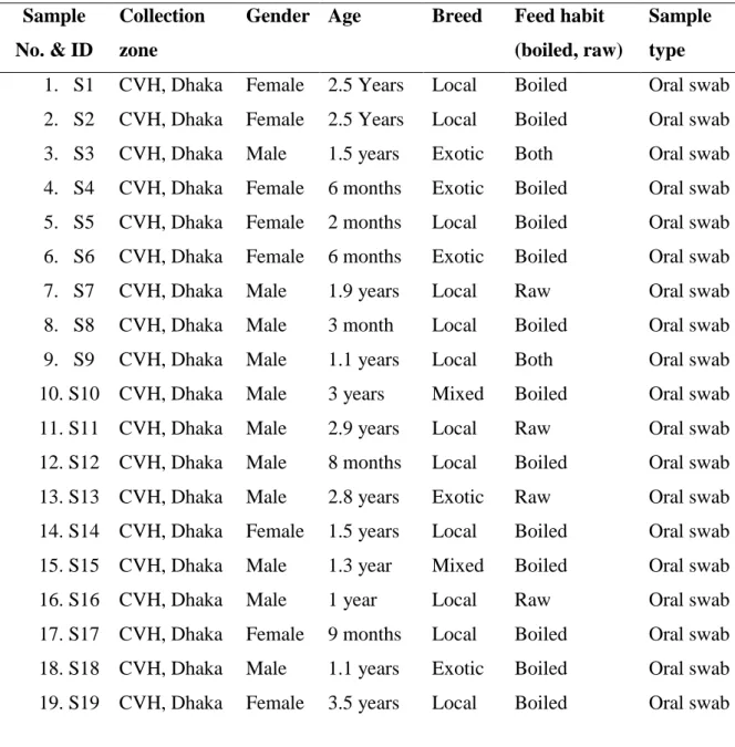
Total 40 oral swab samples were collected from pet cat handled at Central Veterinary Hospital, Dhaka, Bangladesh.
Table 1. Oral swab from pet cat Sample
No. & ID
Collection zone
Gender Age Breed Feed habit (boiled, raw)
Sample type 1. S1 CVH, Dhaka Female 2.5 Years Local Boiled Oral swab 2. S2 CVH, Dhaka Female 2.5 Years Local Boiled Oral swab 3. S3 CVH, Dhaka Male 1.5 years Exotic Both Oral swab 4. S4 CVH, Dhaka Female 6 months Exotic Boiled Oral swab 5. S5 CVH, Dhaka Female 2 months Local Boiled Oral swab 6. S6 CVH, Dhaka Female 6 months Exotic Boiled Oral swab 7. S7 CVH, Dhaka Male 1.9 years Local Raw Oral swab 8. S8 CVH, Dhaka Male 3 month Local Boiled Oral swab 9. S9 CVH, Dhaka Male 1.1 years Local Both Oral swab 10. S10 CVH, Dhaka Male 3 years Mixed Boiled Oral swab 11. S11 CVH, Dhaka Male 2.9 years Local Raw Oral swab 12. S12 CVH, Dhaka Male 8 months Local Boiled Oral swab 13. S13 CVH, Dhaka Male 2.8 years Exotic Raw Oral swab 14. S14 CVH, Dhaka Female 1.5 years Local Boiled Oral swab 15. S15 CVH, Dhaka Male 1.3 year Mixed Boiled Oral swab 16. S16 CVH, Dhaka Male 1 year Local Raw Oral swab 17. S17 CVH, Dhaka Female 9 months Local Boiled Oral swab 18. S18 CVH, Dhaka Male 1.1 years Exotic Boiled Oral swab 19. S19 CVH, Dhaka Female 3.5 years Local Boiled Oral swab
15
20. S20 CVH, Dhaka Female 2 years Local Boiled Oral swab 21. S21 CVH, Dhaka Female 3 months Local Both Oral swab 22. S22 CVH, Dhaka Male 8 months Exotic Boiled Oral swab 23. S23 CVH, Dhaka Male 2 years Local Boiled Oral swab 24. S24 CVH, Dhaka Male 10 months Local Raw Oral swab 25. S25 CVH, Dhaka Female 1.5 years Local Both Oral swab 26. S26 CVH, Dhaka Female 1.7 years Local Both Oral swab 27. S27 CVH, Dhaka Male 1.5 years Exotic Boiled Oral swab 28. S28 CVH, Dhaka Female 1.9 years Mixed Both Oral swab 29. S29 CVH, Dhaka Male 9 months Exotic Boiled Oral swab 30. S30 CVH, Dhaka Male 6 months Exotic Boiled Oral swab 31. S31 CVH, Dhaka Female 1.1 year Exotic Both Oral swab 32. S32 CVH, Dhaka Female 2 years Exotic Boiled Oral swab 33. S33 CVH, Dhaka Female 2 month Local Both Oral swab 34. S34 CVH, Dhaka Male 1.7 years Mixed Both Oral swab 35. S35 CVH, Dhaka Female 2 years Local Raw Oral swab 36. S36 CVH, Dhaka Male 3 years Local Boiled Oral swab 37. S37 CVH, Dhaka Female 1.7 years Local Raw Oral swab 38. S38 CVH, Dhaka Female 1.1 year Local Boiled Oral swab 39. S39 CVH, Dhaka Female 9 months Local Raw Oral swab 40. S40 CVH, Dhaka Male 9 months Exotic Boiled Oral swab
3.1.2 Bacteriological media 3.1.2.1 Agar media
Different agar media were used for bacteriological study, such as, Nutrient agar, MacConkey (MC) agar, Eosin Methylene Blue (EMB) agar, Mannitol Salt (MS) agar, Salmonella-Shigella (SS) agar, SIM test agar media and Simmons Citrate (SC) Agar and Muller Hinton (MH) agar.
16 3.1.2.2 Liquid media
The liquid media used in this study were Nutrient broth, Peptone broth, Methyl-Red and Voges-Proskauer broth (MR-VP broth) and Sugar media (glucose, dextrose, maltose, lactose and mannitol).
3.1.3 Chemicals and reagents
The chemicals and reagents used for this study were 0.1% Peptone water, Phosphate buffered saline (PBS), reagents for Gram’s staining (Crystal Violate, Gram’s iodine, Safranin, Acetone alcohol), 3% Hydrogen peroxide, Phenol red, Methyl red, 10%
Potassium hydroxide, Kovac’s indole reagent (4-dimethylamino-benzaldehyde, concentrated HCL), Mineral oil, Normal saline and other common laboratory chemicals and reagents.
3.1.4 Glass wares and other appliances
The following glass wares and appliances were used during the course of the experiment. Test tubes (with or without Durham’s fermentation tube and stopper), petri dishes, conical flasks, pipettes (1 ml, 2 ml, 5 ml, 10 ml ) & micro-pipettes (10 µl, 100 µl, 200 µl, 1000 µl) slides and cover slips, hanging drop slides, immersion oil, compound microscope, bacteriological loop, sterilized cotton, cotton plug, test tube stand, water bath, bacteriological incubator, refrigerator, sterilizing instruments, thermometer, ice carrier, hand gloves, spirit lamp, match lighter, laminar air flow, hot air oven, centrifuge tubes and machine, PCR machine, thermos scientific nano drop spectrophotometer, UV trans illuminator, Gel documentation machine, electronic balance, syringe and needle, tray, forceps, scalpel, scissors etc.
3.1.5 Antimicrobial discs
Commercially available antimicrobial discs (OXOID Limited, Canada) were used to test the drug sensitivity and resistance pattern. This method is allowed for the rapid detection of drugs efficacy against the test organisms by measuring the diameter of the zone of inhibition.
17
The following antimicrobial agents with their disc concentration were used to test the sensitivity and resistance pattern of the selected E. coli, Salmonella spp., S. aureus and S. epidermidis isolates from oral swab of cat.
Table 2. Antibiotics with disc concentrations
Sl. No. Antimicrobial agents Disc concentration ( µg)
1. Gentamycin (GEN) 10
2. Erythromycin (E) 15
3. Tetracycline (TE) 30
4. Co-Trimoxazole (COT) 25
5. Ampicillin (AMP) 25
6. Streptomycin (S) 10
7. Azithromycin (AZM) 15
3.2 Methods
3.2.1 Brief description of the experimental design
The entire experiment was categorized into two principal steps:
I. The first step included selection of sources, collection of samples, and isolation, identification and characterization of microorganisms on the basis of their colony morphology, motility, biochemical characteristics and molecular identification (only for E. coli)
II. In the second step, the current status of drug sensitivity and resistance pattern of the isolated bacteria were determined.
18
Figure 1: Layout of the experiment Selection of Cat
Collection of sample (oral swab)
Preparation of culture from the swab sample in PBS (1ml)
Total Viable count through 10 fold dilution (1-8)
Multiplication of bacteria in Nutrient Broth by overnight incubation at 370C for 24 hrs.
Isolation of the bacteria on the basis of cultural properties (using EMB, BGA, SS, MC agar media)
Identification of bacteria by Gram’s staining
Biochemical characterization of the isolates using sugar fermentation test (Glucose, Dextrose, Lactose, Maltose, and Mannitol), Indole and MR-VP tests
Molecular identification of E. coli
Antibiotic sensitivity profiling of isolated bacteria against common antibiotics by disc-diffusion method
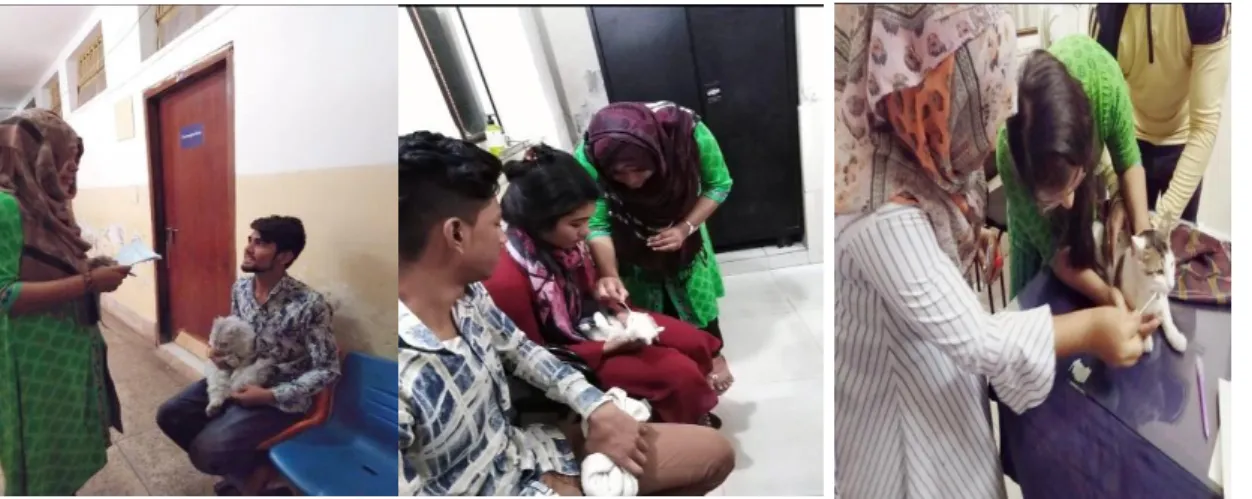
19 3.2.2 Collection and transportation of samples
40 oral swab samples of cats were collected using sterile swab stick in 2 ml Eppendorf tube filled with 1 ml PBS from the cat from Central Veterinary Hospital (CVH), Dhaka.
The collected samples were immediately carried to the laboratory maintaining proper cool chain.
Figure 2: Sample collection from Central Veterinary Hospital (CVH) 3.2.3 Preparation of bacteriological culture media
3.2.3.1 Nutrient broth
Nutrient Broth was prepared by Suspending 25.0 grams in 1000 ml purified/distilled water. Heat was applied to dissolve the medium completely. Sterilizing was done by autoclaving at 15 lbs pressure (121°C) for 30 minutes. The broth was filled in test tubes and incubated at 37ºC for overnight to check their sterility, and stored at 4ºC in the refrigerator until used.
3.2.3.2 Nutrient agar
Nutrient agar was prepared by dissolving 28.0 grams of dehydrated nutrient agar (HiMedia, India) in to 1000 ml distilled water, and was sterilized by autoclaving at 121ºC under 15 lb pressure per square inch for 15 minutes. Then the agar was dispensed into petri dishes, and was incubated overnight at 37ºC to check their sterility, and stored at 4ºC in the refrigerator until used.
20 3.2.3.3 MacConkey agar
49.53 grams of Bacto MacConkey agar (HiMedia, India) was suspended in to 1000 ml distilled water and was heated to dissolve the medium completely. It was then poured in to sterile petri dishes, and was allowed to solidify. After solidification of the media, the plates were then incubated overnight at 37ºC to check their sterility, and stored at 4ºC in the refrigerator until used.
3.2.3.4 Eosine Methylene Blue (EMB) agar
36.0 grams powder of EMB agar base (HiMedia, India) was suspended in 1000 ml of distilled water. The suspension was heated to dissolve the powder completely. The medium was autoclaved for 30 minutes to make it sterile. After autoclaving the medium was put in to water bath at 45ºC. From water bath 10-20 ml of medium was poured in to medium sized sterile petri dishes to make EMB agar plates. After solidification of the medium in the plates, the plates were incubated at 37ºC for overnight to check their sterility, and stored at 4ºC in the refrigerator until used.
3.2.3.5 Salmonella-Shigella agar
60 grams SS agar (HiMedia, India) powder was suspended in 1000 ml distilled water, and heated to dissolve the medium completely. The medium was sterilized by autoclaving. Then the medium was put in to water bath of 45ºC to decrease. After solidification of the medium in the petri dishes, the petri dishes were allowed for incubation at 37ºC for overnight to check their sterility, and then stored at 4ºC in a refrigerator for further use.
3.2.3.6 Mannitol salt (MS) agar
11.1 grams MS agar base (Hi-media, India) powder was suspended in 100 ml of distilled water and heated to dissolve the powder completely. The medium was autoclaved for 30 minutes under 15 lbs pressure. Then the medium was put into water bath maintaining 45°C and poured in to petri dishes to make MS agar plates. After solidifying the medium, the plates were kept in the incubator at 37°C for overnight to check their sterility, and then stored at 4ºC in a refrigerator for further use.
21 3.2.3.7 Mueller Hinton agar
38.0 grams MH agar powder was suspended in 1000 ml distilled water and heated to dissolve the medium completely. After the sterilization by autoclaving at 15 lbs pressure at 121°C for 15 minutes. Cooling was done to 45-50°C.Then it was mixed well and poured into sterile Petri dishes. After solidification of the medium in the petri dishes, the petri dishes were allowed for incubation at 37ºC for overnight to check their sterility and then stored at 4ºC in a refrigerator for future use.
3.2.3.8 Phosphate Buffered Saline (PBS)
To prepare phosphate buffered saline, 8.0 gm of sodium chloride (NaCl), 2.89 gm of disodium hydrogen phosphate (Na2HPO4.12H2O), 0.2 gm of potassium chloride (KCl) and 0.2 gm of potassium hydrogen phosphate (KH2PO4) were suspended in 1000 ml of distilled water. The solution was heated to dissolve completely and pH was adjusted with the help of pH meter. The solution was then sterilized by autoclaving and stored at 4ºC for future use.
3.2.3.9 Simmons Citrate (SC) agar
5.0 gm sodium chloride (NaCl), 2.0 gm sodium citrate (dehydrate), 1.0 gm ammonium dihydrogen phosphate, 1.0 gm dipotassium phosphate, 0.2 gm magnesium sulfate (heptahydrate) were dissolved in 1000 ml distilled water. The pH was adjusted to 6.9. Then agar and bromothymol blue were added. Gently heat with shaking until agar is dissolved. The media were dispensed 5.0 ml into each test tubes. Autoclave at 121°C under 15 lbs pressure for 15 minutes. Cooling in slanted position (slant and butt).
The uninoculated medium will be a deep forest green due to the pH of the sample and the bromothymol blue. During inoculation, the surface of the medium is lightly inoculated by streaking and, where slopes are used, the butt of medium is inoculated by stabbing.
3.2.3.10 SIM (Sulfide, Indole, Motility) media
SIM media was prepared by suspending 36.23 grams in 1000 ml distilled water. Heat to dissolve the media completely. Dispensed in tubes. Sterilized by autoclaving at 15 lbs pressure at 121°C for 15 minutes and stored at 4ºC for future use.
22
3.2.3.11 Methyl Red and Voges–Proskauer (MR-VP) broth
MR-VP broth was prepared by suspending 3.4 grams of MR-VP media (HiMedia, India) in 250 ml of distilled water, distributed in 5.0 ml in each test tube and then autoclaved. After autoclaving, incubated overnight at 37ºC to check their sterility, and then stored at 4ºC for future use.
3.2.3.12 Sugar solutions
To prepare the media, fermentable sugars were added with 1% peptone water. Peptone water was prepared by adding 1.0 gram of Bacto peptone (Difco, USA) and 0.5 grams of sodium chloride in 100 ml distilled water, boiled for 5 minutes, adjusted to pH 7.6 by phenol red (0.02%) indicator, cooled and then filtered through filter paper. The solutions were then dispensed in 5.0 ml amount into cotton plugged test tubes containing invertedly placed Durham’s fermentation tubes. Then the sugars, glucose, dextrose, maltose, lactose, and mannitol for the fermentation were prepared separately as 10 percent solutions in distilled water. A little heat was necessary to dissolve the sugar. These were then sterilized by autoclaving for 15 minutes. The sugar solutions were sterilized in Arnold’s steam sterilizer at 100ºC for 30 minutes for three consecutive days. An amount of 5.0 ml of sterile sugar solution was added aseptically in each culture tubes containing sterile peptone water. The sugar solutions were incubated at 37ºC for 24 hours to check sterility.
3.2.4 Isolation of bacteria 3.2.4.1 Preparation of sample
The raw samples were kept in 1.0 ml PBS, further, any chemical was not used/added for sample preparation.
3.2.4.2 Serial dilution for bacterial culture (10 fold dilution method)
Serial dilution of the stock sample was done for reduction the bacterial concentration for easy total viable count (TVC). This process was conveyed by taking 8 (1-8) Eppendorf tube filled with 450µl of PBS. 50µl of stock sample was transferred from the stock tube (1ml) to the Eppendorf tube next to the stock tube. Then 50µl of diluted sample is transferred from the first Eppendorf tube to the next. Successive dilution
23
should be made in the same way to the last tube and from the last tube 50µl of diluted sample was discarded. From all the tube, 25µl of liquid sample was poured to the nutrient agar media by spreading technique for the total viable count.
3.2.4.3 Primary culture of microorganism
Primary growth of all kinds of bacteria present in the collected samples was performed in nutrient broth. The samples were inoculated in to nutrient broth, and incubated overnight at 37ºC for the growth of the organisms.
3.2.4.4 Isolation in culture media
After primary culture of the organism, a small amount of inoculums from Nutrient broth was streaked on the MacConkey, Salmonella-Shigella and Mannitol Salt agar to observe the colony morphology of the isolates. Characteristic colony morphology of the organisms selected for subculture on selective media, such as, Eosine Methylene Blue agar and MacConkey agar for E. coli, Salmonella-Shigella agar and MacConkey agar for Salmonella spp., and Mannitol Salt agar for S. aureus and S. epidermidis.
Morphological characteristics (shape, size, surface texture, edge and elevation, color, opacity etc.) of the suspected colonies on different agar media were carefully recorded.
3.2.5 Microscopic identification of the suspected colonies by Gram’s staining method
Gram’s staining of the pure culture was performed according to method described by (Cheesbrough, 2006). Briefly, a single colony was picked up with a bacteriological loop, smeared on a glass slide and fixed by gentle heating. Crystal violate was then applied onto smear to stain for two minutes and then washed with running tap water.
Few drops of Gram’s iodine were then added for few seconds. After washing with water, Safranin was added as counter stain and allowed to stain for 2 minutes. The slides were then washed with water, blotted and dried in air and then examined under light microscope (100X) using immersion oil
3.2.6 Identification of isolates by different biochemical tests
Several biochemical tests were performed for confirmation of E. coli, Salmonella spp., S. aureus and S. epidermidis.
24 3.2.6.1 Carbohydrate fermentation test
The carbohydrate fermentation test was performed by inoculating 0.2 ml of nutrient broth culture of the isolated organisms into the tubes containing different sugar media (five basic sugars such as glucose, dextrose, maltose, lactose, and mannitol) and incubated for 24 hours at 37ºC. Acid production was indicated by the color change from red to yellow, and gas production was noted by the accumulation of gas bubbles in the inverted Durham’s tube (Cheesbrough, 2006).
3.2.6.2 Methyl Red test
The test was conducted by inoculating single colony from the pure culture of the test organism in 5 ml sterile MR-VP broth. After 5 days incubation at 37ºC, 5 drops of methyl red solution was added and observed for color formation. Development of red color was positive and indicated an acid pH of 4.5-6 resulting from the fermentation of glucose. Development of yellow color indicated negative result (Cheesbrough, 2006).
3.2.6.3 Voges-Proskauer (V-P) test
The test E. coli organisms were grown in 3 ml of sterile MR-VP broth at 37ºC for 48 hours. Then 0.6 ml of 5% alpha-napthol and 0.2 ml of 40% potassium hydroxide containing 0.3% creatine was added per ml of broth culture of the test organism. Then shaking well and allowed to stand for 5-10 minutes to observe the color formation.
Positive case was indicated by the development of a bright orange red color. In negative cases, there was no development of pink color (Cheesbrough, 2006).
3.2.6.4 Oxidase test
Moisten the paper with a sterile distilled water. Pick the colony to be tested with wooden or platinum loop and smear in the filter paper. Observe inoculated area of paper for a color change to deep blue or purple within 10-30 seconds.
3.2.6.5 Catalase test
For this study 3.0 ml of catalase reagent (3% H2O2) was taken in a test tube. Single colony from the pure culture of the organisms was taken with a glass rod and merged in the reagent. The glass slide was observed for bubble formation. Formation of bubble