i
A Look into Parkinson’s Disease and the Implications of Gene Therapy as A Novel Clinical Approach
By
Md. Rakibul Islam 16146056
A thesis submitted to the School of Pharmacy in partial fulfillment of the requirements for the degree of
Bachelor of Pharmacy (Hons.)
School of Pharmacy Brac University
February 2022
© 2022. Brac University All rights reserved.
Declaration
It is hereby declared that
1. The thesis submitted is my own original work while completing degree at Brac University.
2. The thesis does not contain material previously published or written by a third party, except where this is appropriately cited through full and accurate referencing.
3. The thesis does not contain material which has been accepted, or submitted, for any other degree or diploma at a university or other institution.
4. I have acknowledged all main sources of help.
Student’s Full Name & Signature:
Md. Rakibul Islam 16146056
iii
Approval
The project titled “A Look into Parkinson’s Disease and the Implications of Gene Therapy as A Novel Clinical Approach” submitted by Md. Rakibul Islam (16146056) of Summer, 2021 has been accepted as satisfactory in partial fulfillment of the requirement for the degree of Bachelor of Pharmacy (Hons.) on February 28, 2022.
Examining Committee:
Supervisor:
(Member)
_______________________________
Ms. Marzia Alam Lecturer School of Pharmacy
Brac University
Program Coordinator:
(Member)
_______________________________
Namara Mariam Chowdhury Lecturer
School of Pharmacy Brac University
Deputy Chairperson:
(Member)
_______________________________
Professor Dr. Hasina Yasmin Deputy Chairperson School of Pharmacy
Brac University
Departmental Head:
(Dean)
_______________________________
Professor Dr. Eva Rahman Kabir Dean
School of Pharmacy Brac University
Ethics Statement
The study does not involve any kind of animal or human trial.
v
Abstract
Parkinson’s Disease (PD) is considered as one of the highest occurring neurodegenerative disorders. Motor complexities are the major symptoms of this disease which mainly includes bradykinesia, rigidity and tremor. Moreover, the non-motor symptoms can be included as mental and psychological complexities. Injury to the dopamine pathways is considered as the primary pathological reason of PD. Although the older population represents the majority of the patients, 5- 10% of the patients are relatively younger. Currently, dopamine replacement therapy with levodopa is the most common mode of treatment for alleviating the symptoms and retaining the functional capabilities. Here, gene therapy can be a viable treatment approach for such neurodegenerative disorders since it serves by knocking down the pathological genes. Even though few questions are yet to be answered before incorporating gene therapy in Parkinson’s disease, this novel therapeutic method has the potential to reshape the future of neurologic therapeutics.
Keywords: Parkinson’s Disease; Neurodegenerative Disorder; Levodopa; Gene Therapy;
Dopamine Replacement Therapy.
Dedication
Dedicated to my family and my respected supervisor
vii
Acknowledgement
To begin with, I am grateful to Allah Subhanahu Wa Ta'ala who has bestowed countless blessings, good health, immense patience, courage and strength upon me to complete my undergraduate project.
I am indebted to my family for their unconditional love and support throughout my entire life.
I am forever thankful to my younger brother Md. Rashedul Islam who has always been there to motivate me during the most difficult phases of my life.
I am thankful to my respected supervisor, Ms. Marzia Alam, Lecturer, School of Pharmacy, Brac University, for her continuous support and kind suggestions to guide me to complete my project. Without her directions and helpful criticism, it would have been impossible for me to finish my project work.
It has been a real honor to work under the guidance of Prof. Dr. Eva Rahman Kabir, Chairperson, School of Pharmacy, Brac University. I am also thankful to the School of Pharmacy, Brac University for giving me the opportunity of completing my project work as well as the B. Pharm program.
Last but not the least, I would like to express my gratitude to my fellow project attendees- Tanzila Haque, Madhurza Mitra Mazumder and Ankita Islam who were always very helpful to me in resolving all the project related queries.
Table of Contents
Declaration ... ii
Approval ... iii
Ethics Statement ... iv
Abstract ... v
Dedication ... vi
Acknowledgement ... vii
Table of Contents ... viii
List of Tables ... xi
List of Figures... xii
List of Acronyms ... xiii
Chapter 1 Introduction ... 1
1.1 Global Scenario of Parkinson’s Disease ... 2
1.2 Epidemiology of Parkinson’s Disease ... 3
1.3 Treatment Strategies ... 5
1.4 Impact of Racial Identity ... 6
Chapter 2 Methodology ... 7
Chapter 3 Pathogenesis of Parkinson’s Disease ... 8
3.1 Impact of Aging ... 8
3.2 Impact of Genetic Susceptibility ... 9
3.2.1 Linked Gene 01 (PARK1) ... 10
ix
3.2.2 Linked Gene 02 (PARK2) ... 11
3.2.3 Linked Gene 03 (PARK5) ... 11
3.2.4 Linked Gene 04 ((PARK7)... 11
3.2.5 Linked Gene 05 (PARK 6) ... 12
3.3 Impact of Environmental Vulnerability ... 13
Chapter 4 Clinical Features and Diagnosis of Parkinson’s Disease ... 14
4.1 Clinical Features ... 14
4.2 Diagnosis of Parkinson’s Disease... 16
Chapter 5 Contemporary Treatment Methods for Parkinson’s Disease ... 22
5.1 Treatment for Neuroprotection ... 23
5.2 Symptomatic Treatment of Motor Symptoms ... 25
5.3 Symptomatic Treatment of Non-Motor Symptoms ... 28
Chapter 6 Gene Therapy in Parkinson’s Disease ... 31
6.1 Standard Procedures of Gene Therapy ... 31
6.2 Vectors Used in Gene Therapy... 32
6.3 Gene Therapy through Plasmid Transfection ... 33
6.4 Major Breakthroughs of Gene Therapy for Parkinson’s Disease ... 34
6.5 Site of Action for the Common Strategies of Gene Therapies ... 35
6.6 Routes of action of the prospective treatment candidates ... 36
6.7 Delivery Routes of Gene Therapy into the Brain and Spinal Cord ... 37
6.8 Gene Therapy Method 1: Improving the synthesis of dopamine ... 38
6.9 Gene Therapy Method 2: Trophic factor delivery ... 39
6.10 Gene Therapy Method 3: Gene therapy for neuromodulation ... 40
Chapter 7 Future Prospects of Gene Therapy in Parkinson’s Disease ... 43
Chapter 8 Conclusion... 45
References... 46
xi
List of Tables
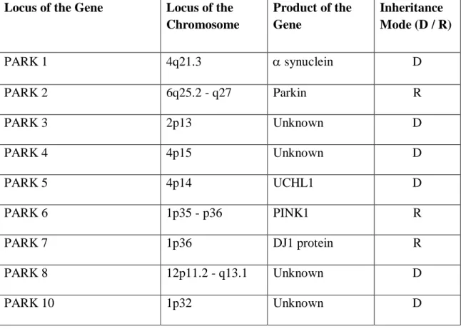
Table 1: Linked Genes in Familial form of Parkinson’s Disease ... 10
Table 2: The Diagnostic Criteria of Parkinson’s Disease by Queen Square Brain Bank ... 18
Table 3: Comparison between the major classes of drugs used in Parkinson Disease ... 22
Table 4: Delivery Routes of Gene Therapy into the central nervous system ... 37
List of Figures
Figure 1: The Regions of the Brain Associated with Parkinson's Disease... 1
Figure 2: Breakdown of Abnormal Proteins by the Ubiquitin-Proteasome Pathway ... 12 Figure 3: Illustration of No Facial Expression and Rigid Posture of a Patient with Parkinson’s Disease... 14 Figure 4: Major breakthroughs associated with gene therapy ... 34 Figure 5: Approximate site of action for the common strategies of gene therapies ... 35 Figure 6: Routes of action of the prospective treatment candidates currently going through clinical trials ... 36
xiii
List of Acronyms
AAV Adeno-Associated Virus
AADC Aromatic Amino Acid Decarboxylase
AON Antisense Oligonucleotides
CAR T-Cell Chimeric Antigen Receptor T Cell
CNS Central Nervous System
DBS Deep Brain Stimulation
DLB Dementia with Lewy Bodies
F- DOPA Fluoro-3,4-Dihydroxyphenylalnine
GAD Glutamate Decarboxylase
GBA P‐Glucocerebrosidase (Gcase) Gene
GCH1 GTP Cyclohydrolase-1
GDNF Glial Cell-Line Derived Neurotrophic Factor
GPe External Globus Pallidus
GPi Internal Globus Pallidus
HUGO Human Genome Organization
kB Kilo-Base (1000 base pairs of DNA or RNA) LRRK 2 Leucine Rich Repeat Kinase 2
LV Lentivirus
MRI Magnetic Resonance Imaging
PINK1 PTEN-Induced Kinase 1
PET Study Positron Emission Tomography Test SNc Substantia Nigra Pars Compacta
SNCA Synuclein Alpha
SPECT Study Single-Photon Emission Computerized Tomography Test
STN Subthalamic Nucleus
TH Tyrosine Hydroxylase
UCHL1 Ubiquitin C-terminal Hydrolase L1
vGAT Vesicular GABA-Transporter
1
Chapter 1 Introduction
Parkinson’s disease (PD) is often considered as the second most prevalent neurodegenerative disorder that affects the central nervous system. In terms of movement disorder, it could easily be considered as the most common neurodegenerative disorder. James Parkinson was the first person to explain this disorder in his famous thesis of ‘‘Essay on the shaking palsy’’ in the year of 1817. He also explained the major signs of the disease that involves the motor symptoms which is considered as the prime attribute of Parkinson’s disease as well as bradykinesia, rigidity and tremor. Later, the disorder was denoted after his name. Moreover, he also described the mental symptoms of Parkinson’s disease. (Tysnes & Storstein, 2017)
Figure 1: The Regions of the Brain Associated with Parkinson's Disease (Adapted from (Mandal, 2019))
According to Tysnes & Storstein (2017) the disorder is distinguished in terms of its neuropathology by the incorporation of Lewy bodies that contains alpha ()- synuclein within the substantia nigra pars compacta. It leads to a depletion of the progression of voluntary movements through the reduction of dopaminergic neurons in the substantia nigra's pars compacta. Moreover, the progression of Parkinson’s disease involves an extensive increase of
- synuclein. Furthermore, the non-motor symptoms involved in this disorder have also been brought to light for the past two decades. However, it is commonly marked as a movement disorder since the motor signs which are considered as the basic indication of this disease are more dominant than the non-motor symptoms. (Tysnes & Storstein, 2017)
1.1 Global Scenario of Parkinson’s Disease
A significant portion of the elderly population is affected by movement disorders. More than one million people in the United States are affected by idiopathic Parkinson’s disease which makes about 1% of the total older population (older than 60 yrs.) is likely to develop this disease. (Sudhakar & Richardson, 2018)
Parkinson’s disease is also considered as the second most common movement disorder that occurs due to neurodegenerative conditions. It has been estimated that this disorder has affected around 5.8 million people around the globe. Moreover, the disease presents itself with the increasing mean age of western world population with a projection of its disease prevalence to rise. (Axelsen & Woldbye, 2018). Moreover, the current population of patients affected by Parkinson’s disease is expected to double by the year 2040 which makes it the fastest growing neurodegenerative disease. (Polissidis et al., 2020)
In terms of the western population, they are aging quite fast due to a low birth rate in those parts of the world. As a result, the current frequency of Parkinson’s disease which is estimated to be 1 in 800 is anticipated to increase. Moreover, some evidence strongly suggests that
3
women are 1.5 times less expected to develop this disease. Nevertheless, this difference in gender associated disease prevalence is mostly restricted to the western population (≥ 70 years) and it is not found to be consistent across different studies. While the most frequent cause of death is pneumonia, it is quite difficult to correctly identify the mode of death in most of the cases. (Lees et al., 2009).
1.2 Epidemiology of Parkinson’s Disease
The frequency of Parkinson’s disease increases steeply with age. It shows a frequency of 17·4 in 100,000 people for an age group of 50 to 59 years while 93·1 in 100,000 people for an age group of 70 to 79 years. While the risk of developing Parkinson’s disease is around 1·5%, the mortality rate is 2 to 1. Moreover, the onset of this disease presents itself with a median age of 60 years and the average duration of disease exposure (from diagnosis to death) is around 15 years. (Lees et al., 2009).
A detailed study of the evaluation of Parkinson’s disease concerning the oldest age-group of patients has been reported by the NYPUM study conducted by the Umeå University of Sweden.
The NYPUM study included the annual incidence rate of the disease to be 18.8 cases per 100,000 of people while it found the average age of the initial occurrence of the disease to be at 70.6 years. The study also found the early-onset of the disease to be very infrequent in population-based studies which accounts only for the 4% of the affected population that exhibited the sign of Parkinson’s disease before the age of 50. (Tysnes & Storstein, 2017) The young population that is affected by Parkinson’s disease accounts for 5-10% of the patients. (Samii et al., 2004). According to Tysnes & Storstein (2017), among the 265 known patients, only 3% were younger than 50 years of age. They typically exhibit the initial symptoms between the age of 21 and 40. In terms of the juvenile population, initial symptoms might be visible before the age of 20 years. The disease shows no variability in terms of
people’s racial and ethnic identity. One of the studies indicated the annual occurrence of Parkinson’s disease to be somewhat around 13 cases per 100,000. (Samii et al., 2004).
The available study reports on the epidemiology of Parkinson’s disease shares methodological differences which makes it very difficult to make a direct comparison between them to make an estimation of the disease prevalence. And these study reports vary significantly concerning the incidence of the disease. Surely, the main difference lies in their methodology but the ascertainment of the disease along with the incorporation of the diagnostic criteria also play a key role. Due to this, some study reports suggest the annual incidence rate to be less than 100 while other studies suggest it to be more than 20 per 100,000 people. One aspect that might be considered as the underlying reason behind the variation in annual incidence rate is the under- diagnosing of Parkinson’s disease among the most elderly data set of the population. (Tysnes
& Storstein, 2017).
Therefore, a key question can be raised in that sense is whether there has been some definite increase in the Parkinson’s disease’s prevalence and incidence rate. According to Tysnes &
Storstein (2017), an increased incidence of neurodegenerative disorders such as Parkinson’s disease and Amyotrophic Lateral Sclerosis (ALS) was reported in Norway during the year of 2000. However, such an increase could be a result of the overall increase of the geriatric population. On the other hand, several recent epidemiology data indicates an actual increase of this disease, especially in male population. The additional risk of Parkinson’s disease in male population was assumed to be related with the substantial changes in men’s day to day habit (e.g., smoking) over the last few decades of twentieth century. Also, the severe air pollution due to the increased amount of vehicle emission over the past few decades could also be associated to the overall increase of Parkinson’s disease’s occurrence. (Tysnes & Storstein, 2017).
5
1.3 Treatment Strategies
The treatment strategies of this neurodegenerative disorder have made some key advancement over the last few decades. However, there are few limitations of the current treatment strategy as they mostly focus on alleviating the major symptoms but do not improve the original causative disease pathology. For example, dopaminergic drugs (e.g., Levodopa) are very common to control the symptoms over a long period of time. However, such repeated use of dopaminergic drugs results in a reduced therapeutic effect over time and adverse effects as well while the underlying pathology of the disease is not treated at all. In recent days, an alternative treatment strategy for this disorder emerged since the incorporation of AAV (Adeno- Associated Virus) vectors allowed the exploration of gene therapy for Parkinson’s disease.
(Niethammer et al., 2018)
Here, gene therapy typically involves the transfer of Antisense Oligonucleotides, DNA, RNA, or Enzymes that can modify such nucleic acids. Here, these substances are carried into definite brain areas so that the expression of one or more specific genes can be controlled. Many recent studies based on this new treatment strategy are conducted in patients with this disorder that presented promising results in terms of providing neurotrophic assistance, restoring the synthesis of dopamine or coordinating different functional nodes of the brain which would allow a balanced communication among these nodes. (Merola et al., 2020)
Moreover, the recent advancement in the field of molecular biology and virology allowed the idea of gene transfer to proceed forward in a more efficient and feasible way. In this case, gene therapy is a viable approach for PD since it involves brain’s pathophysiology which is extensively outlined and primarily dominated by the nigrostriatal dopaminergic neurons. Also, several genes have been identified in recent years that play key roles in the genetic mutations which are associated with the inherited type of Parkinson’s disease. (Mochizuki, 2007)
1.4 Impact of Racial Identity
It is not quite often when Parkinson’s disease is linked with someone’s racial identity.
Moreover, writings and many historical instances of previous generations establish this disorder as unlikely to be a mere postindustrial phenomenon. Although the causative reasons behind PD were unknown for many decades from 1817, genetic and pathological links have recently been found which unveils many whole new aspects of it. (Lees et al., 2009).
7
Chapter 2 Methodology
An extensive literature review was conducted on Parkinson’s disease along with gene therapy for being a viable treatment option for Parkinson’s disease. Here, secondary research methods were incorporated which mainly involved research articles. Articles from renowned journals and websites such as, Nature, The Lancet, PubMed, MDPI and Elsevier were analyzed in search of relevant information regarding the possible implications of gene therapy in Parkinson’s disease. These sources of relevant evidence and statistics allowed the compilation of useful information within the boundary of this project.
Additionally, suitable key terms were utilized to gather the research articles, such as Parkinson’s disease, Gene Therapy, Neurodegenerative Diseases, Substantia Nigra, and so on.
In this case, almost 100 articles have been initially sorted and among them 17 articles were analyzed in a conscientious manner to develop this review paper. Moreover, the Mendeley software was employed as the reference manager to uphold the credit of the work of the original authors.
Chapter 3
Pathogenesis of Parkinson’s Disease
The primary pathological reason in the development of this disorder is considered as the injury to the dopamine pathways from the pars compacta of the substantia nigra located in the midbrain to the caudate nucleus and putamen or striatum. The Lewy neurites and the intraneuronal lewy bodies are considered as the hallmark of Parkinson’s disease. Here, the loss of 80% of the striatal dopamine and 50% of the nigral neurons marks the evident presence of the clinical signs associated with the disease. In this case, the lewy bodies are not only limited within the substantia nigra but also found in the amygdala, locus ceruleus, vagal nucleus, cortex and the peripheral nervous system. Therefore, some of the non-motor symptoms could be associated with the presence of lewy bodies and neurites in those non-motor regions of the brain. (Samii et al., 2004).
Apart from some specific patients who shares a known genetic mutation or Methyl-Phenyl- Tetrahydropyridine (MPTP) exposure, the cause of this neurodegenerative disorder is still considered as to be unknown since it could probably be a consequence of the combination of several factors such as aging, environmental exposures and genetic susceptibility. (Samii et al., 2004). Also, some physiological conditions might be responsible for being the secondary causes of this disease which can be included as encephalitis, hypertension, cerebrovascular disease or severe head injury. (Lees et al., 2009).
3.1 Impact of Aging
Aging is one of the most common factors of the major neurodegenerative diseases as well as Parkinson’s disease. Regarding the pathology involved with aging it is found to be linked with the depigmentation of neurons present in the pars compacta of the substantia nigra. Moreover, up to 16% of the asymptomatic elderly people exhibited incidental Lewy bodies during the
9
autopsy. Also, a minute age-dependent decrease of F-DOPA (Fluoro-Dihydroxyphenylalnine) uptake has been found through the F-DOPA PET (Positron Emission Tomography) studies.
Similarly, age-dependent decrease of striatal dopamine transporters has been found through the SPECT (Single-Photon Emission Computerized Tomography) study. However, the SPECT study didn’t exhibit any difference in-between caudate and putamen which diverges from the pattern. It should be noted that the occurrence of this disorder might increase with age but the mere facilitation of aging doesn’t set the disease in motion. (Samii et al., 2004).
3.2 Impact of Genetic Susceptibility
Most of the patients with this neurodegenerative condition do not exhibit familial traits. Around 15% of the patients who possess first-degree relatives with Parkinson’s disease, do not exhibit any distinctive mode of inheritance. So far, nine loci of genes are found to be related with both the autosomal form of dominant and recessive parkinsonism. Nevertheless, familial patterns are accounted for due to exposures of the general environment. (Samii et al., 2004).
In terms of the monozygotic twins, extensive concordance was not found in most of the studies.
However, the PET study reports suggest a greater rate of concordance compared to the clinical methods. Surprisingly, these monozygotic twin studies showed broad concordance for the initiation of the disease before the mean age of onset (50 years) while it exhibited subtle concordance when the disease onset is after 50 years of age. Here, such finding can be perceived as a significant link between the genetic susceptibility and the early onset of Parkinson’s disease. (Samii et al., 2004).
Furthermore, the genetic contribution in Parkinson’s disease is brought to light through the identification of five genes and four different gene loci (Table 1) in Parkinson’s disease that involve familial traits. In this case, anomalies within a single gene account for a very few numbers of cases while the identification of those five genes and four different gene loci
allowed a more accurate understanding of the possible manifestation of neurodegeneration in familial as well as the sporadic form of Parkinson’s disease. (Samii et al., 2004).
Table 1: Linked Genes in Familial form of Parkinson’s Disease (Adapted from (Samii et al., 2004))
3.2.1 Linked Gene 01 (PARK1)
It is named as Synuclein Alpha (SNCA) by the Human Genome Organization (HUGO). It was recorded that the coding for the protein of synuclein exists within large American families of Italian Origin. Here, the synuclein gene exists with a missense mutation. Similarly, this situation was also recorded in some other families of Italian and Greek origin which could be indicative of the founder effect. In addition to the first mutation, a small German Pedigree exhibited a second mutation within the synuclein gene. Although the function of the protein of synuclein is not known it is considered as one of the most vital components of the Lewy
Locus of the Gene Locus of the
Chromosome
Product of the Gene
Inheritance Mode (D / R)
PARK 1 4q21.3 synuclein D
PARK 2 6q25.2 - q27 Parkin R
PARK 3 2p13 Unknown D
PARK 4 4p15 Unknown D
PARK 5 4p14 UCHL1 D
PARK 6 1p35 - p36 PINK1 R
PARK 7 1p36 DJ1 protein R
PARK 8 12p11.2 - q13.1 Unknown D
PARK 10 1p32 Unknown D
Dominant – D; Recessive – R
11
bodies. The name of synuclein protein suggests that it is found in the synaptic terminals as well as the nuclei. (Samii et al., 2004).
3.2.2 Linked Gene 02 (PARK2)
It is associated with the early onset of Parkinson’s disease (juvenile) which is autosomal recessive in nature. The cases that involve early onset of Parkinson’s disease, nearly half of them are caused due to parkin mutations of different kinds in autosomal recessive forms of this disorder. The likelihood of this disease further increases due to the occurrence of heterozygous mutations in the protein of parkin. Moreover, these mutations result in the loss of cells in the substantia nigra pars compacta as well as the locus coeruleus without the presence of Lewy bodies. In this regard, the hypothesis that considers the omission of ubiquitin-proteasome system as the common idiosyncrasy for the pathogenesis of this neurodegenerative disorder becomes relevant since parkin is detected as a ubiquitin-protein ligase that ubiquitinates synuclein. (Samii et al., 2004).
3.2.3 Linked Gene 03 (PARK5)
It is named UCHL1 by the HUGO which codes for the deubiquitinating enzyme of Ubiquitin Carboxyl-Terminal Hydrolase L1 which affects the proteasomal degradation through the marking of abnormal proteins. Such markings involve the breakdown of the bond between the ubiquitin molecules. The only occurrence of this mutation was first identified in two German siblings which marks the impact of this mutation somewhat to be provocative. (Samii et al., 2004).
3.2.4 Linked Gene 04 (PARK7)
The PARK7 gene is associated with the early onset (juvenile) which is autosomal recessive in nature. It plays an important role by balancing the oxidative stress by coding for the protein of DJ1. Here, the PARK7 gene is positioned around the same region as the PARK6 gene on
chromosome 1p36. However, the positions are clearly distinctive since no overlap can be observed between their markers. (Samii et al., 2004).
3.2.5 Linked Gene 05 (PARK 6)
The PARK6 gene is associated with the mutations involved in PTEN Induced Kinase 1 (PINK1) which is found within the mitochondria. Here, PTEN stands for Phosphatase and Tensin Homolog which is deleted on chromosome ten. (Samii et al., 2004).
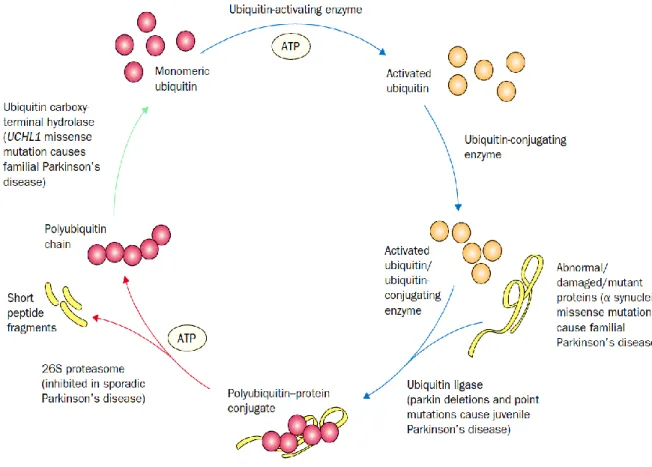
Figure 2 depicts the pathway of identifying and labelling the abnormal, damaged or mutated proteins (blue arrows) through the attachment of ubiquitin molecules. The process is known as ubiquitination and it works as the signaling for the ATP dependent degradation. The process is indicated with red arrows in Figure 2 which is conducted by the 26S proteasome complex.
Figure 2: Breakdown of Abnormal Proteins by the Ubiquitin-Proteasome Pathway (Adapted from (Samii et al., 2004))
13
Furthermore, the green arrow depicts the process of deubiquitination which allows the recovering of the ubiquitin from the polyubiquitin chain. However, failure in such a mechanism might result in parkinsonism. (Samii et al., 2004).
3.3 Impact of Environmental Vulnerability
A study published in 1983 suggested that patients might develop severe levodopa-responsive parkinsonism after being exposed to a substance called Methyl-Phenyl-Tetrahydropyridine (MPTP). It is because the substance can cross the blood-brain barrier easily and then gets converted into MPP+ through astrocytes. The positively charged ion works as a neurotoxin within the central nervous system. Moreover, these ions are attached to the dopaminergic cells which inhibits the mitochondrial complex 1 within the respiratory chain. The MPTP is considered as the prime substance from the surroundings to develop such levodopa-responsive parkinsonism. (Samii et al., 2004).
On the other hand, some substances from the environment exhibit preventative properties against Parkinson’s disease. One of such substances can be included as carbon monoxide and it might be associated with cigarette smoking which makes cigarette smoking inversely related with the possibility of developing this disorder. It is assumed that carbon monoxide along with the other constituents of cigarettes provides some sort of neuro-protective effect. Here, carbon monoxide plays a probable role of destroying the free radicals. Moreover, caffeine is also believed to be inversely related with the possibility of developing Parkinson’s disease in a way which is not clear yet. (Samii et al., 2004).
Chapter 4
Clinical Features and Diagnosis of Parkinson’s Disease
4.1 Clinical Features
The disease commonly includes the loss of dexterity or sometimes slight impairment in foot movement. The beginning of the disease is slow but steady and the initial symptoms might be unnoticed in most of the patients. In a few cases patients also exhibit fatigue and rigidity. There are some changes that are not visible to the patient but to his or her family and friends. The changes might be included as, a depressed rigid face and appearance, lack of arm swing while flexing the arm, slow and dull speech. In elderly patients, the signs and symptoms are often overlooked while ascribing them to old age, depression and rheumatism. (Lees et al., 2009).
Figure 3: Illustration of No Facial Expression and Rigid Posture of a Patient with Parkinson’s Disease (Adapted from (Lees et al., 2009))
15
Moreover, there might be a gap of 2 to 3 years between the onset of initial symptoms and the diagnosis. Sometimes patients as well as their families were able to relate to the occurrence of relevant motor symptoms that are subtle but persistent for more than a decade. Furthermore, patients tend to lose the sense of rhythm in their day-to-day activities. Also, the patient might lose the quality of their handwriting. Such changes in handwriting are gradual but persistent. It includes the writing to be increasingly smaller and narrowed after a few lines of their write-up where the lines are sloped in an upward direction. (Lees et al., 2009).
In a few cases patients report the loss of their smell. However, in most of the cases the patients are not aware of their hyposmia until they are tested. Aside from the loss of smell, patients also experience sleep disturbances that include disoriented movement of arms and legs, shouting out during sleep and falling down from the bed during sleep. All of these symptoms of sleep disturbances add up to Rapid Eye Movement (REM) sleep disorder. During the severe phases of REM sleep disorder, the patient might be administered with clonazepam. (Lees et al., 2009).
According to Lees et al. (2009), the complications that arise by the first two years of the disease sometimes cause a different diagnosis. Such obscure complications can be included as swallowing difficulties, urinary incontinence, amnesia, lack of consciousness, falling and fainting. Moreover, the late stages of this disease can be marked by the pill rolling tremor of hands, along with a stony face. Moreover, the speech becomes dull, accelerated and indistinctive (Figure 3). The patients start to get disoriented in terms of their movement such as their freezing of steps and moving the entire body as one-piece during walking while displaying Parkinson's gait. Also, the dexterous movements become slow as well as desynchronized and such inabilities in motor functions lead to falls. Patients find it difficult to dress, bath, lie down in bed or eat food by themselves. That’s why the patients require support to perform their basic daily activities during the late stages of Parkinson’s disease. Moreover, the terminal phase of this disease comes with drooling of saliva, constipation, difficulty in
chewing along with swallowing, urinary retention and frequent urge of urination. Therefore, the terminal patients often require the help of Percutaneous Endoscopic Gastrostomy and Urinary Catheterization. (Lees et al., 2009).
The patients who exhibit a low response to L-dopa, acute Parkinson's gait and speech disorders typically present a higher risk of dementia. In this case, the patient’s age plays the most important role in terms of developing dementia. Moreover, there are some dysfunctions which are much more common in Parkinson’s disease compared to Alzheimer’s disease which can be included as executive dysfunction, visuospatial complexities, lack of attention and alertness.
However, Parkinson’s disease exhibits some very similar pathological features to the clinical syndromes of Lewy Body Dementia (LBD) including dementia, bradykinesia, rigidity, visual hallucinations and delirium which sometimes makes LBD indistinguishable from Parkinson’s disease. (Lees et al., 2009).
4.2 Diagnosis of Parkinson’s Disease
At first the disease was considered as a movement disorder. The disorder was initially thought to present three fundamental symptoms which include rigidity, tremor and bradykinesia.
Throughout the years postural changes as well as the instability was found to be the fourth fundamental symptom. However, PD is very difficult to confirm through the histopathological criteria of this disorder at autopsy. The fact was well established from the 1990s that a significant portion of cases which were clinically diagnosed with Parkinson’s disease do not illustrate the histopathological criteria during autopsy and such cases add up to 20% of the clinically defined cases of Parkinson’s disease. Although the diagnostic criteria provided by the UK Brain Bank has helped to increase the accuracy over the years, uncertainty is still present with the diagnosis of this disorder. (Tysnes & Storstein, 2017).
17
Though rigidity, tremor and bradykinesia are still considered as the fundamental signs of PD, the clinical diagnostic criteria have been revised recently. Here, the basic symptoms of bradykinesia must be accompanied by the tremor and/or rigidity. In this case, postural instabilities are not considered anymore. According to Tysnes & Storstein (2017), the new diagnostic criteria include supportive criteria, absolute exclusion criteria and red flags to define the disorder. Moreover, a diagnosis that could confirm a certain case as ‘Clinically Established Parkinson’s Disease’ requires compliance with a minimum of two supportive criteria, nonoccurrence of absolute exclusion criteria as well no red flags.
Here, the effects of dopaminergic therapy, the motor and non-motor features, rest tremor, presence of dyskinesia induced by levodopa, sympathetic denervation of heart and olfactory loss are included in the supportive criteria of Parkinson’s disease. Moreover, the criteria of absolute exclusion include anomalies of the cerebellum, cognitive alteration in the frontotemporal region, vertical gaze palsy, administration of anti-dopaminergic therapy, no response to levodopa, normal Dopamine Transporter (DaT) scan and ataxia. Furthermore, the red flags can be included as early onset of gait impairment with no progression, early onset of bulbar palsy, respiratory impairment and acute autonomic failure. Also, it includes early dystonia of the neck (Antecollis), bilateral symmetric parkinsonism, recurrent falls due to poor balance and pyramidal tract dysfunction. Furthermore, the red flags also consider the absence of the typical non-motor symptoms of this disease including hyposmia and sleep impairment.
Ultimately, the updated diagnostic criteria aim for attaining a better relation of the clinical diagnosis of Parkinson’s disease with the neuropathological confirmation of the disorder through improving the diagnostic accuracy. (Tysnes & Storstein, 2017).
In this case, the Queen Square Brain Bank criteria (Table 2) is widely used to describe the diagnostic measures of PD and it has made some minor research adjustments recently. The adjustments include MR imaging instead of the CT scan in step-2. Moreover, the elimination
of cases that involve more than one first-degree family member or relative is also disputed.
Moreover, the late advent of visual hallucinations and the early onset of the reduced ability to detect odors (hyposmia) could also be indicative of this neurodegenerative disorder. (Lees et al., 2009).
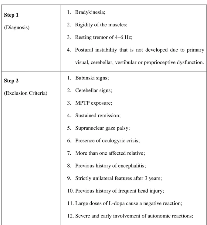
Table 2: The Diagnostic Criteria of Parkinson’s Disease by Queen Square Brain Bank (Adapted from (Lees et al., 2009))
Step 1 (Diagnosis)
1. Bradykinesia;
2. Rigidity of the muscles;
3. Resting tremor of 4–6 Hz;
4. Postural instability that is not developed due to primary visual, cerebellar, vestibular or proprioceptive dysfunction.
Step 2
(Exclusion Criteria)
1. Babinski signs;
2. Cerebellar signs;
3. MPTP exposure;
4. Sustained remission;
5. Supranuclear gaze palsy;
6. Presence of oculogyric crisis;
7. More than one affected relative;
8. Previous history of encephalitis;
9. Strictly unilateral features after 3 years;
10. Previous history of frequent head injury;
11. Large doses of L-dopa cause a negative reaction;
12. Severe and early involvement of autonomic reactions;
19
13. Dementia with memory, language, and praxis problems with early onset;
14. Cerebral tumor or communicating hydrocephalus is present on a CT scan;
15. Treatment with antipsychotic medications when the symptoms first appear;
16. Previous history of recurrent strokes with a gradual progression of the Parkinson symptoms.
Step 3
(Positive Criteria of Supportive Prospective)
1. Hyposmia;
2. Unilateral onset;
3. Rest tremor present;
4. Visual hallucination;
5. Progressive disorder;
6. Chorea induced by L-dopa;
7. A ten-year or longer clinical course;
8. Response to L-dopa for 5 years or more;
9. Excellent response (70–100%) to L-dopa;
10. Persistent asymmetry affecting the side onset most.
The vascular complications of Parkinson’s disease might arise from a previous history of transient ischemic attacks and hypertension. Typically, vascular parkinsonism presents itself without any rest tremor and normal olfactory functions while the response to L-dopa remains poor. Moreover, a substantial change can be observed by the MRI scan within the ischaemic subcortical white-matter. On the other hand, in some rare cases of vascular parkinsonism with subacute onset, hemiparesis is the major factor arising from lacunar stroke within the caudate
nucleus, putamen or globus pallidus. Ultimately, the cerebrovascular diseases that coexist in older patients along with this disease can modify the clinical expression of PD. (Lees et al., 2009)
Though Parkinson’s disease’s cases are often confused with multiple system atrophy of parkinsonism, the cases of such atrophy are very rare. For instance, such atypical cases of parkinsonism represent only one twentieth of Parkinson’s disease. Also, there is occurrence of urinary incontinence, syncope and erectile dysfunction in men during multiple system atrophy which typically occurs during the late 60s of the patients. In addition to this, swallowing complications, slowness and rigidity of movement, speech difficulties and gait complications are seen by the later years. In a few cases, the patient might lose the ability to sweat. However, it gets tough to tell the difference between Parkinson's disease and multiple system atrophy when the patient shows a positive response to Levodopa even with dyskinesias while autonomic failure is very subtle. There are few patients who exhibit autonomic cardiovascular failure along with Parkinson’s disease. (Lees et al., 2009).
Another condition that shares close resemblance to Parkinson’s disease can be included as the parkinsonism of Progressive Supranuclear Palsy in terms of the presentation of the disease. It commonly occurs during the late 70s or 80s of the patients where autonomic failure is not present. Although some patients show early onset of notable bradyphrenia along with the slowness of vertical saccades in progressive supranuclear palsy, the axial and bulbar signs are not as prominent as in Parkinson’s disease. Here, the common aspect of progressive supranuclear palsy parkinsonism and multiple system atrophy parkinsonism is, both of them exhibit rapid progression compared to Parkinson’s disease. Also, both the disorders share an average length of time from commencement to death is roughly 9 years. (Lees et al., 2009).
21
The common misdiagnosis of PD occurs with essential tremors of large magnitude which commonly starts during old age and continues into the resting state. In this case, the tremor is mostly notable during activities particularly when the hands are being outstretched and there is no slowness of movement. In case of dystonic tremor and atypical tremor, the patient might exhibit some stiffness of muscle at the wrist and unable to swing one arm at the time of walking.
However, bradykinesia could be hard to detect sometimes along with the presence of tremor and dystonia. (Lees et al., 2009).
The diagnosis of Parkinson’s disease does not require any additional inspection in most of the cases, and the clinical grounds cover most of the aspects of the diagnosis. However, the uncertainty that might arise could be resolved by a second opinion not by further inconclusive inspections. The most common contradiction might arise from vascular parkinsonism, progressive supranuclear palsy or multiple system atrophy. However, proper implication of olfactory tests could be helpful to dispute such contradictions since normal olfaction is rarely presented in this disease. (Lees et al., 2009).
Chapter 5
Contemporary Treatment Methods for Parkinson’s Disease
The available treatment options of Parkinson’s disease allow the patient to maintain their quality of life to some extent along with preserving their functional capability. However, there is no available cure to this progressive disorder. Over the last four decades, dopamine replacement therapy with levodopa has been the only viable approach to treat the symptoms of Parkinson’s disease. Although few supplementary dopaminergic drugs have recently been incorporated in treating the symptoms of this disease, levodopa still remains the “gold standard” option in this field. However, many people prefer to go with the alternative drugs during the initial stages of the treatment since levodopa has shown motor complications in several cases. Although integrating dopamine agonists and type-b of monoamine oxidase inhibitors could be useful in treating the symptoms of Parkinson’s disease while exerting a lower level of motor complications, they cannot reach the potency level of levodopa.
Furthermore, the complexity lies within the determination of the timing of the treatment approaches such as, the selection of suitable therapeutic agents, combination or sequence of these agents, onset of treatment as well as the timing of necessary surgical intervention and parenteral therapy during advanced stages. (Schapira, 2007).
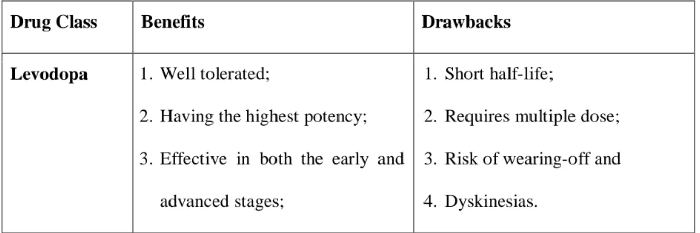
Table 3: Comparison between the major classes of drugs used in Parkinson Disease (Adapted from (Schapira, 2007))
Drug Class Benefits Drawbacks
Levodopa 1. Well tolerated;
2. Having the highest potency;
3. Effective in both the early and advanced stages;
1. Short half-life;
2. Requires multiple dose;
3. Risk of wearing-off and 4. Dyskinesias.
23 4. Low risk of cognitive-
instabilities.
Dopamine Agonists
1. Well tolerated;
2. Long half-life;
3. Low risk of dyskinesias;
4. Little risk of wearing-off;
5. Dosing requires low frequency;
6. Effective in both the early and advanced stages;
7. New routes of delivery systems, e.g., skin patch (Rotigotine).
1. Less potent than levodopa;
2. Cardiac valve fibrosis with ergot agonists;
3. Higher risk of cognitive- instabilities than levodopa.
Monoamine Oxidase Inhibitors
1. Easy to use;
2. Well tolerated;
3. Probable neuroprotection;
4. Requires only one dose per day;
5. Effective in both the early and advanced stages;
1. Benefits are not optimum;
2. Selegiline gets metabolized into amphetamine.
5.1 Treatment for Neuroprotection
Coenzyme Q10, Vitamin E and Monoamine oxidase type B (MAO-B) inhibitors such as selegiline could potentially be used to counterbalance the progression of the disease through neuroprotection. However, during some trials conducted in different research centers it was found that Vitamin E is not beneficial for the patients with early onset of this disease. Moreover, selegiline was initially perceived as a neuroprotective agent but it is more effective in providing
symptomatic relief in terms of the motor symptoms by delaying the start of the levodopa treatment. Furthermore, the neuroprotective effects associated with the MAO-B inhibitors is somewhat debatable. Also, one of the preliminary studies suggest that coenzyme Q10 has the potential to reduce the intensity of the progression of this disease when given in high doses but it is yet to be confirmed by though dome large-scale studies and longer follow-up. Here, glutathione can be included since it is intravenously used at many centers. However, the effectiveness in symptomatic relief as well as the neuroprotection exerted by glutathione needs to be recognized by controlled studies. (Samii et al., 2004).
In addition, during the early onset of this disease the functional imaging techniques conducted on the dopaminergic system suggested that dopamine agonists are effective in slowing down the loss of nigrostriatal neurons compared to the patients who started with levodopa under few controlled trials. Since the interpretation of the imaging results is tricky and debatable, dopamine agonists are yet to be confirmed as potent neuroprotective agents. (Samii et al., 2004) Moreover, some study suggests that neurotrophic factors could be utilized for treating the symptoms and for neuroprotection. However, the neurotrophic factor derived from the glial cell line could not improve the motor complications when as were incorporated in a double- blind trial while the factors were administered by an implanted intracerebroventricular catheter.
Surprisingly, a different open label study involving the same neurotrophic factor showed motor improvements and enhancement in daily activities when the infusion of neurotrophic factor is administered directly into putamen. And so, the poor results of the double-blind trial could be indicative of the neurotrophic factor being unable to reach the target tissues. However, no solid evidence is available to support the effectiveness of neuroprotective agents at the cellular level in terms of slowing down the progression of Parkinson’s disease. (Samii et al., 2004).
25
5.2 Symptomatic Treatment of Motor Symptoms
The treatment that deals with the motor symptoms is started when the motor complexities cause disability for the patient or when the symptoms become bothersome. In terms of the patients with young age, anticholinergic agents can be used to treat the tremors when it persists as the major symptom. However, this type of therapeutic agent presents itself with a number of side- effects which makes them disadvantageous for the older patients. Sometimes therapeutic agents are used despite being less potent. For example, Amantadine is employed during the initial therapy of this disease. Even though it exerts weak antiparkinson effect, it is effective to reduce motor fluctuations and dyskinesias induced by levodopa. (Samii et al., 2004).
Still, dopamine agonist or levodopa is considered as the most suitable treatment option for early onset of PD. It’s because the dopamine agonist rarely causes dyskinesia when it is utilized through monotherapy. That’s why treatment starts with dopamine agonists during the early onset of this disease. However, levodopa is considered as the ideal treatment option during the initial therapy of PD since it is less expensive than the dopamine agonist and it is relatively safer for the elderly patients. In this case, the agonist monotherapy can be employed to effectively control the disease for the first couple of years even if they are less potent than levodopa in terms of antiparkinson effects. Here, the dopamine agonists might vary in terms of their potency or effectiveness since there is no direct comparison available for them. Also, differences might exist in their side effect profile by their relevant therapeutic doses. However, some serious complications such as fibrosis of cardiac, retroperitoneal and pulmonary valve could be avoided by the newer dopamine agonists or the non-ergot dopamine agonists since these complications result from a long-term treatment with ergot dopamine agonists. (Samii et al., 2004).
Complications of Dopamine Agonists: The dopamine agonists commonly exhibit a number of undesired effects that include fluid retention in the leg, nausea, reduced blood pressure, hallucinations in older patients with cognitive deficiency, drowsiness and random sleep attacks.
In this case, Domperidone- a dopamine antagonist can be used to treat nausea which does not penetrate the blood-brain barrier or doesn’t make the disease worse either. Hence, the dopamine agonists should be utilized based on their tolerance since differences within their characteristics might be associated with different unwanted pharmacodynamic effects. (Samii et al., 2004).
Complications of Levodopa: Levodopa is undoubtedly the fundamental component of the treatment of Parkinson’s disease since it has the highest potency in terms of the antiparkinson effects. And the majority of the patients who get their treatment started with dopamine agonists require the additional inclusion of levodopa within a period of 5 years. Moreover, levodopa is usually combined with carbidopa to make the common formulations. It is necessary so that the dopa-decarboxylase is unable for peripheral conversion to dopamine. The unwanted effects exerted by levodopa are somewhat similar to the side effects of dopamine agonists. However, there are few unwanted pharmacodynamic effects that are infrequent with levodopa such as, hallucinations, fluid retention in the leg and drowsiness. In this case, the levodopa induced nausea might be controlled by incorporating extra carbidopa or domperidone. (Samii et al., 2004).
One of the most common problems associated with the long-term use of levodopa can be included as motor instabilities. It’s because the patients start to feel sluggish and tremulous as levodopa’s effect wears off. Later, the patient might experience concurrent shifts between mobility and immobility. In fact, 25 to 50% of the patients taking levodopa even by a low dose might develop such motor instabilities. The frequency of these complications in early onset ranges to a higher proportion of 90% at the 5 years period. (Samii et al., 2004).
27
The complications of dyskinesia develop due to a long-term exposure to levodopa and the dyskinesia might present itself with several patterns. In this case, dyskinesia is most commonly exhibited as peak-dose chorea, a type of dyskinesia that develops when the concentration of levodopa in the blood is at its peak level. However, dystonia can also present itself combinedly with chorea. Here, another pattern of dyskinesia can be included as Diphasic dyskinesia. It exhibits dyskinesia at the start and/or the end of a particular dose cycle. And when the pattern involves painful dystonia occurring at the end of a levodopa dose cycle is known as off-period dystonia. (Samii et al., 2004).
Since levodopa has a short half-life of about 90–120 min, it could be the key reason behind the motor fluctuations. These fluctuations can be alleviated by maintaining the therapeutic effects for a longer period of time, improving the absorption of the doses and altering the dose timings.
The absorption of levodopa is reduced due to a high protein intake. Hence, it could limit levodopa’s capacity to cross the blood-brain barrier. Therefore, if the protein intake could be adjusted with the timing of the doses by maintaining an adequate interval, it might be helpful to reduce the motor instabilities. Moreover, the therapeutic effects of levodopa can be maintained by developing a controlled release dosage form of it. However, it can make the absorption profile of levodopa obscure. (Samii et al., 2004).
In this case, dopamine agonists can be helpful to increase the efficacy of levodopa by reducing the off-time. The levodopa induced dyskinesia can be treated by reducing the dose of levodopa.
However, reducing levodopa’s dose can set off the loss of control of the symptoms associated with parkinsonism. Dopamine agonists such as Amantadine can be used to negate such reactions by exerting antagonistic effects on the N-methyl-D-aspartate glutamate receptor (NMDARs). Moreover, the dyskinesia is assumed to be caused by the pulsatile dopaminergic stimulation. Hence, this process can be exploited to reduce the peaks and troughs by utilizing
the adjunctive agents. Thus, it can potentially be employed as a strategy for treating or reversing dyskinesias. (Samii et al., 2004).
5.3 Symptomatic Treatment of Non-Motor Symptoms
The major symptoms of non-motor complications along with the autonomic dysfunctions of this disease can be included as constipation, orthostatic or postural hypotension, urinary complications, sexual dysfunctions, depression, sleep disorder and psychosis. Suitable treatment strategies of the major non-motor symptoms are included below:
Constipation: Constipation can be alleviated by increasing the intake of fluid and fiber. In this case, stool softeners, stool softeners, suppositories, enemas and fiber supplements such as psyllium can be used to further improve the condition.
Orthostatic Hypotension: Orthostatic hypotension can be resolved by enhancing the intake of salt and fluid. Careful reduction of the antiparkinson drugs might also be helpful. Here, fludrocortisone can be used to maintain the electrolyte-fluid balance and midodrine to treat the hypotension further. (Samii et al., 2004).
Urinary complications: Anticholinergic medications and -adrenergic blockers can be used to alleviate urinary complications such as urine urgency. Oxybutynin and tolterodine might be the preferred anticholinergic agents while prazosin and terazosin are the adrenergic-blocking agents. However, the adrenergic-blocking drugs can worsen the hypotension while the anticholinergics might aggravate constipation. (Samii et al., 2004).
Sexual and Erectile Dysfunctions: Sexual and erectile dysfunctions in men could be treated with sildenafil where close monitoring of blood pressure is a must.
Depression: Another non-motor complication of this disorder can be included as depression.
Although there is no data to suggest which drug should be considered as the best option,
29
depression can be treated by the selective serotonin reuptake inhibitors. Since tricyclic antidepressants might aggravate orthostatic hypotension, they should be used with caution.
Moreover, venlafaxine can be considered as a potent solution for depression in hypotensive patients since it increases blood pressure. (Samii et al., 2004).
Sleep Disorder: Sleep disorder can also be a non-motor complication and the associated complications can include drowsiness, moving the limb on a regular basis, sleep attacks, rapid eye movement (REM) sleep behavior disorder along with sleeplessness at night due to rigidity and bradykinesia. Here, narcolepsy is likely to develop due to the dopamine agonists. In this case, stimulants can be helpful and the agonist might need to cut-off. The periodic limb movements and sleeplessness can be treated with levodopa before going to the bed which will give a prolonged duration of action. Also, the REM sleep behavior disorder can be treated effectively with clonazepam at low-dose. (Samii et al., 2004).
Psychosis: The occurrences of psychosis is assumed to be drug-induced in most of the cases.
In this case, hallucinations are more likely to develop by dopamine agonists than levodopa.
Such conditions can be alleviated by reducing the dose of levodopa as well as discontinuing the agonist or anticholinergic agent. Sometimes neuroleptic agents might need to be included in the process to obtain better results. According to a randomized, double-blind and controlled study, Clozapine is the most potent neuroleptic agent for PD. However, patients need to be careful on their granulocytes count since Clozapine might potentially be associated with fatal agranulocytosis. Therefore, quetiapine is considered as the best solution in this case since it avoids the risk of agranulocytosis while it does not exhibit much extrapyramidal adverse effects like the risperidone and olanzapine. (Samii et al., 2004).
Furthermore, many of the open label studies suggest central cholinesterase inhibitors as a potential treatment option for dementia and psychosis. A randomized, double-blind and controlled study suggests rivastigmine to be effective in Lewy body dementia. Donepezil is
another cholinesterase inhibitor that improves vigilance in patients. However, at this point of time there is not enough information to suggest any of the cholinesterase inhibitors as the irrefutable best option. (Samii et al., 2004).
31
Chapter 6
Gene Therapy in Parkinson’s Disease
The idea of incorporating gene therapy in finding the cure or treatment for different disorders can be dated back to the 1960s. However, the technical difficulties were there associated with the delivery of the gene into target tissues. The goal is to ensure maximum therapeutic efficacy without getting any of the unwanted side effects. According to Forsayeth et al. (2010), Parkinson’s disease has been a potential candidate in terms of implicating gene therapy since the development of vectors.
6.1 Standard Procedures of Gene Therapy
The standard procedure of gene therapy can be included as delivering the genetic material for introducing the exogenous genes or to modify the endogenous gene’s expression. Gene therapy can also be utilized to introduce a transgene so that cellular function can be enhanced. In a common case scenario, a sequence that encodes the human wildtype isoform representing an enzyme is delivered. In order to counterbalance an under-expressed gene or a mutated non- functional gene by a functional gene, this therapeutic strategy can be utilized. Similarly, such therapeutic strategies can also be utilized to express growth factors and other similar proteins to enhance the survival of the cell. (Hitti et al., 2019).
Also, the genetic therapeutic approach can be extended further in terms of a useful tool to knockdown pathological genes. In this case, antisense oligonucleotides (AON) can be utilized which involves creating a complementary sequence of 20 nucleotides to the mRNA of interest.
After that gene knockdown takes place through the hybridized AON along with the transcript of interest. For a different technique of gene knockdown, viruses can be utilized for delivering the small interfering RNA (siRNA) or microRNA. (Hitti et al., 2019).
Moreover, such therapeutic techniques can be incorporated for a number of physiological conditions which also includes neurological disorders like Parkinson’s disease. Typically, the technique of gene therapy involves engineered viruses to play the role of delivery vectors. In this case, recent advancements in the molecular engineering of viral vectors allow them to carry viral DNA in a minute amount. Here, the minute amount of viral DNA is necessary for generating engineered viral particles or for the viral packaging and not allowing the expression of viral proteins. (Hitti et al., 2019).
6.2 Vectors Used in Gene Therapy
Vectors used in human gene therapy can be developed from a number of viruses which includes, Adeno-Associated Virus (AAV), Herpesvirus, Adenovirus and Lentivirus. Here, each of the viruses comes with some advantages as well as disadvantages. For instance, adenovirus and herpesvirus can provoke an immune response inside the body after they’re delivered since they are pathogenic in humans. Therefore, most of the time these kinds of pathogenic viruses are used for oncologic applications since an immune response that will target tumor cells might be helpful for a better therapeutic efficacy. Alternatively, the AAV and lentivirus do not provoke an immune response but they might exert a long-term expression which makes them suitable for non-oncologic applications. Here, lentivirus can be described as a retrovirus with single stranded RNA which can insert its genetic materials within the host genome. The main advantage of this property lies in its ability to express the transgene or the exogenous gene for a longer period of time. (Hitti et al., 2019).
However, this comes with the risk of disrupting the endogenous gene which is known as insertional mutagenesis. On the other hand, the AAV can be described as a virus with single stranded DNA which doesn’t affect the endogenous genes mainly by keeping its DNA mostly extrachromosomal. Although the AAV falls behind lentivirus in terms of integration, the AAV
33
transduction can still exhibit the transgene or the exogenous gene for a longer period of time through a stable extrachromosomal construct. Moreover, these two vectors also vary in terms of their carrying capacity of the genetic material. Here, the lentiviruses are able to carry a genetic material of around 8-9 kB (kilo Base). On the other hand, the AAV is only able to carry genetic material less than 5 kB. Therefore, the AAV is not suitable in some particular applications. (Hitti et al., 2019)
6.3 Gene Therapy through Plasmid Transfection
Another technique of gene transfer can be included as the ‘plasmid transfection’ which involves the delivery of a plasmid or a circular DNA. In this case, the plasmids are delivered by nanoparticles, electroporation, liposomes or by some other physical means. However, plasmid transfection is not considered as a viable option for the treatment of this neurodegenerative disorder since viral transduction allows a much more sustained expression of transgene which is also faster to perform. (Hitti et al., 2019).
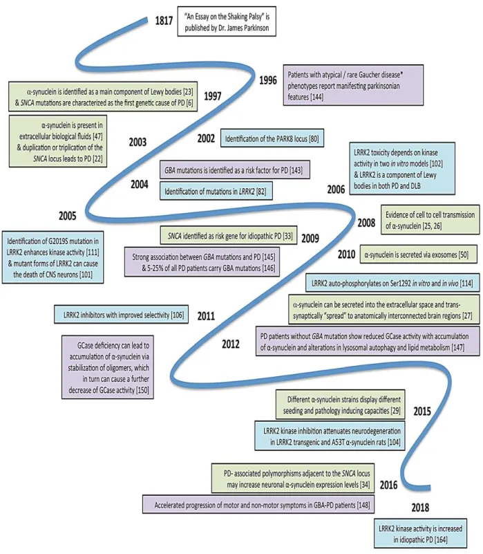
6.4 Major Breakthroughs of Gene Therapy for Parkinson’s Disease
A visual timeline of the milestone discoveries associated with the genetic therapy for Parkinson’s disease is illustrated below:
* Gaucher disease is a rare genetic condition that involves dysfunction of lysosomal storage due to the mutations of biallelic GBA. (Polissidis et al., 2020)
Figure 4: Major breakthroughs associated with gene therapy (Adapted from (Polissidis et al., 2020))
35
6.5 Site of Action for the Common Strategies of Gene Therapies
The site of actions for the common strategies of gene therapies are illustrated below:The pathways depicted in Figure-5 do not represent any exact anatomical location. Rather, they illustrate the connections. Here, both LV–TH AADC GHC1 and AAV2–hAADC enhances the availability of striatal dopamine. In this case, the LV–TH AADC GHC1 increases the availability of both tetrahydrobiopterin and L-dopa. The AAV2–GAD (Adeno-Associated Virus Type 2- Glutamate Decarboxylase) increases GABAergic (Gamma-Aminobutyric acid) blockade of the STN (Subthalamic Nucleus) by increasing the decarboxylation of glutamate.
(Berry & Foltynie, 2011).
Figure 5: Approximate site of action for the common strategies of gene therapies (Adapted from (Berry & Foltynie, 2011))
6.6 Routes of action of the prospective treatment candidates
A visual illustration of the routes of action of the prospective treatment candidates is given below:
Figure 6: Routes of action of the prospective treatment candidates currently going through clinical trials (Adapted From (Polissidis et al., 2020))