A Review of the Prevalence of Acinetobacter That Contains the blaNDM-1 Gene
By Ashikan Rabbi
15126020 Atoshi Debnath Tooli
16136011
A thesis submitted to the Department of Mathematics and Natural Sciences in partial fulfillment of the requirements for the degree of
B.Sc. in Microbiology
Department of Mathematics and Natural Sciences Brac University
May 2022
© 2022. Brac University All rights reserved.
ii
Declaration
It is hereby declared that
• The thesis submitted is my/our original work while completing a degree at Brac University.
• The thesis does not contain material previously published or written by a third party, except where this is appropriately cited through full and accurate referencing.
• The thesis does not contain material that has been accepted or submitted for any other degree or diploma at a university or other institution.
• We have acknowledged all main sources of help.
Student’s Full Name & Signature:
Ashikan Rabbi 15126020
Atoshi Debnath Tooli 16136011
iii
Approval
The thesis titled “A Review of the Prevalence of Acinetobacter That Contains the blaNDM-1 Gene” submitted by
1. Ashikan Rabbi (15126020) 2. Atoshi Debnath Tooli (16136011)
of Spring 2022 has been accepted as satisfactory in partial fulfillment of the requirement for the degree of B.Sc. in Microbiology on April 26, 2022.
Examining Committee:
Supervisor:
(Member)
_______________________________
Professor Fahim Kabir Monjurul Haque, PhD
Assistant Professor, Department of Mathematics and Natural Sciences
Brac University
Program Coordinator:
(Member)
_______________________________
Mahbubul H. Siddique, PhD
Assistant Professor and Coordinator, Mathematics &
Natural Sciences Brac University
Departmental Head:
(Chair)
_______________________________
Professor A F M Yusuf Haider, PhD
Chairperson, Department of Mathematics and Natural Sciences
Brac University
iv
Ethics Statement
Hereby, We, Ashikan Rabbi and Atoshi Debnath Tooli, consciously assure that for the manuscript “A Review of the Prevalence of Acinetobacter That Contains the
blaNDM-1Gene” the following is fulfilled:
1) This material is the authors' original work, which has not been previously published elsewhere.
2) The paper is not currently being considered for publication elsewhere.
3) The paper reflects the authors' own research and analysis truthfully and completely.
4) The paper properly credits the meaningful contributions of co-authors and co-researchers.
5) The results are appropriately placed in the context of prior and existing research.
6) All sources used are properly disclosed (correct citation). Copying of text must be indicated by using quotation marks and giving proper references.
7) All authors have been personally and actively involved in substantial
work leading to the paper and will take public responsibility for its content.
v
Abstract
The blaNDM-1 gene is responsible for multidrug resistance in a wide variety of organisms including Acinetobacter which are responsible for opportunistic infections in immunocompromised patients. The gene is primarily found in Asia; however it has been detected in various parts of the world. We performed a review of the prevalence of the blaNDM-1 gene in members of the Acinetobacter species throughout the world. We performed a literature review in PubMed using “ndm-1” and “Acinetobacter”. Eighty eight articles were included in the study and the results showed that blaNDM-1 positive Acinetobacter are most prevalent in Asia (64.7 %) and Africa (33 %), they are primarily found in clinical samples (80.4 %). The studies were mostly conducted in South East Asia, China, Middle East and North Africa and the proportion of blaNDM-1 positive Acinetobacter was usually higher in countries from these regions.
Keywords: Acinetobacter; NDM-1, New Delhi metallo-beta-lactamase
vi
Acknowledgement
We would like to thank Professor Fahim Kabir Monjurul Haque, PhD, Assistant Professor, Department of Mathematics and Natural Sciences and Mahboob Hossain, PhD, Professor, Department of Mathematics and Natural Sciences for guiding us through our thesis and helping us in every possible way.
We would thank our parents for supporting us all our lives.
I would individually like to thank my close friend Tabassum Binte Matiur for helping me from the start and supporting me in any way possible. I would also like to thank Aneeka Nawar for pushing me and helping me understand the basics of a systematic review. Lastly I would like to thank my brother for looking out for me and guiding me this whole time.
vii
Table of Contents
Declaration ... ii
Approval ... iii
Ethics Statement ... iv
Abstract ... v
Acknowledgement ... vi
Table of Contents ... vii
List of Tables ... viii
List of Figures ... ix
List of Acronyms ... x
Introduction ... 1
Materials and methods ... 2
Literature search ... 2
Inclusion and exclusion criteria ... 2
Data extraction and definition ... 3
Results ... 8
Prevalence by continent ... 8
Prevalence by country ... 9
Prevalence by source ... 13
Prevalence by resistant Acinetobacter ... 14
Prevalence by year ... 18
Discussion... 19
Conclusion ... 21
References: ... 21
viii
List of Tables
Table 1: List of selected articles ... 7 Table 2: Number of blaNDM-1 positive organisms by country ... 11 Table 3: Percentage of resistant Acinetobacter that are NDM-1 positive ... 16
ix
List of Figures
Figure 1: Flow chart of article selection for the review ... 4 Figure 2: Pie chart of blaNDM-1 positive organisms by continent ... 8 Figure 3: Line graph to compare the number of blaNDM-1 positive organisms with the
number of papers found for each continent ... 9 Figure 4: Chart showing number of blaNDM-1 positive organisms by country ... 10 Figure 5: Scatter plot of blaNDM-1 positive organisms by country against the number of articles reviewed for each country ... 11 Figure 6: Heat map of Number of positive Acinetobacter by country ... 12 Figure 7: Pie chart of blaNDM-1 positive organisms by source of sample ... 13 Figure 8: Line graph of the number of blaNDM-1 positive organisms against the number of papers reviewed based on the source of the sample ... 14 Figure 9: Chart showing the log of the number of resistant organisms detected alongside the number of NDM-1 positive organisms among them by country ... 15 Figure 10: Chart showing the log of the number of resistant organisms detected alongside the number of NDM-1 positive organisms among them by source ... 17 Figure 11: Chart showing the number of NDM-1 positive organisms detected by year
alongside the number of papers counted for each year ... 18
x
List of Acronyms
Bla Beta-lactamase
NDM New Delhi metallo-beta-lactamase
K. pneumoniae Klebsiella pneumoniae
E. coli Escherichia coli
A. baumannii Acinetobacter baumannii
CRAB Carbapenem Resistant Acinetobacter baumannii
WHO World Health Organization
Acinetobacter spp. Acinetobacter species
BD Bangladesh
KSA Kingdom of Saudi Arabia
S. Korea South Korea
PCR Polymerase chain reaction
WGS Whole genome sequencing
PFGE Pulse field gel electrophoresis
MLST Multi – Locus Sequence Typing
ICU Intensive care unit
env. Environment
Introduction
The blaNDM-1 gene was first detected in patients in Sweden who had previously traveled to New Delhi, India back in 2009 [1]. Ever since, the gene has spread to almost every part of the world including countries in Europe, Africa and the Americas [2]. The spread of the gene is of major concern as it is a carbapenemase gene which can give rise to resistance against a wide variety of antibiotics [3]. Carbapenems are effective against resistant bacteria that can produce β – lactamase. However, metallo - β – lactamases (MBLs) can break down Carbapenems [4]. Bacteria containing the blaNDM-1 gene can hydrolyze all β – lactam antibiotics except aztreonam [5]. The blaNDM-1 gene poses a major threat due to its multidrug resistant nature and its ability to spread via plasmid conjugation [6]. Therefore, we have limited options when it comes to treatment of infections caused by blaNDM-1 positive organisms. The blaNDM-1 gene can be found in a number of infectious and non-infectious bacteria such as Klebsiella pneumoniae, Escherichia coli and Acinetobacter baumannii [7]. It has been isolated from clinical samples such as urine, blood and respiratory samples. It has also been detected in environmental samples such as water samples and animal feces [16]
[56].
Acinetobacter are Gram-negative saprophytic organisms that are non-fermentative facultative
anaerobes. They are commonly found in soil, wastewater, vegetables and human and animal skins [8]. Members of the Acinetobacter species of bacteria are opportunistic pathogens that can cause infections in immunocompromised patients [9]. Despite their low virulence, multidrug resistant Acinetobacter infections are of major concern in hospital settings as they target patients who are already sick and due to their resistance, treatment becomes very difficult [10]. CRAB (Carbapenem-resistant A. baumannii) has been added to the “critical group” of bacteria by the World Health Organization (WHO) that poses the greatest threat to
2
human health and requires more research [10]. The blaNDM-1 gene can be transferred to Acinetobacter from both members of the Acinetobacter spp. and other species [12] [13].
Due to the threat posed by multi drug resistant Acinetobacter infections and the multi-drug resistant nature of the blaNDM-1 gene, we wanted to look into the prevalence of blaNDM-1
positive Acinetobacter organisms. The purpose of this review was to analyze the spread of the blaNDM-1 gene among Acinetobacter throughout the world and in different settings as these organisms can cause opportunistic infections that would be difficult to cure due to their multi drug resistant nature.
Materials and methods
Literature search
A literature search was performed for the prevalence of the blaNDM-1 gene in members of the Acinetobacter spp. The search was performed through the PubMed electronic database
between March 2021 and November 2021. The keywords used in the search process were:
ndm-1, and, Acinetobacter.
Inclusion and exclusion criteria
Articles were selected for review based on title, followed by a read through the abstract and finally if it appeared to be relevant, we went through the full article. Articles that were included had to specify the method of identification of blaNDM-1 gene in organisms, the origin of the organisms and the number of Acinetobacter containing the blaNDM-1 gene. Conversely the exclusion criteria were articles that did not specify the exact source of the samples, not mentioning the number of samples tested or the number of samples that had blaNDM-1 positive Acinetobacter. Systematic reviews, duplicates of already reviewed articles and any article that did not have a full text available were also excluded.
3
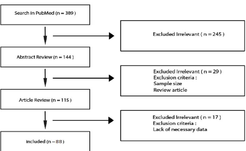
Data extraction and definition
The information obtained from each of the chosen articles include: author’s name, period of study, year of publication, type of sample, number of sample, source of sample, method of detection of blaNDM-1 gene, number of Acinetobacter and number of blaNDM-1 positive Acinetobacter. In some cases not all of the information could be obtained, however for all the
articles source of the sample, method of detection of the blaNDM-1 gene and the number of blaNDM-1 positive Acinetobacter was mentioned. In some cases the study worked with resistant organisms only and that has been noted in the data. A condensed version of the chosen articles along with some of the data collected is shown in Table 1. Some papers are listed twice as they worked with two or more different types of samples. The information for each type of sample is listed separately.
4
Figure 1: Flow chart of article selection for the review
5 First author Period of
study
Year of publicatio
n
Country of origin
Method of identification
Number of Acinetobacte
r
Number of blaNDM-1
Acinetobacte r
Type of sample
Chen Y. [14] 2009 - 2010 2011 China PCR 2109 4 Clinical sample
Islam M. A. [7] 2010 - 2010 2012 BD PCR 18 3 Clinical sample
Mataseje L. F. [15] 2009 - 2010 2012 Canada PCR 9 0 Enterobacteriaceae
Murali S. [18] 2011 - 2011 2012 India PCR 1 1 Donor Eye
Wang Y. [16] 2010 - 2010 2012 China PCR 1 1 Animal swabs
Yang J. [17] 2008 - 2009 2012 China PCR 3114 27 Clinical sample
Farzana R. [20] 2010 - 2011 2013 BD PCR 15 4 Clinical sample
Hasan B. [24] 2010 - 2011 2013 Pakistan PCR 90 1 Clinical sample
Mesli E. [19] 2008 - 2012 2013 Algeria PCR 113 5 Clinical sample
Revathi G. [23] 2009 - 2010 2013 Kenya &
Rwanda PCR 16 1 Clinical Sample
Yanik K. [25] - 2013 Turkey PCR 132 0 Clinical sample
Zhang C. [22] 2010 - 2010 2013 China PCR 42 13 Water sample
Zhang R. [21] 2010 - 2013 2013 China PCR 1067 7 ICU env.
Ageevets V. A. [32] 2011 - 2013 2014 Russia PCR - 1 Clinical sample
Bakour S. [26] 2011 - 2013 2014 Algeria PCR 47 11 Clinical sample
Cicek A. C. [35] 2011 - 2012 2014 Turkey PCR 101 0 Clinical sample
Jones R. N. [29] 2011 2014 Europe PCR 472 0 Clinical sample
Kulkova N. [33] 2011 - 2012 2014 Slovakia PCR 9 0 Blood culture
Lauderdale T. L. [34] 2010 2014 Taiwan PCR 408 1 Clinical sample
Pasteran F. [31] 2012 2014 Paraguay PCR 2 2 Clinical sample
Peirano G. [27] 2010 - 2013 2014 Canada PCR 1 0 Clinical sample
Rafei R. [30] 2012 2014 Lebanon PCR 4 4 Clinical sample
Zheng F. [28] 2010 - 2010 2014 China PCR 169 2 Clinical sample
Ahmed M. A. [38] 2012 - 2013 2015 Egypt PCR 150 59 Clinical sample
Memish Z. A. [40] 2012 - 2012 2015 KSA PCR 79 1 Clinical sample
Novovic K. [43] 2012 - 2014 2015 Serbia PCR 28 0 Clinical sample
Quiñones D [37]. 2010 - 2012 2015 Cuba PCR 500 1 Clinical sample
Rafei R. [41] 2011 - 2013 2015 Lebanon PCR 116 5 Clinical sample
Shrestha S. [42] 2013 - 2014 2015 Nepal MLST 246 51 Clinical sample
Sung J. Y. [39] 2006 - 2013 2015 S. Korea PCR 21 2 Clinical sample
Tran H. H. [44] 2010 - 2012 2015 Vietnam PCR 31 3 Hospital env.
sample
Zenati K. [36] 2011 - 2013 2015 Algeria PCR 67 32 Hospital env.
sample
Adler A. [48] 2014 - 2015 2016 Israel PCR 313 16 Clinical sample
Bouguenoun W. [46] 2014 - 2014 2016 Algeria PCR 3 2 Clinical sample
Bouguenoun W. [46] 2014 - 2014 2016 Algeria PCR 6 5 Hospital env.
sample
Cetinkol Y. [51] - 2016 Turkey PCR 50 0 Clinical sample
Kateete D. P. [52] 2007 - 2009 2016 Uganda PCR 29 0 Clinical sample
Kateete D. P. [52] 2007 - 2009 2016 Uganda PCR 11 0 Hospital env.
sample
Mathlouthi N. [49] 2015 - 2015 2016 Libya PCR 36 8 Clinical sample
Ramoul A. [47] 2010 - 2013 2016 Algeria PCR 43 7 Clinical sample
Timofte D. [50] 2014 - 2015 2016 Romania PCR - 0 Clinical sample
Tran D. N. 45] 2010 – 2014 2016 Vietnam PCR 582 23 Clinical sample
El-Mahdy T. S. [59] 2014 - 2014 2017 KSA PCR 10 3 Clinical sample
Gomaa F. A. M. [57] 2014 - 2015 2017 Egypt PCR 56 13 Clinical sample
Hammami S. [64] 2014 - 2014 2017 Tunisia PCR 5 0 Clinical sample
Hasan M. J. [55] 2013 - 2013 2017 BD PCR 22 2 Clinical sample
Islam M. A. [56] 2012 - 2012 2017 BD PCR - 13 Water sample
6
Joshi P. R. [61] 2014 - 2015 2017 Nepal PCR 44 6 Clinical sample
Manohar P. [58] 2014 - 2015 2017 India PCR 5 0 Clinical sample
Mellouk F. Z. [54] 2013 - 2015 2017 Algeria PCR 7 2 Clinical sample
Morakchi H. [100] 2013 - 2013 2017 France -
Algeria PCR 14 0 Pigeon stool
Pirii L. E. [62] 2015 2017 Romania WGS 1 0 Burn patient
Romero J. L. [63] 2013 -2014 2017 Spain PCR 4 0 Sea food
Uwingabiye J. [60] 2015 - 2015 2017 Morocco PCR 47 9 Clinical sample
Uwingabiye J. [60] 2015 - 2015 2017 Morocco PCR 36 18 Hospital env.
Sample
Yagoubat M. [53] 2014 - 2015 2017 Algeria PCR - 0 Clinical sample
Yagoubat M. [53] 2014 - 2015 2017 Algeria PCR 8 5 Hospital env.
sample Agoba E. E. [71] 2015 - 2015 2018 South
Africa PCR 24 1 Clinical sample
Banerjee T. [68] 2012 - 2016 2018 India PCR 100 34 ICU env.
Cheikh H. B. [74] 2013 - 2016 2018 Tunisia PCR 101 1 Clinical sample
Dziri O. [73] 2015 - 2016 2018 Tunisia PCR 3 0 Clinical sample
Faccone D. [65] 2013 - 2015 2018 Argentina PCR 10 1 Clinical sample
Gentilini F. [70] 2014 - 2015 2018 Italy PCR 6 1 Pets
Jaidane N. [75] 2013 - 2015 2018 Tunisia PCR 246 7 Clinical sample
Jain M. [66] 2016 - 2017 2018 India PCR 28 3 Clinical sample
Jain M. [66] 2016 - 2017 2018 India PCR 8 0 ICU env.
Kuntaman K. [69] 2015 - 2016 2018 Indonesia
- Japan PCR 75 6 Clinical sample
Leungtongkam U.
[72] 2013 - 2015 2018 Thailand PCR 339 31 Clinical sample
Maamar E. [76] 2014 - 2015 2018 Tunisia PCR 13 2 Clinical sample
Rahman M. [67] 2012 - 2012 2018 India PCR 106 20 Clinical sample
Abouelfetouh A. [78] 2010 and
2015 2019 Egypt PCR 74 9 Clinical sample
Al - Hamad A. [81] 2014 2019 KSA PCR 21 1 Clinical sample
Al - Hamad A. [81] 2014 2019 KSA PCR 74 1 Hospital env.
Sample Anane Y. A. [84] 2016 - 2017 2019 South
Africa PCR 52 0 Fish body and
water Anane Y. A. [84] 2016 - 2017 2019 South
Africa PCR 48 0 Slaughterhouse
env.
Gomez L. G. [86] 2016 2019 Venezuela
Plasmid profile, PFGE, MLST
8 3 Clinical sample
Khalid S. [79] 2017 - 2017 2019 India PCR - 4 Clinical sample
Kumar S. [80] 2013 - 2016 2019 India PCR 97 25 Clinical sample
Lee Y. [85] 2018 2019 Taiwan PCR 188 0 Clinical sample
Qamar M. U. [83] 2015 - 2016 2019 Pakistan PCR 29 4 Clinical sample
Rakhi N. N. [77] 2016 2019 BD PCR 4 2 Clinical sample
Shah M. W. [82] 2015 - 2016 2019 KSA PCR 135 2 Clinical sample
Anane Y. A. [95] 2016 - 2017 2020 South
Africa PCR 100 2 Clinical sample
Benamrouche N. [87] 2012 - 2016 2020 Algeria PCR 92 5 Clinical sample
Kalasseril S. G. [90] 2017 - 2018 2020 India PCR 2 0 Hospital env.
Sample
Kongthai P. [96] 2013 - 2015 2020 Thailand PCR 339 1 Clinical sample
Lukovic B. [94] 2018 - 2018 2020 Serbia PCR 280 7 Clinical sample
Manandhar S. [93]
2012 - 2018 2020 Nepal PCR 383 79 Clinical sample
Moubareck C. A.
[97] 2015 - 2016 2020 UAE PCR 341 1 Clinical sample
7
Rao M. [91] 2011 - 2016 2020 Malaysia WGS 13 3 Clinical sample
Sanou S. [88] 2016 - 2016 2020 Burkina
Faso PCR 4 1 Clinical sample
Sharma M. [89] 2013 - 2014 2020 India PCR 150 28 Clinical sample
Tada T. [92] 2015 - 2018 2020 Myanmar WGS 45 5 Clinical sample
Monnheimer M. [98] 2017 - 2018 2021 Ghana PCR 45 19 Clinical sample
Soliman A. M. [99] 2014 - 2015 2021 Japan PCR 1 0 Vegetable
Table 1: List of selected articles
BD, Bangladesh; S. Korea, South Korea; KSA, Kingdom of Saudi Arabia; UAE, United Arab Emirates; env., environment.
8
Results
Prevalence by continent
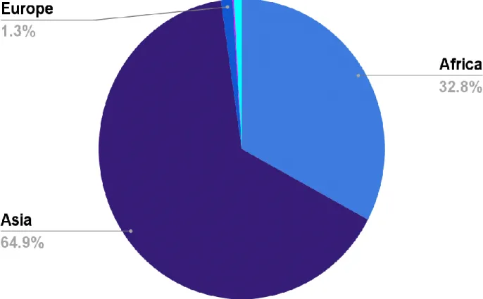
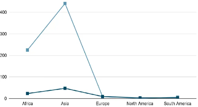
When looking at the number of NDM-1 positive organisms found by continent, out of a total of 684 NDM-1 positive organisms, 441 were from Asia, 225 were from Africa, 9 from Europe, 6 from South America and lastly 1 from North America. The numbers were obtained from 87 papers (one paper was excluded as the location was not limited to one continent) of which 47 were based in Asia (Turkey was counted under Asia), 25 were from Africa, 9 were from Europe, 3 were from North America and 3 were from South America. Figure 2 shows a pie chart for the distribution of the detected organisms among the 5 continents and figure 3 is a line graph to show the relation between the numbers of papers compared to the number of organisms detected.
Figure 2: Pie chart of blaNDM-1 positive organisms by continent
9
Prevalence by country
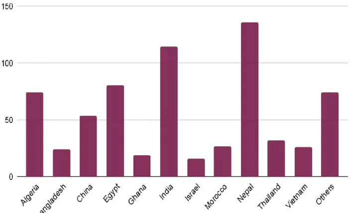
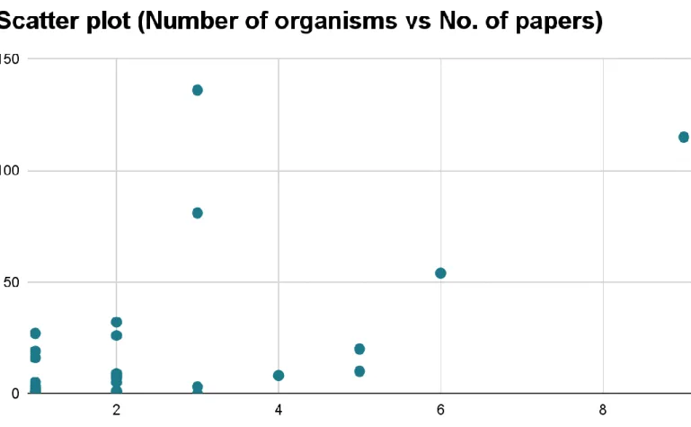
We found data for 37 different countries after excluding papers where multiple countries were involved. The country with the greatest number of articles was India with 9 separate papers followed by Algeria which had 8 papers. However, the highest number of NDM-1 positive Acinetobacter was found in Nepal at 136 followed by India with 115. Figure 4 shows the total number of NDM-1 positive organisms found for each country based on the data reviewed. Any country with ten or less organisms detected falls under the “Others” column.
This includes 26 different countries. Among them Tunisia had the most with 10 detected organisms, followed by Lebanon and Libya both with 9 detected organisms each. Six of the twenty-six countries had zero NDM-1 positive Acinetobacter which were Canada, Japan, Slovakia, Spain, Turkey and Uganda. A scatter plot (figure 5) is also included to show the
Figure 3: Line graph to compare the number of blaNDM-1 positive organisms with the number of papers found for each continent
10
relation between number of organisms detected and the number of papers for each country.
Table 2 shows the full list for all the countries.
Figure 4: Chart showing number of blaNDM-1 positive organisms by country
11 Country of
origin
No. of organisms
Country of origin
No. of organisms
Country of origin
No. of organisms
Algeria 74 South Korea 2 Slovakia 0
Argentina 1 KSA 8 South Africa 3
Bangladesh 24 Lebanon 9 Spain 0
Burkina Faso 1 Libya 8 Taiwan 1
Canada 0 Malaysia 3 Thailand 32
China 54 Morocco 27 Tunisia 10
Cuba 1 Myanmar 5 Turkey 0
Egypt 81 Nepal 136 UAE 1
Ghana 19 Pakistan 5 Uganda 0
India 115 Paraguay 2 Venezuela 3
Israel 16 Romania 1 Vietnam 26
Italy 1 Russia 1 Total 678
Japan 0 Serbia 7
Table 2: Number of blaNDM-1 positive organisms by country
Figure 5: Scatter plot of blaNDM-1 positive organisms by country against the number of articles reviewed for each country
12
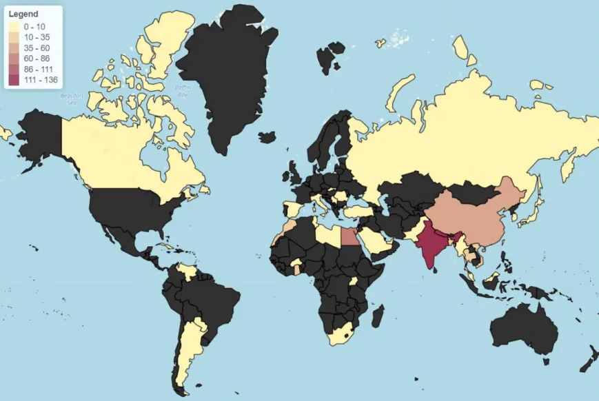
Figure 6: Heat map of Number of positive Acinetobacter by country
13
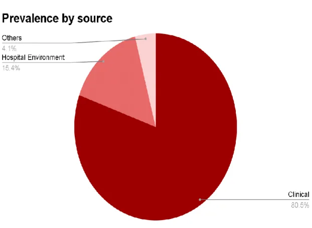
Prevalence by source
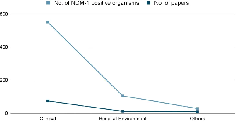
The source of the organism can be broadly classified into three categories. These are clinical specimens, samples from hospital environment and lastly other samples which include samples from water (excluding water from hospital sources), food, plants and animals. The pie chart in figure 6 shows the distribution of 683 NDM-1 positive organisms based off the source of the organism. The data for the number of papers for each source is also given, however some papers which tested samples from different sources have been counted for all types of samples they tested. For example, papers that tested both a hospital environment and clinical samples were counted for both hospital environment and clinical samples. A line graph of the number of NDM-1 positive organisms detected to the number of papers is presented in figure 7.
Figure 7: Pie chart of blaNDM-1 positive organisms by source of sample
14
Prevalence by resistant Acinetobacter
It is difficult to find the percentage of Acinetobacter that are NDM-1 positive from the data since the total number of Acinetobacter detected is not mentioned for some studies. For the studies where the number of Acinetobacter detected was mentioned, some only worked with resistant organisms while other did not so we cannot count them under the same category. We therefore broke down the data into the number of resistant Acinetobacter that turned out to be NDM-1 positive. Resistant organisms mainly constitute Acinetobacter that were carbapenem resistant or multidrug resistant. The chart below (figure 8) shows the log of the number of resistant Acinetobacter that are NDM-1 positive for each country alongside the log of the number of resistant Acinetobacter tested. We used the logarithmic values as it was easier to visualize and compare. This is because the sample size for each country varies greatly. This
Figure 8: Line graph of the number of blaNDM-1 positive organisms against the number of papers reviewed based on the source of the sample
15
means it would not be accurate to compare the proportion of resistant organisms with the proportion of NMD-1 positive organisms in the chart, but we can at least get an idea of how the values vary from country to country. Values for Lebanon, Paraguay and Venezuela were excluded as the numbers of resistant organisms detected in these countries were less than 10 which were not enough to provide an accurate representation. The chart also does not show the countries where resistant Acinetobacter were found but none of the organisms were NDM-1 positive. This includes Japan and Romania. The highest percentage of NDM-1 positive organisms among resistant Acinetobacter was found in Algeria (35.94%) followed by Bangladesh (34.78%) and then Morocco (32.53%). List of the percentage for each country is given in table 3.
Figure 9: Chart showing the log of the number of resistant organisms detected alongside the number of NDM-1 positive organisms among them by country
16
Country of origin No. of resistant Acinetobacter detected
Percentage of resistant Acinetobacter that are NDM-1 positive (%)
Algeria 64 35.94
Bangladesh 23 34.78
China 169 1.18
Egypt 74 12.16
India 152 18.42
Israel 313 5.11
KSA 105 4.76
Libya 44 20.45
Malaysia 13 23.08
Morocco 83 32.53
Pakistan 29 13.79
South Africa 224 1.34
Tunisia 360 2.78
UAE 167 0.6
Vietnam 582 3.95
Table 3: Percentage of resistant Acinetobacter that are NDM-1 positive
If we consider the number of NDM-1 positive organisms compared to the number of resistant organisms with respect to the source we get the results shown in figure 9. We used the log of the values once again as it is difficult to compare them directly due to the large size of clinical samples. Therefore the proportion is not as significant as it appears. If we break down the values by percentage, out of 2166 resistant clinical isolates 6.65% (144) were NDM-1 positive. Out of 120 resistant Acinetobacter samples from hospital environments, 20% (24) were NDM-1 positive. Lastly, out of 108 resistant environmental samples, 0.93% (1) was NDM-1 positive.
17
Figure 10: Chart showing the log of the number of resistant organisms detected alongside the number of NDM-1 positive organisms among them by source
18
Prevalence by year
Prevalence by year is difficult to represent accurately as most of the studies took place over two or more years. Outside of studies that were only conducted in a single year, we also counted studies where the number of months in a particular year was double that of any other year for the study. For example, if a study took place from December 2014 till August 2015, we counted the study for 2015 as the number of months in 2015 was more than double that spent in 2014 for this study. Figure 10 shows a chart of this data alongside the number of papers from which the data was collected.
Figure 11: Chart showing the number of NDM-1 positive organisms detected by year alongside the number of papers counted for each year
19
Discussion
Based on the articles reviewed, members of the Acinetobacter spp. that are NDM-1 positive are most commonly found in Asia followed by Africa. From our results more than half of the identified NDM-1 positive Acinetobacter were from Asia and about one third were from Africa. The rest of the world constitutes less than three percent of the results. By country, the highest numbers of NDM-1 positive Acinetobacter were from Nepal and then India. The data was compared with the number of articles found for each country. While more articles did tend towards more organisms, there was no clear relation between the two factors. However, this does not imply that Nepal has the largest prevalence of NDM-1 positive Acinetobacter as we couldn’t take other factors into account such as number of organisms tested. It is difficult to consider the number of Acinetobacter tested as some studies only worked with resistant Acinetobacter while some others did not mention whether the Acinetobacter were resistant or
not. Thus the data should be considered merely as an indicator for where the NDM-1 gene may be more prevalent.
In terms of source, majority of the organisms were from clinical samples with less than twenty percent of the detected organisms being from other sources. Less than five percent of the organisms were not from a hospital. When comparing this data with the number of papers found for each source there does appear to be a positive correlation between the number of papers for a particular source and the number of NDM-1 positive Acinetobacter found for the that particular source. Since very few samples were taken from non-hospital settings, it is hard to determine the proportion of organisms outside of hospital settings that may be NDM- 1 positive. However, based off the sample size that was available it does appear the proportion of Acinetobacter that are NDM-1 positive is higher in hospitals compared to other sources.
20
When considering the proportion of resistant Acinetobacter that were NDM-1 positive, we find that the percentage is higher for countries in South East Asia and Northern Africa. In most cases the percentage was lower when the number of tested samples were greater, however this also varied with region. The highest percentages were from Algeria, Bangladesh and Morocco were around a third of the resistant organisms were also NDM-1 positive.
However in all three of these countries the numbers of resistant organisms detected were less than 100. For countries were over 100 resistant organisms were tested, the percentage was around 5 percent or lower, excluding India where out of 152 resistant organisms around 18 percent were NDM-1 positive.
If we look at the percentage of resistant Acinetobacter that are also NDM-1 positive on the basis of setting, we find that the largest percentage was for hospital environment at twenty percent, followed by clinical specimen at around six percent. The percentage for non-hospital settings was less than one percent. However, if we take the number of organisms into account, the sample size is a lot larger for clinical specimens at over 2000 while the number of samples for both hospital environment and non-hospital settings were just over 100. It is likely that the percentage for hospital environment would be lower if we had a larger sample size.
Lastly we broke down the data in a year by year basis. This was made difficult by the fact that most studies took place over a multiyear time period. We had to exclude a lot of studies as a result. Alongside studies that took place over a single year, we also counted papers where the study period in a particular year was at least double the study period in any other year.
According to the results the most NDM-1 positive organisms were detected between 2012 and 2015. The number of papers published in this time frame was also more than other years.
21
While there are more NDM-1 positive organisms detected in years where more papers were published, it is hard to say for certain if the two factors are co-related.
Conclusion
There needs to be more research into Acinetobacter with the NDM-1 gene, especially in regions around Asia and Africa where it is more prevalent. In the age of globalization where people travel far and often it is very easy for the blaNDM-1 gene to spread. Treatment for infections caused by NDM-1 positive Acinetobacter is very limited and can be life threatening. Thus it is important for us to monitor the spread of this gene in various organisms in order to control its spread. While the primary source of NDM-1 positive Acinetobacter is clinical specimens, we cannot ignore other sources either. Even though
clinical specimens are the most likely to cause opportunistic infections, environmental cases of NDM-1 positive Acinetobacter can contribute to the spread of the NDM-1 gene to other organisms in the same species as well as other species through plasmid conjugation.
References:
1. Yong D, Toleman MA, Giske CG, et al. Characterization of a new metallo-β- lactamase gene, blaNDM-1, and a novel erythromycin esterase gene carried on a unique genetic structure in Klebsiella pneumoniae sequence type 14 from India, Antimicrob Agents Chemother, 2009, vol. 53 (pg. 5046-54) https://doi.org/10.1128/AAC.00774- 09
2. Khan, A. U., Maryam, L., & Zarrilli, R. (2017). Structure, Genetics and Worldwide Spread of New Delhi Metallo-β-lactamase (NDM): a threat to public health. BMC microbiology, 17(1), 101. https://doi.org/10.1186/s12866-017-1012-8
3. Kumarasamy, K. K., Toleman, M. A., Walsh, T. R., Bagaria, J., Butt, F., Balakrishnan, R., Chaudhary, U., Doumith, M., Giske, C. G., Irfan, S., Krishnan, P.,
22
Kumar, A. V., Maharjan, S., Mushtaq, S., Noorie, T., Paterson, D. L., Pearson, A., Perry, C., Pike, R., Rao, B., … Woodford, N. (2010). Emergence of a new antibiotic resistance mechanism in India, Pakistan, and the UK: a molecular, biological, and epidemiological study. The Lancet. Infectious diseases, 10(9), 597–602.
https://doi.org/10.1016/S1473-3099(10)70143-2
4. Boyd, S. E., Livermore, D. M., Hooper, D. C., & Hope, W. W. (2020). Metallo-β- Lactamases: Structure, Function, Epidemiology, Treatment Options, and the Development Pipeline. Antimicrobial agents and chemotherapy, 64(10), e00397-20.
https://doi.org/10.1128/AAC.00397-20
5. Khan, A. U., Maryam, L., & Zarrilli, R. (2017). Structure, Genetics and Worldwide Spread of New Delhi Metallo-β-lactamase (NDM): a threat to public health. BMC microbiology, 17(1), 101. https://doi.org/10.1186/s12866-017-1012-8
6. Xiang, T., Chen, C., Wen, J., Liu, Y., Zhang, Q., Cheng, N., Wu, X., & Zhang, W.
(2020). Resistance of Klebsiella pneumoniae Strains Carrying blaNDM-1 Gene and the Genetic Environment of blaNDM-1. Frontiers in microbiology, 11, 700.
https://doi.org/10.3389/fmicb.2020.00700
7. Islam, M. A., Talukdar, P. K., Hoque, A., Huq, M., Nabi, A., Ahmed, D., Talukder, K. A., Pietroni, M. A., Hays, J. P., Cravioto, A., & Endtz, H. P. (2012). Emergence of multidrug-resistant NDM-1-producing Gram-negative bacteria in Bangladesh. European journal of clinical microbiology & infectious diseases : official publication of the European Society of Clinical Microbiology, 31(10), 2593–2600.
https://doi.org/10.1007/s10096-012-1601-2
8. Asif, M., Alvi, I. A., & Rehman, S. U. (2018). Insight into Acinetobacter baumannii:
pathogenesis, global resistance, mechanisms of resistance, treatment options, and
23
alternative modalities. Infection and drug resistance, 11, 1249–1260.
https://doi.org/10.2147/IDR.S166750
9. Dijkshoorn, L.,Nemec, A., andSeifert, H. ( 2007) An increasing threat in hospitals:
multidrug-resistant Acinetobacter baumannii. Nat. Rev. Microbiol. 5, 939–951.
https://doi.org/10.1038/nrmicro1789
10. George M. Eliopoulos, Lisa L. Maragakis, Trish M. Perl, Acinetobacter baumannii:
Epidemiology, Antimicrobial Resistance, and Treatment Options, Clinical Infectious Diseases, Volume 46, Issue 8, 15 April 2008, Pages 1254–1263, https://doi.org/10.1086/529198
11. Vázquez-López, R., Solano-Gálvez, S. G., Juárez Vignon-Whaley, J. J., Abello Vaamonde, J. A., Padró Alonzo, L. A., Rivera Reséndiz, A., Muleiro Álvarez, M., Vega López, E. N., Franyuti-Kelly, G., Álvarez-Hernández, D. A., Moncaleano Guzmán, V., Juárez Bañuelos, J. E., Marcos Felix, J., González Barrios, J. A., &
Barrientos Fortes, T. (2020). Acinetobacter baumannii Resistance: A Real Challenge for Clinicians. Antibiotics (Basel, Switzerland), 9(4), 205.
https://doi.org/10.3390/antibiotics9040205
12. Windy D. Tanner, Robyn M. Atkinson, Ramesh K. Goel, Mark A. Toleman, Lowell Scott Benson, Christina A. Porucznik, James A. VanDerslice, Horizontal transfer of the blaNDM-1 gene to Pseudomonas aeruginosa and Acinetobacter baumannii in biofilms, FEMS Microbiology Letters, Volume 364, Issue 8, April 2017, fnx048, https://doi.org/10.1093/femsle/fnx048
13. Chatterjee, S., Mondal, A., Mitra, S., & Basu, S. (2017). Acinetobacter baumannii transfers the blaNDM-1 gene via outer membrane vesicles. The Journal of antimicrobial chemotherapy, 72(8), 2201–2207. https://doi.org/10.1093/jac/dkx131
24
14. Chen, Y., Zhou, Z., Jiang, Y., & Yu, Y. (2011). Emergence of NDM-1-producing Acinetobacter baumannii in China. The Journal of antimicrobial chemotherapy, 66(6), 1255–1259.
a. https://doi.org/10.1093/jac/dkr082
15. Mataseje, L. F., Bryce, E., Roscoe, D., Boyd, D. A., Embree, J., Gravel, D., Katz, K., Kibsey, P., Kuhn, M., Mounchili, A., Simor, A., Taylor, G., Thomas, E., Turgeon, N., Mulvey, M. R., & Canadian Nosocomial Infection Surveillance Program (2012).
Carbapenem-resistant Gram-negative bacilli in Canada 2009-10: results from the Canadian Nosocomial Infection Surveillance Program (CNISP). The Journal of antimicrobial chemotherapy, 67(6), 1359–1367.
a. https://doi.org/10.1093/jac/dks046
16. Wang, Y., Wu, C., Zhang, Q., Qi, J., Liu, H., Wang, Y., He, T., Ma, L., Lai, J., Shen, Z., Liu, Y., & Shen, J. (2012). Identification of New Delhi metallo-β-lactamase 1 in Acinetobacter lwoffii of food animal origin. PloS one, 7(5), e37152.
https://doi.org/10.1371/journal.pone.0037152
17. Yang, J., Chen, Y., Jia, X., Luo, Y., Song, Q., Zhao, W., Wang, Y., Liu, H., Zheng, D., Xia, Y., Yu, R., Han, X., Jiang, G., Zhou, Y., Zhou, W., Hu, X., Liang, L., & Han, L. (2012). Dissemination and characterization of NDM-1-producing Acinetobacter pittii in an intensive care unit in China. Clinical microbiology and infection : the official publication of the European Society of Clinical Microbiology and Infectious Diseases, 18(12), E506–E513.
a. https://doi.org/10.1111/1469-0691.12035
25
18. Murali, S., Jambulingam, M., Tiru, V., Kulanthai, L. T., Rajagopal, R., Padmanaban, P., & Madhavan, H. N. (2012). A study on isolation rate and prevalence of drug resistance among microorganisms isolated from multiorgan donor and donor corneal rim along with a report on existence of bla NDM-1 among Indian population. Current eye research, 37(3), 195–203.
a. https://doi.org/10.3109/02713683.2011.643270
19. Mesli, E., Berrazeg, M., Drissi, M., Bekkhoucha, S. N., & Rolain, J. M. (2013).
Prevalence of carbapenemase-encoding genes including New Delhi metallo-β- lactamase in Acinetobacter species, Algeria. International journal of infectious diseases : IJID : official publication of the International Society for Infectious Diseases, 17(9), e739–e743. https://doi.org/10.1016/j.ijid.2013.02.024
20. Farzana, R., Shamsuzzaman, S., & Mamun, K. Z. (2013). Isolation and molecular characterization of New Delhi metallo-beta-lactamase-1 producing superbug in Bangladesh. Journal of infection in developing countries, 7(3), 161–168.
https://doi.org/10.3855/jidc.2493
21. Zhang, R., Hu, Y. Y., Yang, X. F., Gu, D. X., Zhou, H. W., Hu, Q. F., Zhao, K., Yu, S. F., & Chen, G. X. (2014). Emergence of NDM-producing non-baumannii Acinetobacter spp. isolated from China. European journal of clinical microbiology &
infectious diseases : official publication of the European Society of Clinical Microbiology, 33(5), 853–860. https://doi.org/10.1007/s10096-013-2024-4
22. Zhang, C., Qiu, S., Wang, Y., Qi, L., Hao, R., Liu, X., Shi, Y., Hu, X., An, D., Li, Z., Li, P., Wang, L., Cui, J., Wang, P., Huang, L., Klena, J. D., & Song, H. (2013).
26
Higher isolation of NDM-1 producing Acinetobacter baumannii from the sewage of the hospitals in Beijing. PloS one, 8(6), e64857.
a. https://doi.org/10.1371/journal.pone.0064857
23. Revathi, G., Siu, L. K., Lu, P. L., & Huang, L. Y. (2013). First report of NDM-1- producing Acinetobacter baumannii in East Africa. International journal of infectious diseases : IJID : official publication of the International Society for Infectious Diseases, 17(12), e1255–e1258.
a. https://doi.org/10.1016/j.ijid.2013.07.016
24. Hasan, B., Perveen, K., Olsen, B., & Zahra, R. (2014). Emergence of carbapenem- resistant Acinetobacter baumannii in hospitals in Pakistan. Journal of medical microbiology, 63(Pt 1), 50–55.
a. https://doi.org/10.1099/jmm.0.063925-0
25. Yanık, K., Emir, D., Eroğlu, C., Karadağ, A., Güney, A. K., & Günaydın, M. (2013).
Karbapeneme dirençli gram-negatif izolatlarda New Delhi metallo-beta-laktamaz-1 (NDM-1) varlığının PCR ile araştırılması [Investigation of the presence of New Delhi metallo-beta-lactamase-1 (NDM-1) by PCR in carbapenem-resistant gram-negative isolates]. Mikrobiyoloji bulteni, 47(2), 382–384.
a. https://doi.org/10.5578/mb.5033
26. Bakour, S., Touati, A., Bachiri, T., Sahli, F., Tiouit, D., Naim, M., Azouaou, M., &
Rolain, J. M. (2014). First report of 16S rRNA methylase ArmA-producing Acinetobacter baumannii and rapid spread of metallo-β-lactamase NDM-1 in Algerian
27
hospitals. Journal of infection and chemotherapy : official journal of the Japan Society of Chemotherapy, 20(11), 696–701.
a. https://doi.org/10.1016/j.jiac.2014.07.010
27. Peirano, G., Ahmed-Bentley, J., Fuller, J., Rubin, J. E., & Pitout, J. D. (2014). Travel- related carbapenemase-producing Gram-negative bacteria in Alberta, Canada: the first 3 years. Journal of clinical microbiology, 52(5), 1575–1581.
https://doi.org/10.1128/JCM.00162-14
28. Zheng, F., Sun, J., Cheng, C., & Rui, Y. (2015). Molecular characteristics of carbapenem-resistant gram-negative bacteria in southern China. Microbial drug resistance (Larchmont, N.Y.), 21(2), 178–185.
a. https://doi.org/10.1089/mdr.2014.0085
29. Jones, R. N., Flonta, M., Gurler, N., Cepparulo, M., Mendes, R. E., & Castanheira, M.
(2014). Resistance surveillance program report for selected European nations (2011). Diagnostic microbiology and infectious disease, 78(4), 429–436.
https://doi.org/10.1016/j.diagmicrobio.2013.10.008
30. Rafei, R., Dabboussi, F., Hamze, M., Eveillard, M., Lemarié, C., Mallat, H., Rolain, J.
M., Joly-Guillou, M. L., & Kempf, M. (2014). First report of blaNDM-1-producing Acinetobacter baumannii isolated in Lebanon from civilians wounded during the Syrian war. International journal of infectious diseases : IJID : official publication of the International Society for Infectious Diseases, 21, 21–23.
https://doi.org/10.1016/j.ijid.2014.01.004
28
31. Pasteran, F., Mora, M. M., Albornoz, E., Faccone, D., Franco, R., Ortellado, J., Melgarejo, N., Gomez, S., Riquelme, I., Matheu, J., Ramon-Pardo, P., & Corso, A.
(2014). Emergence of genetically unrelated NDM-1-producing Acinetobacter pittii strains in Paraguay. The Journal of antimicrobial chemotherapy, 69(9), 2575–2578.
https://doi.org/10.1093/jac/dku139
32. Ageevets, V. A., Partina, I. V., Lisitsyna, E. S., Ilina, E. N., Lobzin, Y. V., Shlyapnikov, S. A., & Sidorenko, S. V. (2014). Emergence of carbapenemase- producing Gram-negative bacteria in Saint Petersburg, Russia. International journal of antimicrobial agents, 44(2), 152–155.
a. https://doi.org/10.1016/j.ijantimicag.2014.05.004
33. Kulkova, N., Babalova, M., Sokolova, J., & Krcmery, V. (2015). First report of New Delhi metallo-β-lactamase-1-producing strains in Slovakia. Microbial drug resistance (Larchmont, N.Y.), 21(1), 117–120.
a. https://doi.org/10.1089/mdr.2013.0162
34. Lauderdale, T. L., Hsu, M. C., Mu, J. J., Chang, F. Y., Lai, J. F., Tan, M. C., Wang, H. Y., & Chang, S. C. (2014). NDM-1-producing Acinetobacter soli from Taiwan. Diagnostic microbiology and infectious disease, 80(2), 168–169.
https://doi.org/10.1016/j.diagmicrobio.2014.06.008
35. Cicek, A. C., Saral, A., Iraz, M., Ceylan, A., Duzgun, A. O., Peleg, A. Y., & Sandalli, C. (2014). OXA- and GES-type β-lactamases predominate in extensively drug- resistant Acinetobacter baumannii isolates from a Turkish University Hospital. Clinical microbiology and infection : the official publication of the European Society of Clinical Microbiology and Infectious Diseases, 20(5), 410–415.
29
a. https://doi.org/10.1111/1469-0691.12338
36. Zenati, K., Touati, A., Bakour, S., Sahli, F., & Rolain, J. M. (2016). Characterization of NDM-1- and OXA-23-producing Acinetobacter baumannii isolates from inanimate surfaces in a hospital environment in Algeria. The Journal of hospital infection, 92(1), 19–26.
a. https://doi.org/10.1016/j.jhin.2015.09.020
37. Quiñones, D., Carvajal, I., Perez, Y., Hart, M., Perez, J., Garcia, S., Salazar, D., Ghosh, S., Kawaguchiya, M., Aung, M. S., & Kobayashi, N. (2015). High prevalence of bla OXA-23 in Acinetobacter spp. and detection of bla NDM-1 in A. soli in Cuba:
report from National Surveillance Program (2010-2012). New microbes and new infections, 7, 52–56. https://doi.org/10.1016/j.nmni.2015.06.002
38. El-Sayed-Ahmed, M. A., Amin, M. A., Tawakol, W. M., Loucif, L., Bakour, S., &
Rolain, J. M. (2015). High prevalence of bla(NDM-1) carbapenemase-encoding gene and 16S rRNA armA methyltransferase gene among Acinetobacter baumannii clinical Isolates in Egypt. Antimicrobial agents and chemotherapy, 59(6), 3602–3605.
https://doi.org/10.1128/AAC.04412-14
39. Sung, J. Y., Koo, S. H., Kim, S., & Kwon, G. C. (2015). Emergence of Acinetobacter pittii harboring New Delhi metallo-beta-lactamase genes in Daejeon, Korea. Annals of laboratory medicine, 35(5), 531–534.
a. https://doi.org/10.3343/alm.2015.35.5.531
40. Memish, Z. A., Assiri, A., Almasri, M., Roshdy, H., Hathout, H., Kaase, M., Gatermann, S. G., & Yezli, S. (2015). Molecular characterization of carbapenemase
30
production among gram-negative bacteria in saudi arabia. Microbial drug resistance (Larchmont, N.Y.), 21(3), 307–314.
a. https://doi.org/10.1089/mdr.2014.0121
41. Rafei, R., Pailhoriès, H., Hamze, M., Eveillard, M., Mallat, H., Dabboussi, F., Joly- Guillou, M. L., & Kempf, M. (2015). Molecular epidemiology of Acinetobacter baumannii in different hospitals in Tripoli, Lebanon using bla(OXA-51-like) sequence based typing. BMC microbiology, 15, 103.
a. https://doi.org/10.1186/s12866-015-0441-5
42. Shrestha, S., Tada, T., Miyoshi-Akiyama, T., Ohara, H., Shimada, K., Satou, K., Teruya, K., Nakano, K., Shiroma, A., Sherchand, J. B., Rijal, B. P., Hirano, T., Kirikae, T., & Pokhrel, B. M. (2015). Molecular epidemiology of multidrug-resistant Acinetobacter baumannii isolates in a university hospital in Nepal reveals the emergence of a novel epidemic clonal lineage. International journal of antimicrobial agents, 46(5), 526–531. https://doi.org/10.1016/j.ijantimicag.2015.07.012
43. Novovic, K., Mihajlovic, S., Vasiljevic, Z., Filipic, B., Begovic, J., & Jovcic, B.
(2015). Carbapenem-resistant Acinetobacter baumannii from Serbia: revision of CarO classification. PloS one, 10(3), e0122793.
a. https://doi.org/10.1371/journal.pone.0122793
44. Tran, H. H., Ehsani, S., Shibayama, K., Matsui, M., Suzuki, S., Nguyen, M. B., Tran, D. N., Tran, V. P., Tran, D. L., Nguyen, H. T., Dang, D. A., Trinh, H. S., Nguyen, T.
H., & Wertheim, H. F. (2015). Common isolation of New Delhi metallo-beta- lactamase 1-producing Enterobacteriaceae in a large surgical hospital in
31
Vietnam. European journal of clinical microbiology & infectious diseases : official publication of the European Society of Clinical Microbiology, 34(6), 1247–1254.
a. https://doi.org/10.1007/s10096-015-2345-6
45. Tran, D. N., Tran, H. H., Matsui, M., Suzuki, M., Suzuki, S., Shibayama, K., Pham, T. D., Van Phuong, T. T., Dang, D. A., Trinh, H. S., Loan, C. T., Nga, L. T., van Doorn, H. R., & Wertheim, H. F. (2017). Emergence of New Delhi metallo-beta- lactamase 1 and other carbapenemase-producing Acinetobacter calcoaceticus- baumannii complex among patients in hospitals in Ha Noi, Viet Nam. European journal of clinical microbiology & infectious diseases : official publication of the European Society of Clinical Microbiology, 36(2), 219–225.
https://doi.org/10.1007/s10096-016-2784-8
46. Bouguenoun, W., Bakour, S., Bentorki, A. A., Al Bayssari, C., Merad, T., & Rolain, J. M. (2016). Molecular epidemiology of environmental and clinical carbapenemase- producing Gram-negative bacilli from hospitals in Guelma, Algeria: Multiple genetic lineages and first report of OXA-48 in Enterobacter cloacae. Journal of global antimicrobial resistance, 7, 135–140.
a. https://doi.org/10.1016/j.jgar.2016.08.011
47. Ramoul, A., Loucif, L., Bakour, S., Amiri, S., Dekhil, M., & Rolain, J. M. (2016).
Co-occurrence of blaNDM-1 with blaOXA-23 or blaOXA-58 in clinical multidrug- resistant Acinetobacter baumannii isolates in Algeria. Journal of global antimicrobial resistance, 6, 136–141.
a. https://doi.org/10.1016/j.jgar.2016.05.003
32
48. Adler, A., Glick, R., Lifshitz, Z., & Carmeli, Y. (2018). Does Acinetobacter baumannii Serve as a Source for blaNDM Dissemination into Enterobacteriaceae in Hospitalized Patients?. Microbial drug resistance (Larchmont, N.Y.), 24(2), 150–153.
https://doi.org/10.1089/mdr.2016.0330
49. Mathlouthi, N., El Salabi, A. A., Ben Jomàa-Jemili, M., Bakour, S., Al-Bayssari, C., Zorgani, A. A., Kraiema, A., Elahmer, O., Okdah, L., Rolain, J. M., & Chouchani, C.
(2016). Early detection of metallo-β-lactamase NDM-1- and OXA-23 carbapenemase- producing Acinetobacter baumannii in Libyan hospitals. International journal of antimicrobial agents, 48(1), 46–50.
a. https://doi.org/10.1016/j.ijantimicag.2016.03.007
50. Timofte, D., Panzaru, C. V., Maciuca, I. E., Dan, M., Mare, A. D., Man, A., & Toma, F. (2016). Active surveillance scheme in three Romanian hospitals reveals a high prevalence and variety of carbapenamase-producing Gram-negative bacteria: a pilot study, December 2014 to May 2015. Euro surveillance : bulletin Europeen sur les maladies transmissibles = European communicable disease bulletin, 21(25), 10.2807/1560-7917.ES.2016.21.25.30262.
a. https://doi.org/10.2807/1560-7917.ES.2016.21.25.30262
51. Çetinkol, Y., Telli, M., Altunçekiç Yıldırım, A., & Çalgın, M. K. (2016).
Karbapeneme dirençli Acinetobacter baumannii suşlarında kolistin/sulbaktam kombinasyonu etkinliğinin değerlendirilmesi [Evaluation of the efficacy of colistin/sulbactam combination on carbapenem-resistant Acinetobacter baumannii strains]. Mikrobiyoloji bulteni, 50(3), 460–465.
a. https://doi.org/10.5578/mb.26289