Potentiometric Glucose Detection by Paper-based
Electrochemical Sensor on CMOS Chip
Katsuyuki Yamaoka*1, Jun Eguchi2, Shigeyasu Uno3
Departement of Electrical and Electronic Engineering, Ritsumeikan University, 1-1-1 Noji-Higashi, Kusatsu, Shiga, 525-8577, Japan
Corresponding author, e-mail: re0050rk@ed.ritsumei.ac.jp*1, suno@fc.ritsumei.ac.jp3
Abstract
This paper presents a low cost portable medical device for biochemical sensor using CMOS chip and paper-based fluidic channel. We measured a potential produced by enzyme activity of glucose between the working and reference electrode on CMOS chip. A liquid sample is transported by paper-based fluidic channel, which is made of chromatography paper and silicone resin, and consists of the area for filtering a sample (filter layer) and that for reacting enzyme (enyzme layer). The paper-based fluidic channel is used by combining CMOS chip, and the solution with glucose is dropped from top of the paper-based fluidic channel. The concentrations of glucose are detected by potentiometry (open circuit potential time). The experimental results show that the glucose concentration is measured by CMOS chip and paper-based fluidic channel.
Keywords: CMOS LSI chip, paper-based fluidic channel, electrochemical detection, glucose , open circuit potential time
Copyright © 2017 Universitas Ahmad Dahlan. All rights reserved.
1. Introduction
In recent years, poplutation of elderly is increasing at developed countries. In particular, 40% of Japanese population is expected to be over 65 years old [1]. As elder people tend to have difficulty in moving a long distanse, the demand for medical checkup at home is increasing. Meanwhile, low cost portable medical devices would improve healthcare enviroment in developing countries as well. Thus, an inexpensive and portable medical testing equipments are required in most of the countries. These medical testing equipments include a glucose sensor and an alcohol sensor.
The sensor developments have been carried out by using a CMOS chip and electrochemical measurement [2]. The CMOS chip miniaturize to the mm size and integrate electronic circuits. Also, electrochemical measurement is possible to quantify the measurement results. The research is conducted by combining potentiometry as one of the electrochemical measurement method with CMOS chip [3]. The research detected enzyme activity, and the potential is measured by using the circuits [4]. The bottom of microwells and outlet of the solution were formed by an epoxy resin (SU-8), and the pump is used to deliver the solution from the outlet [4, 5]. However, this method requires the external pump in addition to the CMOS chip. It is sufficient for use in the laboratory, but it is not suitable as the small medical testing equipments [6].
In this work, we focused on capillary of chromatography paper (CHRPR) and silicone resin. The capillary of CHRPR enables to deliver a solution, and CHRPR is possible to process miniaturization [7]. In addition, silicone resin do not contaminate the solution [8]. We conducted detection of glucose by potentiometory using CMOS chip and fluidic channel made of CHRPR and silicone resin.
2. Research Method
We used the open circuit potential time (OCPT) as the measurement method. The OCPT is theoretically represented by the Nernst equation:
TELKOMNIKA ISSN: 1693-6930 837
where E is the electrode potential [V], is the standard electrode potential [V], R is the gas constant [ ], T is the temperature [K], n is the number electron, F is the Faraday constant [C/mol], is the oxidant concentration [M], is the reductant concentration [M]. The electrode potential is determined by the standard electrode potential and the ratio of the oxidant concentration and reductant concentration in the solution [9].
In the measurement of glucose, the oxidant is ferricyanide (FERRI, ) and the reductant is ferrocyanide (FERRO, ). FERRO are produced by reducing FERRI, after the glucose is oxidized by the glucose oxidase (GOD) (Figure 1).
Figure 1. The enzyme reaction of glucose
The electrode potential is determined by the concentration ratio of the FERRO and FERRI. Increasing the concentration of glucose decreases the electrode potential because increasing the concentration of glucose produces the FERRO. We used an electrode that was fabricated on the CMOS chip to measure the glucose (Figure 2).
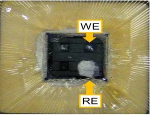
Figure 2. CMOS chip electrode
The CMOS chip (Rohm 0.18m 1P5M standard CMOS process) was designed in our laboratory. The four metal pads on the chip are arranged, and they are wired to the outside of chip. We made working electrode (WE) and refrence electrode (RE) on these metal pads. The WE was made by graphene ink (Sigma Aldrich, 798983), the metal pad is covered with graphene ink, and the CMOS chip is heated for 60 minutes at 300 ℃on a hot plate. In a method for fabricating the RE, the metal pad is covered with the silver-silver chloride ink for the reference electrode (Ag/AgCl, BAS, 011464), and then naturally dried at room temperature for 12 hours (humidity is 60%, temperature is 25℃). After electrodes are formed, the bonding wires are coated by the silicone resin (Momentive Performance Materials Japan LLC, TSE399 C) in order to prevent from the leakage of the solution and disconnection of wiring.
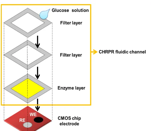
Figure 3. Bird's-eye view of a combination of CMOS chip electrode and CHRPR fluidic channel
The CHRPR fluidic channel consists of two pieces of paper. The first sheet filters the samples (filter layer) and the second sheet contains an enzyme and mediator (enzyme layer). The CHRPR fluidic channel was made of CHRPR (Whatman 1CHR) and silicone resin (TSE3996 C). The fabrication of the CHRPR fluidic channel are used for a robot arm (DESKTOP ROBOT MUSASHI ENGINEERING, INC, SM200SX 3A) and the dispenser (Precision dispensing machine, Musashi engineering, ML-5000X II). We fabricated the patterns of the fluidic channels using the software which controls the robot arm. The robot arm dropped a silicone resin to CHRPR in the syringe, and the silicone solidifies by humidity (humidity is 60%, temperature is 25℃) for one day. The patterns of the CHRPR fluidic channel are showen in Figure 4.
We fabricated an enzyme layer on one of the fluidic channels. The enzyme layer was functionalized by glucose oxidase (GOD, Wako 190units/ g, 074 02401), potassium ferricyanide (FERRI, Wako, 161 03725) and phosphate buffered saline (0.01 mol/L, PBS, Wako, 164 18541).
Figure 4. The patterns of the fluidic channels, (a) Front side, (b) Back side
TELKOMNIKA ISSN: 1693-6930 839
Figure 5. CHRPR fluidic channel, (a) aspects, (b) filter layer, (c) enzyme layer
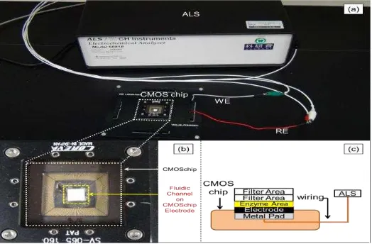
In the measurement, the CHRPR fluidic channel was placed on the CMOS chip electrode. The CMOS chip electrode was connected to the electrochemical measurement analyzer (ALS, ALS/CH Instruments Electrochemical Analyzer Model 610DR) (Figure 6).
Figure 6. (a) Connecting the CMOS chip and the electrochemical measurement analyzer, (b) Placing CHRPR fluidic channel on the CMOS chip electrode, (c) The schematic view of the
CMOS chip, CHRPR fluidic channel and electrochemical measurement analyzer,
3. Results and Analysis
The glucose solution was dropped on top of the filter layer. The concentrations of glucose in the solution are 0.5mM, 1.0mM, 2.0mM, 5.0mM and 10mM. The glucose solutions contain glucose (MP Biomedicals, 492 61 5) in the PBS. The sample ume was 20L and the measurement time was 150s.
The experimental results shown in Figure 7 indicate that the glucose concentration is measured using the CMOS electrodes and the CHRPR fluidic channel. The decreases of the potential with increasing the concentration of glucose were confirmed by the results at 150s. The decrease is attributed to the fact that FERRO was produced by enzyme reaction of glucose and the concentration of FERRO was higher than FERRI. In this way, Potential takes negative value according to the Nernst equation Equation (1), and the decrease of potential was confirmed. In addition, the reproducebility was represented by 1.0mM, 2.0mM, 5.0mM and 10mM.
detection accuracy of 0.5mM. The experimental slope approaches theoretical one by improving detection accuracy of 0.5mM. The of the experimental results was 0.929, so reproducebility is gained by the experiment because the is close to unity. The minimum glucose concentration of detection by the experiment was 1mM. The concentration of glucose for diagnostics of diabetes is 11mM. Thus, the results of the experiment suggest the ability to apply for medical check-up.
Figure 7. OCPT for three concentrations of glucose (Horizontal axis is time [s] and vertical axis is potential [V])
Table 1. Average potential in 150s
Concentrations of glucose [mM] Average potential [V]
0.5 0.03738
1.0 0.02339
2.0 0.05054
5.0 0.08991
10 0.10280
Figure 8. Calibiration curve of average potential and errorbars in 150s
(Horizontal axis is logarithm of glucose concentrations [M] and vertical axis is potential [V])
4. Conclusion
TELKOMNIKA ISSN: 1693-6930 841
Acknowledgements
This work was supported by JSPS KAKENHI Grant Number 26289111. This work was supported by VLSI Design and Education Center (VDEC), the University of Tokyo in collaboration with Synopsys, Cadence Design Systems, and Mentor Graphics. The VLSI chip in this study has been fabricated in the chip fabrication program of VLSI Design and Education Center (VDEC), the University of Tokyo in collaboration with Rohm Corporation and Toppan Printing Corporation.
References
[1] Ministry of Internal Affairs and Communinatons in Japan.
[2] Paolo Livi, et al. Monolithic Integration of a Silicon Nanowire Field Effect Transistors Array on a Complementary Metal Oxide Semiconductor Chip for Biochemical Sensor Applications Analytical Chemistry. Chem. 2015; 87: 998 −9990.
[3] Wei Jhe Ma, et al. A Portable Low Power Acquisition System with a Urease Bioelectrochemical Sensor for Potentiometric Detection of Urea Concentrations. Sensor. 2016; 16: 474.
[4] Yusuke Kanno, et al. Potentiometric bioimaging with alarge-scale integration (LSI-based electrochemical device for detection of enzyme activity. ELSEVIER, Biosensors and Bioelectronics. 2016; 77: 70-714.
[5] David Welch, Jennifer Blain Christen, et al. Seamless integration of CMOS and microfluidics using flip chip bonding. Journal of Micromechanics and Microengineering. 2013; 23(3).
[6] Tao Dong, et al. Recent Developments in Optical Detection Technologies in Lab-on-a-Chip Devices for Biosensing Applications. Sensor. 2014; 14: 15458-15479. doi:10.3390/s140815458
[7] J Sherma, B Fried. Handbook of Thin-Layer Chromatography Third Edition. 2003: 1.
[8] Arshad, et al. Investigating Flashover Behaviour of Silicone Rubber Insulators under Contaminated Conditions IEEE, 10.1109/CEIDP.2015.7352108
[9] PB Luppa, et al. Clinically relevant analytical techniques, organizational concepts for application and future perspectives of point-of-care testing. ELSEVIER, Biotechnology Advances. 2016; 34: 139–160. [10] Andres W Martinez, Scott T Phillips, George M Whitesides, et al. Three dimensional microfluidic
![Figure 7. OCPT for three concentrations of glucose (Horizontal axis is time [s] and vertical axis is potential [V])](https://thumb-ap.123doks.com/thumbv2/123dok/252664.504563/5.595.134.459.363.603/figure-ocpt-concentrations-glucose-horizontal-axis-vertical-potential.webp)
