Evaluation
of
EBV-LMPI
as
prognostic
indicator
of nasopharyngeal
carcinoma
in
fndonesian patients
A.N.Kurniawan*, Ria Kodariah.,
Meryanne Elisabeth-,Averdi
RoezinT, SoehartatiGondhowiardjot
Abstrak
Empat puluh delapan kasus karsinoma nasofartng yang teLah mendapat radioterapi, diperilca ekspresi EBERnya pada sediaan histologik dengan cara hibridisasi
in
situ . Didapatkan 69Vo kasus yang positif untuk EBER. Sediaan histoLogikdai kasus yang EBER
positif diperiksa ekspresiLMPl-
nya dengan cara imunohistokimia dengan hasil 68Vo yang positif. Data stadium klinik ke 48 kasus dan responsradiasi
dari 32 kasus diperoleh dari rekam medik. Data ketahanan hidup sampai 2 tahundari
16 pasien diperoleh dengan cara surat menyurat dan hubungan telpon. Secara statistik ditemukan hubungan bermakna antara klasifil<nsi histopatologik menurut 'Working Formulation' dan respons radiasi. Hubungan bermaknajuga
ditemukan antara elcspresi LMPL dengan data ketahanan hidup pasien-Hal ini
menunjukkan bahwaLMPI
yang telah diketahui mempunyai pengaruh terhadap sel limfoid dan epitel, secara klinikopatologik terbukti dapat berfungsi sebagai saLah satu indikator prognostik. (Med J Indones 2002; 11:8I'6)
Abstract
Forty eight cases of nasopharyngeal carcinoma patients who had been treated with radiotherapy were analysed
for
the expressionof
EBER in the nucleusof
the tumor cells byin situ hybridiTation technique. EBER was
expressedin
69Voof
the cases. Histologic specimens positivefor
EBER were then examinedfor
the expression of EBV-LMP1 by immunohistochemistry, which showed 689o positîvity. Clinical staging of 48 cases and radiation response bf 32 cases were collected. Data of up to 2 years of survival of 16 cases were obtained through postal and phone communication. Statistical analysis showed th.at the Working Formulation histologic classification of NPC correlated significantly with radiation response. The expression of EBV-LMPI was correlated significantLy tvith the survivalof the patients. Thus
EBV-LMPI
which has been shown to possess inJluenceon
lymphoid and epithelial cells, clinicopathologically proved to have a function as prognosric indicator. (Med J Indones 2002; 1l: 81-6)Keywords'. nasopharyngeal carcinomà, histopathologic types, EBER, EBV- Impl, survival
Nasopharyngeal
carcinoma
(NPC) is
the 4'h
mostfrequent malignancy
in
Indonesia.'
NPC
has been
shown
to
havea
strongrelationship
with
EpsteinBarr
virus
@BV)
infection.
Indeed,EBV
has been proven to be expressedin al[
three typesof NPC.2
Expressionof
EBV
genomein NPC
cells could be
demonstratedby
the
occurrence
of
Epstein
Barr-encoded
small
fu\A
(EBER)
with
in sira hybridization
technique. TheEBV
genome containsBNLF-l
gene. Oneof
the productsof
this
gene,the
LMPI
protein,
has been shownto have
oncogenic potential
by
transfbrming epithelial cel[s in
vitro.3
Clinically
it
wasshown that
the expression andnon-expression
of LMP1
in
tumor
cells
showed different tendencyof
tumorgrowth
and metastasis.aAs
radiotherapy
is
the
standard
treatment
for NPC
patients,
the
outcome
of
this
trealment modality
is measuredeventually
as thesurvival
of
the patients.The
purpose
of
this
study
is
to
evaluate
the role of
LMPl in NPC
patients
who
were
treated with
radiotherapy compared
wilh
their
survival
unlil
2 years after treatment.METHODS
Forty eigbt
cases
of
Indonesian
NPC patients
who
cameto
the Department
of
Ear, Nose
andThroat Dr
Depctrtment of Anatomic Pathology' , Ear, Nose and ThroatT-!
Cipto
Mangunkusumo
National Central Hospital for
biopsy
enteredthis
study.The pathologic
diagnosisof
the
biopsy
specimen wereconfirmed
at the Departmentof Anatomic
Pathology.
These caseswere
seenin
theyear
1995, 1996
and
1997.
Subsequentto
diagnosis, the patientsunderwent radiotherapy
at theDepartment
of Radiology,
Division of
Radiotherapy, wherefollow-up
study
for
a limited time (at
leastfor
two
months) wasdone. Afterwards
housecalls
were
attempted to record whether the patients werestill
alive or not.The histopathologic
specimenswere
analyzedfor
the expressionof EBER,
using the
standard procedureof
in
situ
hybridization technique.
The
expression
of
EBER
in
histologic
specimen
was examined
right
after
the
completion
of
each reaction.
EBER
was registered as'positive' if
purple granules were foundin
the
nucleusof the tumor cells.
EBER
was considered'negative'
if
there were no granules seenin
thb nucleus.Specimens
from
EBER
positive cases
were then
treated by
immunohistochemistry using
the
LMP1
antibody
(DAKO).
LMPl
was considered'positive'
if
the antigen-antibody complex was
seen
as
yellow-chocolate granules
on
the
plasma
membrane'and
intracytoplasmic,
while no
granules meant 'negalive'
result.
Radiation
responseof
the
caseswere
assessedwithin
2
months after completion
of
radiation, The
responseswere categorized as complete
response(CR),
partial
response
(PR)
and no response(NR).
Survival
data were collectedby
stampedretum mailing
letters and
by
phone calls, based upon personal data aswritten in
the hospital medical record.The histologic
diagnosis,clinical
staging, radiotherapydata and
survival
data were
collected
and
tabulated.Statistical analysis using chi-square test,
with
Yates'correction
where necessary, andMantel-Haenszel
chi-square
test, was done
lo
look
for
correlation
of
thepathologic characteristics and the radiation
responses andsurvival.
Ethical
clearance
to use the patients' material
was obtained.RESULTS
The gender
distribution
females,
which
made
thewas:
35
males
and
13ratio
of
2.7
:
l.
The
ageMed J Indones
distribution
of
the cases was asfollows:
the
mean ageof
thepatients were
44.5
years,the youngest
was
17years
old
and the
oldest was
70
years
old.
Thehistologic types
of NPC used in
this
stuCy is
theWorking Fdrmulation
classification,s'6
which
divides
NPC
into
3 types:keratinizing
squamouscell
carcinoma(KS),
typeA
carcinoma, and typeB
carcinoma.Table 1. Histotogic types ol NPC
Histologic types # cases Vo
Keratinizing Sq. cetl carcinoma (KS) Type A catcinoma
Type B carcinoma
Undi fferentiated carcinoma *
9
28
9 2
l8
58 18 6Total
* Specimen unretrievable for reclassification into the Working Formulation
Clinical
staging
of
the
cases
as
assessed
beforeradiotherapy is
seenin Table
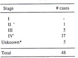
2.Table
2.
Clinical staging (TNM)Stage # cases
100
48
I
II'
UI TV'Unknown*
I
5 3'7
)
Total
*
Data not retrievable lrom the medica.l recordRadiation'response
data which were collected from
the medical record were available
in
32 cases,
sofurther
analysis
could be
done
only
upon
these
32 cases.The radiation responses in ttrese 32 cases were as
follows:
Table 3. Radiation response of NPC cases
Radiation response # cases 48
Complete response (CR) Partial response (PR)
No
response (NR)l2
l8 2
[image:2.612.320.472.403.517.2]VoL 11, No 2, April- June 2002
The
expression
of
EBER
in
histologic
specimensshowed
that
some
of
the
tumor
cells
containedabundant
and dark
granuies,
while
others
were less
dense
and lighær.
Both
were
regarded
as
positive
cases.
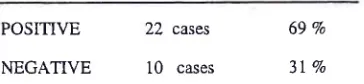
Results of the ISH test were recorded as follows :
Table
4. EBER positivity
of NPC
casesEBV-LMP I as prognostic indicator of
NPC
83Table
7.
EBER,LMPI,
radiation response and histologic typesHistopathologic lypes
KS
TypeA
TypeBs)
)
POSITIVE
NEGATIVE
22
casest0
cases69%
31 7o
EBER
positive
3negative
3LMPl
positive
2negative
I Radiation responseCR
PR
NR
I
4
I
l4
59 5
8
ll
0
4
I
J J 1
Mantel-Haenszel
X2 = 0.0363
(NS)
X2 = 5.9993 p < .025
POSITIVE
NEGATIVE
15 cases
7
casesImmunohistochemical reaction
with
antibody to LMPl
was done
oîthe22
caseswhich
showed positiveEBER
staining.The result
of the
22 cases is shownin Table
5.Table 5. LMP1 positivity of EBER -positive NPC cases
From
this table
it
could be
seen
that there was
nosignificant correlation
of
the eKpressionof EBER
andLMP1 with
the histopathologic
typesof
NPC. On
theother hand, there was significant conelation
of
the radiation responsewith
the 3 histologic typesof
NPC.Further
analysis
was
done
to
seethe correlation of
LMPI
expression with staging of
the
disease(Table
8).
Table 8.
LMPI
expression versru stagingClinical Stage
LMPI
positiveLMPI negative
X2 test for trend = 0.006 (NS) ; p = 6.66
There was
no
significant conelation between
thepositivity
of
LMP1 with
the staging
of
the
tumor,
although
the numberof
cases analysed were small.The expression
of LMPI
was analysed to seeits effect
on
radiation response and survival (Table 9).
68 Vo
32 Vo
Obtaining
the
survival data
of
the
patients
weresuccessful
in
only l6
patients. Information
wasrestricted
to
survival
status
within
2
years
after
completion of treatment (Table 6).
Table
6. Survival
data of NPC patientsIV
m
I
1l
6 4
0 0
I
DEAD
ALIVE
[2 cases
4 cases
TOTAL I 6 cases
Analysis
of
EBER and LMPl
eKpression
andradiation
responseaccording
to
histopathologic
types are shownin Table
7.Table 9.
LMPI
versus outcome of therapyRadiation Response Survival
v2
CR PR x2
NS
Dead Alive
5.013
<.05 LlvlPl positive [image:3.612.303.545.122.282.2] [image:3.612.45.227.219.261.2]84
Kurniawan et alResult
of
this
analysis
showed
that
LMPI
did
notcorrelate
well
,with
radiation
response,
but
wassignificantly
correlated
with
survival
of
the
patients.Out
of
16 caseswhich
hadsurvival
data,the
numberof
patients evaluated
in
this
Table
was
only
11because
the
other
5
cases
did not
have LMPI
examination
asthey
wereEBER
negative.DISCUSSION
This
study
has
several constraints
due
to
theincomplete data retrieved
from
the medical
record(only
32 out
of 48
study
cases hadradiation
responsedata) and the
scarcity
of follow
up dataof
the patientsmore than
2
months post radiation.
Only
1/3
(16
outof 48)
of
patients could be
tracedof their
being
aftercompletion
of
treatment.
This fact
posesa
limitation
of
analysis
of
the
data, although
the
histopathologic
and
immunohistochemistry
andmolecul4r
studycould
be done
completely.
Epstein
Barr virus
has
been
linked
with
severalmalignancies,
such
as
Burkitt's
lymphoma,
naso-pharyngeal carcinoma,
B
cell
lymphoma,
T.
cell
lymphoma, Hodgkin's
disease and gastriccarcinoma.'
Besides,
it
is
also
a
causal agent
for
infectious
mononucleosis.
Most of
the malignanciesoccur
yearsafter
viral
infection
in
the
cells.
In
NPC,
latentinfection of EBV
has been detectedin vivo
astype
II
latency,
expressingEBNAI, LMP1, LMP2A,
LMP2B
and EBERs.t
As
EBERT
are
expressed
at high
abundance,
approximately
107copies
per
cell
in
all
latently infected cells, EBERs can
be
consistently detectedin
NPC
specimensby
Northern
blot
and insita hybridization. The latter
technique can be done onparaffin
embedded specimen.Thus, the
expressionof
EBER
in
a
histologic specimen
denotes
the
sureexislence
of
EBV
genome
in
the
cell. This
is
thereason
why
we examined
theLlvlPl
expressionin
all
EBER positive
casesonly. In
our
series, thepositivity
of EBER
was 68Vo.This figure is lower
than
reportby Murono et ale who
detected 82Voof EBER in
56primary NPC
cases
in
Korea. The
reason
for
thisdifference
is
unclear,
it
may be
causedby
different
probe, different lechnical
skill
or
different strain of
EBV. Non
expression
of
the
EBER
does
not necessarily meanthat
lheEBV
genomeis
not present.Takeuchi et
a1r0who
delectedEBER
positive
in'75Voof
NPC
cases,found out that by
using
PCR
methodwhich
can detect onecopy of
EBV DNA
percell,
theycould
detect
EBV-DNA in
30Vo
of
the EBER
-Med J Indones
negative
NPC
cases.EBER is
a non-translatedmRNA
and the
function
is sofar
unclear.There
is
no
significant correlation
between
EBER
aird
LMP1
and histologic types
of
NPC
(Table
7).This finding
emphasizedthe
fact
that
EBV
has
beenshown to be
found
in
all
three typesof NPC.rr
Result
of this
study
showedthat there
was
statistical
significant corrélation between the histologic
types(the
Working
Formulation)
and theradiation
response(Table
7). This
finding
is
in
accordance
with
ourprevious
report,o
which
showed
the
percentagedifference
of
radiation
responsesof
the
3
types
of
NPC. Data
from our
previous
report
showed that CR
(complete
response)can
be
achieved
in
116(16,6%)
of
KS type,
and
8/19
(42,IVo)
of
type
A
carcinoma,while
type
B
carcinoma
showeda3l7
(42,97o)of
CR. Thus,this classification could
be used asa
prognostic
indicator of
NPC.LMP1
as
a
protein product
of
BNLF1
gene has
amolecular
weight
of
60-66
kD.
It
can
be
detected in
plasma membrane and
partly
in
the cytoplasm
as
it
has
3
domains
:
intracytoplasmic
N-terminus,
trans-membrane
dan intracytoplasmic C-terminus.
LMP1
has several
functions in vitro,
a.o.
to immortalize
thelymphocytes
and severalepithelial cells, to
upregulatethe
bcl-2
in
lymphocytes
andthe expression
of
A20
zinc
finger
protein
in
epithelial cells,
which
may
leadto
reducedapoptosis.
In
vivo
LMPI
has been shownto influence
thegrowth
and tendencyof
metastasisof
NPC.
Hu et
ala reported
that
LfvIPl
positive
tumor
grew faster
and
more
expansively
than
LMPl
negativetumor,
but
had betterprognosis. On
the otherhand,
LMPI
negative
tumor
recurred
in
a
higher
frequency
and
showed tendency
lo
form
metastasis.Our
study
confirmed
thenotion that
LMPl
has someint-luence
on
the
outcome
of
the
patients,
as
thecorrelation
of LMPI
with
survival was
statistically
significant
(Table
9). This is true
despite the
fact of
the
low
number
of survival
dataof
our patients, not
tomention
the otherfactors
thatmay influence
the causeof
deathof
the diseased patienls.However,
for
a short termfollow-up,
there was nosignificant correlation
of
LMPI with
the
radiation
response
(Tabte
9).
Tq
ascertain
the influence
of
LlvlPl
as un on"og.n,t'
lurther
studies have
to
be
done, such
as
closemonitoring
and good recordingof the
iollow-up of
thepatients
after radiation
lreatment
io
assessthe
tumor
growth
and/or metastasis.This'can
be
donein
a goodVol I
I,
No 2,Apil
-
June 2002find a conelation
of LMP1 with
the
proliferation
andapoptotic
index of
tumor.l3CONCLUSION
Analysis
of
correlation
between
histopathologic
characteristics,radiation
response andsurvival
data of IndonesianNPC
cases had been done.The result
has somelimitation
dueto
the small numberof retrievable
data.
Working
Formulation classification
of
NPC
histo-pathology has significant correlation
with radiation
response, thus
having prognostic
importance.LMPl
did
not show significant influence
onradiation
response,however
LMP1
has
a role
as
prognosticfactor
for
survival
of NPC.
ACKNOWLEDGMENT
The
authors
wish
to
thank
Li
Fu Hu, MD,
PhD"Karolinska Institute, Stockholm,
for
the
donation
of
EBER probe
andLMPI
antibody to run
thepathology
tests.
Gratitude
is
also due
to
National
lnstitute of
Health
Research
and Development, Department of
Health, Republic
of Indonesia, for research
grant
for
the implementation
of
the
clinical, pathologic
andsurvival
study. Guidance and
scientific supiort were
accepted
from
Eijkman
Institute
for
Molecular
Biology, Jakarta.
REFERENCES
1.
Cancer in Indonesia 1992. Histopathologic data. Directorate Generalof
Health
Services, Departmentof
Health, Republic oflndonesia; Cancer Registry ofThe lndonesianEBV-LMP
I
as prognostic indicanr ofNPC
85Society
of
Pathologists; Indonesia Cancer Foundation.Jakarta
Pagano JS. The Epstein -Barr Virus and Nasopharyngeal
Carcinoma. Editorial. Cancer 1994; '74 :2397-8.
Hu
LF, Chen
F,
Zheng
X,
Ernberg
I,
Cao
SL, ChristenssonB et
al.
Clonability and tumorigenicityof
human epithelialcells
expressingthe
EBV
encoded membrane protein LMPI. Oncogene 1993; 8 : 1575-83.Hu LF, Chen F, Zhen
Q-l
Zhang Y-W, Luo Y,Zheng X etal. Differences in the growth pattern and clinical course
oi
EBV-LMPI expressing and non-expressing nasopharyngeal carcinomas. EurI
Cancer 1995; 31A : 658-60.Hsu HC, Chen CL, Hsu MW, Lynn TC,
Tu
SM, HuangSC. Pathology of nasopharyngeal crcinoma. Proposal of a new histologic classification correlated
with
prognosis.Cancer 1987; 59 : 945-51.
Kurniawan
AN,
SyafrilA,
SusworoR.
Nasopharyngeal carcinoma.An
altemative classificationas
a
Working Formulation. Med J Univ Indon 1993; 2 : 37 -41 .Pagano JS. Epstein-Ban virus: the first human tumor virus and its role
in
cancer. Proc Assoc Am Physicians 1999; 111 :573-80.Hu
LF.
Nasopharyngeal carcinomaand
Epstein-Barr virus. Stockholm, Karolinska Institutet,1996. Thesis.Murono S,Yoshizaki
T,
Park C-S,
Furukawa
M. Associationof
Epstein-Barrvirus infection
with
p53protein
accumalion
but not bcl-2
protein
innasopharyngeal carcinoma. Histopathol 1999 ; 3 4: 432-8.
Takeuchi
H,
KobayashiR,
HasegawaM,
Hirai
K. Detectionof
latent Epstein-Barrvirus (EBV)
DNA
inparaffin
sections
of
nasopharyngeal carcinomasexpressing no EBV-encoded small RNAs using
in
situ PCR. ArchVirol
1997; 142:1'143 -56.Vasef
MA,
Ferlito
A,
Weiss LM.
Nasopharyngeal carcinoma, with emphasis on its relationship to Epstein-Barr virus. Ann Otol Rhino Laryngol 1997; 106 : 348- 56.Nicholson LJ, Hopwood P, Johannessen
I,
Salisbury JR,Codd J, Thorley Lawson D et al. Epstein-Barr viru's latent membrane protein does
not inhibit
differentiation andinduces tumorigenicity
of
human epithelial
cells.Oncogene 1997 ; 15 : 2'75 - 83.
Vera Sempere FJ, Burgos JS, Botella
MS,
Morera C.Immunohistochemical expression of Bcl-2 oncoprotein in EBV-associated nasopharyngeal carcinoma correlated to histological type and survival. Histopathol 1997; 12l,9 -18.
J
5.
7.
8.
9.
ll.
10
T2
86
Kumiawan et alATTACHMENT TABLE
Med J Indones
NO SEX AGE PA NO, NPC TYPE STAGE RT DOSE RR EBER IMPl SURVIVAL
1 48 95021 17 A 4 + f,b PR +) +) ,)
2 31 592420 4 + fo PR ) Dead
58 503490 B + 70 NR +) +) Dead
VI ôt 9503570 B + lo PR +) +) Dead
F l5 )504008 B + i6 PR ) Dead
VI l9 t504538 KSS + t6 PR +) -)
7 V 1 )507326 A 4 + t6 PR +) )ead
M l8 1604602 c + i6 CR +) +) \live
M 50 1601 900 4 + 10 PR )eaC
10 M 30 9602686 (SS + + lo PR -) ,)
11 M 42 1602000
l
4 + t0 CR Dead12 M 35 9602895 A 4 + lb PR +) +) 2
13 F 30 9603554 <SS 4 + â6 CR +) +) I
14 M OJ 9606207 t\ + bb
]R
) Alive15 F 60 9602029 B + 52.8
]R
+) (+) 216 M 32 9604997 A + 66
]R
+) (+) Alive17 M 49 9504734 A + oo CR +) C) ,|
18 N,,I t0 9603972 B + Db PR (+) (-) Dead
'19 Vl l0 9602682 A + t6 PR ?
20 V +5 1606697 B 3 + lo CR (+) (+) Dead
21 \4 1 160641
I
A + t6 PR +),|
22 M
l6
1604434 KSS 4 + ;6 CR (+) +) Alive23 F z1 )603728 q 4 + l6 PR (+) +) 1
24 M 34 )602797 q 4 + 52.8 PR
) ,|
25 F 55 9602927 q 4 + l6 PR +) +) Dead
zo F 20 3600060 (SS 4 + rtJ
!R
) 227 M 43 9507250 q 3 + 66
]R
+) (+) 228 F 23 9605237 A 3 + bb PR +) (+)
29 M 65 9700248 A 4 + 40 R +) (-) Dead
30 53 97009 1 6 KSS 4 + 66 PR (-)
3t N/ 59 9701426 A 2 + 66
iR
+) (-) fead32
f/
65 9701 51 4 A 4 + ô0 PR (+) +)33 30 r504536 A (-) ') ,)
34 rI 17 )606742 A ? 2 (*) +)
35 vl 54 )604089 ,| ,| ,) ,7
+) +) 2
36 M ;5 )601 87304 ! ? ,} ,) ,) [+) ) 2
37 V 50 16051 67
l
') ,) ? (+) ) 238 ;2 1603393 \5S 4 ,) ? ,, +) (+) 2
39 F +3 r602843 A 4 ,) ? ? ,|
+0 M 56 9603754
{
4 ') ,) ,2+) (+) ')
41 M 30 9604456 3 4 (*) 30 ,) \,ls ?
42 M 19 9502936 4 ,| 7 ,) ,)
43 F 35 950371 9 A 4 (*) 20 +) (-) ,>
44 M 65 9502414 A 4 (+) 50 PR \IS ?
45 M 39 9507031 Undiff - ,) ,| NS ,)
46 lr4 70 9603308 A ,) ,) 2 +) +) ,)
47 F 23 9s06653 A 4 ô0 PR NS ,)
48 [r4 )J 9701 929 KSS 4 +) ôb FR NS Dead
Note:
?
= no data availableNS = marerirl not sufficient
l/lll/ =notdone
RT = radiotherapy
RR = radiation response