In vitro maturation (IVM) as a new technique to treat polycystic ovary
syndrome (PCOS) and induce pregnancy in Indonesia
Soegiharto Soebijanto
Department of Reproductive Endocrinology Obstetrics and Gynecology, Faculty of Medicine, University of Indonesia, Jakarta
Abstrak
Tujuan Dilakukan penilaian terhadap keberhasilan kehamilan pada penanganan dengan In Vitro Maturation (IVM) pada kasus-kasus PCOS (Poly Cystic Ovary Syndrome) sebagai teknik yang pertama dilakukan di Indonesia.
Metode Tulisan ini merupakan laporan kasus dari teknik yang baru dikembangkan di Indonesia. Bahan penelitian adalah 7 kasus dengan PCOS yang jelas, diantaranya 1 pasien dengan riwayat OHSS (Ovary Hyper Stimulation Syndrome) pada prosedur fertilisasi in vitro sebelumnya dan 1 pasien dengan gambaran PCOS, kemungkinan hiperesponder, di Rumah Sakit Family dari bulan Januari sampai bulan Mei 2009. Induksi folikel dengan dosis minimal, primming HCG 10.000 IU pada hari ke 10 dan 40 jam kemudian dilakukan ovum pickup, selanjutnya diinseminasi dan folikel yang terbuahi dilakukan penilaian. Embrio yang bermutu baik ditransfer kedalam uterus. Penilaian kehamilan dilakukan secara biokimiawi, penilaian adanya kantung janin dan denyut jantung.
Hasil Telah dilakukan teknik IVM di RS Family, Jakarta Barat bersama dengan tim TRB RS Family pada tujuh kasus. Dari tujuh pasien ditemukan 156 folikel antral atau rata-rata 22 folikel perpasien, ditemukan 81 oosit, dan setelah dimaturasi diperoleh 61 oosit matur (75%). Pada tiga bulan dilakukan fertilisasi in vitro dan 4 kasus dilakukan ICSI (In Cystoplasmic Sperm Infection). Pada serial kasus ini diperoleh 412 embrio, dan 22 buah embrio ditransfer, dan dari 7 kasus diperoleh 2 kehamilan (9%).
Kesimpulan Teknik In Vitro Maturation (IVM) merupakan alternatif untuk mengatasi masalah infertilitas pada pasien PCOS dengan keunggulan risiko sindrom hiperstimulasi ovarium yang rendah serta biaya yang lebih murah. (Med J Indones 2009; 18: 269-75)
Abstract
Aim To assesse the success of inducing pregnancies in the treatment of PCOS (Poly Cystic Ovary Syndrome) cases with in vitro maturation as a newly application technique in Indonesia.
Methods This paper is a report of 7 cases in Indonesia that used the newly developed technique. There were 7 cases confi rmed PCOS, in which 1 patient with a history of OHSS (Ovary Hyper Stimulation Syndrome) in a previous IVF procedure and 1 patient with PCOS characteristics, suspected hyper responder, in the Family Hospital from January to May of 2009. Follicular induction was performed with a minimum dose, primming with HCG 10.000 IU, on the 10th day and 40 hours later ovum pick up was performed, followed by in vitro maturation. Subsequently, insemination was performed and the inseminated follicle was assessed. Well qualifi ed embryos then transferred them into the uterus. We then performed assessment of pregnancy biochemically, by the presence of embryonic sac and embryonic heart beat.
Results We have performed the IVM (In Vitro Maturation) technique in the Family Hospital, West Jakarta, along with the TRB team of the Family Hospital in seven PCOS cases. From these patients, we have found 156 antral follicles (average of 22 follicles per patient), 82 oocytes, and after maturation, 61 mature oocytes (75 %). In three cases, in vitro fertilization was performed, while in 4 cases ICSI (In Cystoplasmic Sperm Infection) was performed. In these serial cases we obtained 41 embryos, and 22 fertilized embryos were transferred. Of 7 cases, we achieved two successful pregnancies (29%).
Conclusion In vitro maturation is an alternative procedures in solving infertility problems for PCOS patients with lower risk of OHSS and more cost effective than conventional IVF. (Med J Indones 2009; 18: 269-75)
Compared to normal women, PCOS have a larger ten-dency to catch the side effect of OHSS. It is believed that the stimulation of ovulation may induce ovarian and endometrial cancer.1 Besides, stimulation with gonado-tropin and GnRH agonist can be costly.2
Many scientists have performed IVF (in vitro fertiliza-tion) in the normal cycle because of the simplicity of the method, limited side effects, low cost and considering the patient’s comfort. Although the normal cycle pro-vides limited success due to the low number of oocytes gained, the success rate of pregnancy is 46% after the procedure has been repeated 4 times.3 Therefor, it has been considered to perform the IVF technique in the nor-mal cycle with in vitro maturation. By this way, even without costly stimulation the patient still has a good chance of achieving pregnancy.
Nonetheless, this technique should only be applied in certain cases considering the specifi c conditions of each case, to optimize the success of pregnancy. Fur-ther research must be performed to fi nd the optimum conditions for cultivation to improve the implantation rate of oocytes that are matured in vitro.
This article will describe the experience of performing the IVM program in PCOS cases and predicted hyper-responders.
The IVM technique was fi rst developed in 1940.4 It was per-formed until 1960 when a technique was develop to obtain an oocyte from a de Graaf follicle through laparoscopy.5
Cha in 1991, and Trounson in 1994, announced the success of achieving pregnancy through IVM in PCOS cases.6,7 PCOS patients are good candidates for IVM because of the number of antral follicles in this case group. In general, 30 to 35% of PCOS patients will achieve and 10 to 15% in PCOS patients.8,9
Thus, IVM was performed to lower costs, to prevent OHSS in PCOS patients, and to simplify the procedure of IVF.10,11
METHODS
All the cases were collected in the Family Hospital, Ja-karta from April 2008 to April 2009. This is a prelimi-nary report of a prospective study, assessing the rate of in vitro maturation, fertilization, the formation of em-bryo, and ultimately, pregnancy.
The cases used in this study were 7 infertility cases with PCOS, established through the Rotterdam criteria,
and one case with a history of OHSS (ovarian hyper-stimulation syndrome). HSG was performed to observe uterine cavity in cycle before the program was started.
Hormonal measurements were taken for FSH, LH, E2 and testosterone. On the sexual partner we performed sperm analysis and sperm recovery test. We injected the patient with gonadotropin 150 IU in days 8, 9, and 10 of the menstrual cycle, and priming with HCG 10.000 IU. On the 11th day and 40 hours later we performed OPU (Ovum Pick Up)
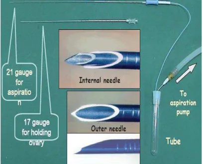
During OPU we measured the diameter of the follicle. OPU was performed using a combination of size 19 g aspiration needle and a set of aspiration needles with the outer needle sized 17 g and inner needle sized 20 g with a suction pressure of 80 to 110 mmHg.
Basic USG
USG was performed on the second day of the men-strual cycle to observe the uterus and to determine the number of BAF (basal antral follicle) in both ovaries. If the patient has an irregular cycle, then progesteron preparation (Provera 5 mg, twice daily for fi ve days) was used to induce menstruation.
The next USG was performed on the 6th and 9th days to measure the thickness of the endometrium and to ensure the presence of a developing follicle. (Figure1)
Injections of 150 IU gonadotropin was given to the pa-tients on days 8, 9, and 10. On the 11th day, we gave an injection of 10.000 IU HCG. In the case of the hyperre-sponder whose follicle was 11 mm on the 9th day, HCG priming was performed on the 10th day.
HCG Administration
Patients were given 10.000 IU hCG 36 hours prior to OPU. OPU was performed between day 10–14, de-pending on the endometrial thickness (must be 6 mm or more) and the follicular diameter. This is very im-portant to prevent spontaneous ovulation.
OPU (Ovum pick up)
One day before the OPU, a medium was prepared: Syn-vitro fl uch, LAG, IVM, FSH solution, hCG solution, stored in 37o Celcius and 5% CO
2. Obtained oocytes were incubated in LAG medium for 2 to 3 hours, then transferred to fi nal IVM maturation medium, consisted of IVM medium, patient serum, and FSH solution. hCG solution was incubated for 24 to 48 hours. The next day, the oocyte was denuded, then stored at ISM1 medium for 2 hours. Oocytes that fulfi ll requirements underwent ICSI (intracytoplasmic sperm injection).
The needles used for OPU were a combination of size 19 g aspiration needle and a set of aspiration needles, outer 17 g needle, inner 20 g needle, with a suction pressure of 80 to 110 mmHg. All visible follicles were extracted while the patient underwent general anesthesia.
Method of locating oocytes
Follicular solution was transferred to a petri dish then examined under a stereo miscroscope. Follicular so-lution was strained through a cellular sieve of 70 um, then diluted by a washing solution, then transferred to a petri dish (falcon 60 x 15 mm). All procedures were performed on a plate of 37o C.
Maturation procedure of oocyte12,13,14
Medium was prepared at least one hour ahead of OPU and placed in an incubator of 30o C. Three petri dishes were required (falcon 35 x 10 mm) consisting of 2–2,5 ml of oocyte washing solution under mineral oil for each patient. For each patient, we also prepared, 50 ml Falcon fi lled with 25-50 ml of oocyte washing solution in an incubator. The oocytes were then gathered in a 10 ml tube fi lled with washing medium mixed with 2-3 ml. 2 IU/ml heparin. Other than the usual medium, we also used 0,9% NaCl mixed with 2 IU/ml of heparin.
Immature oocytes were incubated in a tissue culture dish (falcon 60 x 15 ml, consisting of 1 ml maturation medium with a fi nal concentration of 75 mIU/ml of FSH and 75 mIU/ml of LH in 30o C with a fl ow of 5%
CO2 and 95% air or of three gasses 90 % N2, 5 % CO2, and 5 % O2 with a humidity of 100 %. This medium was prepared at least 2 years prior to OPU.
Ovum pick up was performed 40 hours after hCG ad-ministration under general anesthesia. OPU was per-formed using COOK ovum aspiration needle size 19 g or COOK ovum aspiration needle set consisting of 17 g outer needle and 20 g inner needle. (Figure 2 and 3)
Needle selection depends on the condition of the fol-licle. In patients with a history of OHSS and hyper-responder, where the diameter of most follicles were 9 to 11 mm, we used a 19g needle. But in patients with PCOS, in which most of the diameter of follicles were 6 to 8 mm, we used a needle set of 17 g and 21 g needles. In the latter condition, the follicles were fi rst punctures with the 17 g outer needle to the adjacent of the target
Figure 2. Ovum pick up needle16
follicle, then the 21 g inner needle was inserted into the follicle and the follicular solution was aspirated. For the next follicle, the inner needle was then retracted into the outer needle that was manipulated in such a way that it reaches the side of the next follicle.
In cases where the vaginal walls were considered thick and rigid, we used the outer and inner needle set, be-cause the 19 g needle was very pliable making it dif-fi cult to puncture the vaginal wall. During perforation, pressure to the abdominal wall was applied by a nurse to help fi xate the ovary.
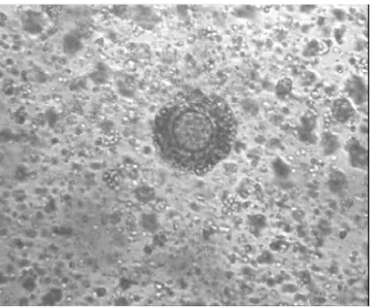
Rinsing of the needles, or tubing, after two or three as-pirations of the follicular solution was very important. Rinsing was performed by using NaCl solution mixed with heparin to prevent coagulation in the lumen of the needle. Aspirated solutions were immediately brought to the laboratory to observe for oocyte cumulus complex. (Figure 4,5, and 6)
RESULTS
The fi rst patient, 31 years old, has been infertile for 8 years and has underwent induction of ovulation 8 times, and failed. Physical examination showed body weight 88 kg, height 160 cm, BMI 34.5, with signs of hirsut-ism. Ultrasonographic examination on the second day of menstruation showed: uterus within normal limits, 40 basal antral follicles (BAF) in both ovaries. Hor-monal examination results showed: FSH 4,5 mIU/ml , LH 3,0 mIU/ml, E2 <20 pg/ml dan testoterone 3,8 ng/ dl. Sperm analysis of the husband were within normal range with sperm recovery of 10 million/ml.
Out of 11 mature oocytes that underwent ICSI, 8 were fertilized, and developed into 8 embryos. Four days af-ter OPU we transferred 4 embryos. The patient did not become pregnant and menstruated.
The second patient was 30 years old, with a history of blighted ovum three years before. Screening revealed a non patent right tube and appearance of policystic ova-ries with more than 20 BAFs. Hormonal examination showed FSH 6,0 mIU/ml , LH 3,9 mIU/ml, E2 <20 pg/ ml. Sperm analysis of the husband were within normal range with sperm recovery of 4.5 million/ml. Upon OPU the follicle was 7.4 mm to 12 mm in diameter. In this patients we obtained 9 immature oocytes and retained 8 oocytes post maturation that were viable for ICSI. Fertilization occured in 5 oocytes and developed into 3 embryos of excellent quality. The three embryos were transferred and given luteal support with vaginal progesteron. The patient became pregnant with one gestational sac, an apparent yolk sac, fetal pole and pulsation. (Figure 7)
Figure 4. oocyte cumulus complex
Figure 6. intra cytoplasmic sperm injection
The third patient was 42 years old with a history of OHSS in a previous IVF procedure, the patient did be-come pregnant but miscarried. Upon examination on the second day of menstruation we found 11 BAFs. Sperm analysis of the husband showed a sperm count of less than 100.000 ml. Upon OPU we obtained 7 im-mature oocytes, to which we induced maturation. Upon maturation, we obtained 6 oocytes that were adequate for ICSI. Fertilization occured in 4 oocytes and devel-oped into 4 embryos. Three embryos were transferred and given luteal phase support with vaginal progester-on. The patient failed to be pregnant.
The fourth patient was 31 year old with a history of abortion. Screening revealed 16 BAFs (PCO), FSH 6,2 mIU/ml, LH 5,4 mIU/ml, E2 23,2 pg/ml. Sperm analy-sis of the husband revealed sperm recovery of less than 100.000 ml. Upon OPU, the follicular diameter was 5 mm, and to 12 mm. From this patients we obtained 19 oocytes, and 13 oocytes post-maturation that were ad-equate for ICSI. Out of those, 12 oocytes became fer-tilized and 11 developed into embryos. Four embryos were frozen and three were transferred and given luteal support using vaginal progesteron. The attempt for pregnancy was not successful.
The fi fth patient was 28 year old with 4 years of pri-mary infertility and a history of failed IVF. Screening revealed bilateral non patent tubes and the appearance of PCO ovaries with 25 BAFs. Sperm analysis of the husband were within normal range with sperm recovery of 3 million/ml. From this patients we obtained 13 immature oocytes, performed maturation, and obtained 12 oocytes ready for ICSI. Fertilization occured in 8 oocytes and 7 of them developed into embryos. Two embryos were transferred. The patient was pregnant and fetal pulsation was recorded on the 6th week of pregnancy.
The sixth patient was 31 years old, with a history of four failed IVFs. The second IVF was successful but was a BO and the latest IVF showed signs of OHSS. The pa-tient had two non patent tubes. After ICSI four oocytes were fertilized and three embryos developed. These three were tranferred, but the patient failed to be pregnant.
The seventh patient was 31 year old who previously had been pregnant but miscarried. She has been married for three years, and has a menstrual cycle of 36 to 48 days. Sperm analysis were within normal limits with sperm recovery of 2 million/ml. We counted 24 BAFs. Upon OPU the follicular diameter was to 9 mm. In this patient we obtained 7 immature oocytes on which we performed maturation. Post maturation we obtained 7 oocytes that were ready for ICSI. Post ICSI, three oocytes were fer-tilized and developed into 3 embryos. These three were transferred, but the patient failed to be pregnant
DISCUSSION
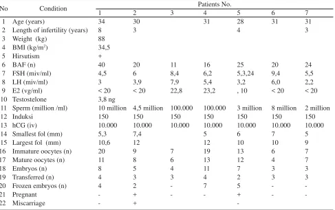
Table 1 shows that out of the seven patients who received the IVM procedures, two patients had obvious PCOS, 1 patient with a history of OHSS in a previous IVF proce-dure. Although hormonally there were no obvious signs of PCOS (less than 23.2 pg/ml), all patients had BAF (more than 11) and all patients fulfi lled the Rotterdam criteria. These are typical of hyperresponders who run the risk of developing OHSS once stimulated through the usual protocol (long or short protocol).
Out of the seven spouses, two cases were oligospermia. Totally in this series there were 156 BAFs, with a mean of 22 follicles and a mean of 12 oocytes. This meant that the OPU was successful in 56% of the cases, which was quite a feat considering that the follicular diam-eters were between 5.3 to 10 mm, and that this was the fi rst OPU attempt on smaller follicles.
Out of 59 mature oocytes, all underwent ICSI, from which 41 (69 %) embryos developed. Out of 7 patients who received transfer, two (28.6%) became pregnant. Although the population size is yet to be expanded, this is an encouraging preliminary compared to the achieve-ments of other countries. Results from other countries based on the cilinical pregnancy rates were: France 18%, Japan 26.8%, Scandinavia (Sweden and Finland) 22%, Vietnam 27.6% and Canada 28%.15, 16 Out of 81 oocytes that were matured in vitro, we harvested 59 (73 %) ma-ture oocytes. This shows that the in vitro maturation technique was quite successful.
This fi nding showed that IVM as an IVF (In Vitro Fer-tilization) procedure was successfully performed in In-donesia. In the future, IVM will be the fi rst choice for patients who are willing to undergo reproductive tech-nology, for the following reasons: 1) simplifi ed proce-dure; 2) cost effi ciency; 3) avoidance of OHSS.
Other advantages of IVM are the preservation of oo-cytes or ovarian tissue in cancer cases, more comfort-able for the patient because it is less pain due to in-jections and the less frequent monitoring of hormonal levels in the blood. Also, it reduces the trauma of hav-ing to endure OHSS in previous IVFs. (Tabel 2)
Acknowledgment
We are especially grateful to the team of Family Hospi-tal (dr. Muchsin Jaffar, dr. Hadi Syarbaini and dr. Yus-lam Edi Fidianto) who have given great assistance in producing this article.
No Condition Patients No.
1 2 3 4 5 6 7
1 Age (years) 34 30 31 28 31 31
2 Length of infertility (years) 8 3 4 3
3 Weight (kg) 88
4 BMI (kg/m2) 34,5
5 Hirsutism +
6 BAF (n) 40 20 11 16 25 20 24
7 FSH (miv/ml) 4,5 6 8,4 6,2 5,3,24 9,4 5,5
8 LH (miv/ml) 3 3,9 7,9 5,4 3,2 6,0 2,2
9 E2 (vg/ml) < 20 < 20 22,8 23,2 , 10 < 20 < 20
10 Testostelone 3,8 ng
11 Sperm (million /ml) 10 million 4,5 million 100.000 100.000 3 million 8 million 2 million
12 Induksi 150 150 150 150 150 150 150
13 hCG (iv) 10.000 10.000 10.000 10.000 10.000 10.000 10.000
14 Smallest fol (mm) 5,3 7,4 5 6 7 5
15 Largest fol (mm) 10,6 12 12 10 10 9
16 Immature oocytes (n) 20 9 7 19 13 6 7
17 Mature oocytes (n) 11 8 6 13 12 4 7
18 Embryos (n) 8 5 4 11 7 3 3
19 Transferred (n) 4 3 3 4 2 3 3
20 Frozen embryos (n) 4 2 - 7 5 -
-21 Pregnant - + - - + -
-22 Miscarriage - +
-Table 1. Characteristic patients and results of IVM
Description n (%)
Follicular puncture 156
Oocyte cummulus complex 81 (52%)
Mature oocyte 61 (75%)
ICSI
Oocyte
Fertilization
Embryo ( good)
59
41 (69%)
22 (54%)
Conventional IVF
Oocyte
Fertilization
3
0
REFFERENCES
1. Nargund G, Waterstone J, Bland J, Philips Z, Parsons J and Campbell S Cumulative conception and live birth rates in natural (unstimulated) IVF cycles.Hum Reprod 2001; 16:259-62
2. Tarlatzis BC, Grimbizis G, Bontis J, Mantalenakis S. Ovar-ian stimulation and ovarOvar-ian tumours: a critical reappraisal. Hum Reprod Update 1995; 1: 284-301
3. Daya S, Gunby J, Hughes EG, Collins JA, Sagle MA and YoungLai EV Natural cycles for in-vitro fertilization: cost-effectiveness analysis and factors infl uencing utcome. Hum Reprod 1995; 10:1719-24
4. Rock J, Menkin MF. In vitro fertilization and cleavage of human ovarian eggs. Science 1946; 100: 105-7
5. Edwards RG, Bavister BD, Steptoe PC. Early stages of fer-tilization in vitro of human oocytes matured in vivo. Nature 1969; 221: 632-5
6. Cha KY, Koo JJ, Choi DH, Han SY, Yoon TK. Pregnan-cy after in vitro fertilization of human follicullar ooPregnan-cytes collection from non stimullated cycles, their culture in vitro and their transfer in a donor oocytes program. Fertil Steril1991;55:109-13
7. Trounson A, Wood C and Kausche A In vitro maturation and the fertilization and developmental competence of oo-cytes recovered from untreated polycystic ovarian patients. Fertil Steril 1994; 62:353–62
8. Donderwinkel PF, Schoot DC, Coelingh Bennink HJ and Fauser BC Pregnancy after induction of ovulation with re-combinant human FSH in polycystic ovary syndrome. Lan-cet 1992; 340:983
9. Chian RC, Buckett WM, Tulandi T and Tan SL () Prospec-tive randomized study of human chorionic gonadotrophin priming before immature oocyte retrieval from unstimulat-ed women with polycystic ovarian syndrome. Hum Reprod 2000; 15:165–7
10. Hadi S, Muchsin J Yuslam EF, Dianing M, Soegiharto S: Laporan Kasus, Pengalaman: Kehamilan dengan In Vitro Maturation (IVM) di FFC RSIA Family. Pertemuan Ilm-iah Tahunan Himpunan Fertilitas dan Endokrinologi Re-produksi Indonesia; 2009 Jan, Semarang. Indonesia. 11. Delvigne A and Rozenberg S Epidemiology and prevention
of ovarian hyperstimulation syndrome (OHSS): a review. Hum Reprod Update 2002; 8:559-77
12. Cha KY, Koo JJ, Choi DH, Han SY, Yoon TK. Pregnancy after in vitro fertilization of human follicullar oocytes col-lection from non stimullated cycles, their culture in vitro and their transfer in a donor oocytes program. Fertil Steril 1991;55:109-13
13. Paulson RJ, Sauer MV, Francis MM, Macaso T, Lobo RA. Factors affecting pregnancy success of human in-vitro fertil-ization in unstimulated cycles. Hum Reprod 1994; 9: 1571 14. Chian RC, Niwa K, Sirard MA. Effect of cumulus cells
pres-ent during differpres-ent periods of culture on maturation in vitro of bovine oocytes. Theriogenology 1994; 41:1499–508 15. Chian RC, Demirtas E, Buckett W, Tan SL. In vitro
matu-ration of oocytes collected from unstimulated ovaries for oocyte donation. Fertil steril 2007; 88: 62-7