The Philippine Journal of Crop Science
Crop Science Society of the Philippines
PO Box 165, UPLB, College, 4031 Laguna, Philippines pjcs.pcs@gmail.com
February 14, 2017
CERTIFICATE OF ACCEPTANCE
This is to certify that the paper of
Suryaminarsih Penta,
Kusriningrum, Ni'matuzahroh, Tini Surtiningsih
and
Tri Mujoko
entitled
“
The Antibiosis of Biological Agents Streptomyces sp., Gliocladium sp., and
Trichoderma harzianum From East Java Indonesia To Fusarium Oxysporum”
is
accepted for publication in The Philippine Journal of Crop Science, and will be
included in our Volume 42: Supplement No.1, June 2017.
Sincerely yours,
THE ANTIBIOSIS OF BIOLOGICAL AGENTS Streptomyces sp. , Gliocladium sp., and
Trichoderma harzianum
FROM EAST JAVA INDONESIA TO FUSARIUM OXYSPORUM
by
Suryaminarsih Penta *), Kusriningrum **), Ni'matuzahroh***),
Tini Surtiningsih***),Tri Mujoko*)
*) Department of Agritechnology, Agricultur Faculty , University of Pembangunan Nasional
"Veteran" East Java
**) Department of Veternary Medical, Airlangga University
***) Department of Biology, Science and Technology Faculty, Airlangga University
Correspondence: arsihpenta@yahoo.co.id
ABSTRACT
This study was undertaken to determine the species of biological agents
Streptomyces sp. from Pare-Kediri tomatoes land, Gliocladium sp., collection of Pandaan
Food Crops and Horticulture Plant Protection, and to know their antagonistic to F.
oxysporum f.sp. lycopersici soil borne pathogens from Wajak village - East Java- Indonesia.
Completely randomized design was used and each treatment was repeated four times.
Biological agents were identified by morphology characteristics and DNA sequensing.
Streptomyces sp. wich was found, was identified as Streptomyces griseorubens and
Gliocladium sp. as Gliocladium virens. The results also showed that S. griseurubens G.
virens and T. harzianum were hiperparasit of F. oxysporum hyphae, food and space
competition potentialy to F. oxysporum in rhizosphere and induced through the formation of
pheripher roots. The third mixture of these biological agents also produced antibiosis in the
rhizosphere that could inhibit F. oxysporum f.sp. lycopersici growth.
1. Introduction
Fusarium oxysporum f.sp. lycopersici is a fungal pathogen of tomato plants wilting.
This fungi lives as a saprophyte and organic matter residual in soil. Combination of several
microbial soil saprophyte with multiantagonis mechanism were more efektifly to pressure
the population and activity of pathogen. Some biological agents were decomposers and
growth hormone producer. Biological agents Streptomyces sp., degrades carbon from crop
residues and decomposes recalcitrat protein to proteolysis(Doi, et al, 2008). Streptomyces
sp. also can enhance the growth of plant height , fruit prodused, count of flowers and tomato
plants (Sastrahidayat, I.R, 1994). Trichoderma sp. and Gliocladium sp. as biological agents,
also serves as biofertilizer that packed in compost as solvent P and K elements. They were
able to promote plant growth . Gliocladium sp., Streptomyces sp., and Trichoderma
harzianum, are used as biological agents to control the pathogen population (Pal and
Gardener, 2006)
Several studies have shown the relationship mechanisms between microbial
pathogens and biological agents that support the process of biological control. Trichoderma
sp. produces lytik enzymes as a chitin cell wall degradation in mycoparasit proces to get
nutrients and improve their own cell wall at the division process (Chater and Chandra, 2006).
S. griseus produces lytic enzymes that capable degrading of fungal cell walls (Anitha and
Rabeeth, 2010). Competition between Fusarium sp. pathogenic strains and Fusarium sp.
non-pathogenic on roots of the host plant is the food competition, Where the two Fusarium
grow and develop on the same side of the root (Olivain et al., 2006). T. harzianum produces
indole-3-acetic acid (IAA), which induces the formation of pheriphere roots, increasing the
content of IAA and root dry weight, induces plant resistance with xylanase inducer
components, and colonizes plant roots (Gruber and Saiboth, 2012: Semangoen, 2000 ).
The toxic metabolic resulting by Trichoderma virens against Phytium sp. is gliovirin, while T.
harzianum produces pyrone antibiotics against Geomannomyces graminis (Anitha and
recycling that is going very complex, complicated, and involves biochemical reactions that
the mechanism are still not able to understand (Benitez et al, 2004). Fusarium wilt disease
can be controlled by using a combination of Streptomyces sp., T. harzianum and
Gliocladium sp. The success of this control is determined by the relationship between
biological agents such antagonism against Fusarium wilt disease of tomato (L. esculentum).
S. griseorubens, G. virens and T. harzianum as biological agents were compatible grow on
PDA media and formed an association that does not harm each other or not produced
secondary metabolites that could inhibit the growth of biological agents each other. A single
biological agents S. griseorubens (S), T. harzianum (T), a mix of two biological agents (SG,
ST, GT) and a mixed of three biological agents (SGT) more inhibited the development of the
colony diameter of F. oxysporum than a single biological agent G. virens in vivo. Giving
mix of two biological agents S.griseorubens and G. virens as well as S. griseorubens and
T. harzianum as well as three biological agents S. griseorubens, G. virens and T. harzianum
to inhibit disease severity of tomato fusarium wilt caused by T. oxysporum f.sp. lycopersici
. The aim of this study was determine the species of biological agents Streptomyces sp.
from Pare-Kediri tomatoes land, Gliocladium sp., collection of Pandaan Food Crops and
Horticulture Plant Protection, and to know their antibiosis to F. oxysporum f.sp. lycopersici
soil borne pathogens from Wajak village - East Java- Indonesia.
2. Materials and Methods
Material research consists of: Streptomyces sp. isolates (from Pare-Kediri land
tomatoes) Gliocladium sp., Trichoderma harzianum (from the collection of Food and
Horticulture Crops Plants Protection - Pandaan) and F. oxysporum from fusarium wilt
deseases tomato plants (from Wajak-Malang village), 80% sand soil and Potato Dextrose
Agar medium (SAP Chemical), Malt extrac medium (Citroen). Primary tools used were
Vortex, Centrifuge (the 8-place E8 centrifuge is an economical fixed speed centrifuge),
Descriptive study was conducted to identify the species of biological agents,. Experimental
study was conducted to determine the inhibition of antibiosis of biological agents against F.
oxysporum. It used a completely randomized design with five treatment types of biological agents,
namely: single biological agents Streptomyces sp. (S), a mixture of Streptomyces sp. and Gliocladium
sp. (SG), a mixture of Streptomyces sp. and T. harzianum (ST), a mixture of Streptomyces sp.,
Gliocladium sp., and T. harzianum (SGT) as well as the control treatment (without biological
agents ), each treatment was repeated four times.
Identification of microorganisms (Sastrahidayar, 1994; , Singh et al, 2002.; Singletown et
al, 1993)
Biological agents were isolated by using Dhingra and Sinclair soil plating method: 1
gram soil of chilli tomato land was weighed with analytical balance, then made the
suspension by dilution 10-4. Furthermore Streptomyces sp. was isolated by preparing of 1 mL suspension and was taken aseptically, it was spreaded on GNA medium. T. harzianum
and Gliocladium sp. from PPFHC - Pandaan was isolated like Streptomyces isolated but the
isolation was done on PDA media. Biological agents obtained, then purified, and propagated
on PDA in Petri dishes.
F. oxysporum were isolated by the excitation of fresh ingredients method. Parts of
the plant stems of fusarium wilt tomato plants infections was cleaned, then sterilized with
70% alcohol, then wind dried, then sliced plant skin with a scalpel. The incision was
inoculated on PDA medium. Pathogenic fungi that grew was isolated and purified. Fungus F.
oxysporum which purely was propagated on PDA. Streptomyces sp., was Identified in the
microbiology laboratory Tropical Diseases Center (TDC) Airlangga University with 16S rRNA
DNA sequencing method. Gliocladium sp. was identified in Laboratory Culture Collection of
Institut Pertanian Bogor by 18S rRNA squensing method. Colonies of T. harzianum was
identified by macroscopic and microscopic observation in Plant Health Laboratory of the
Competition and antibiosis test in the rhizosphere (Olivain et al. 2006)
Ruby varieties of tomato seeds was soaked in a solution of 1.25% sodium hypochorit for 20
minutes and washed three times with sterile water. Seeds was germinated on malt extract
agar medium (10 g / liter) in Petri dish. These seeds was incubated in the dark at 22 º C for 3
days. 1 cm sprouts / seedlings of the same size was used for treatment. 5 mL suspension of
biological agents mixture of Streptomyces sp. Gliocladium sp, and T. harzianum according to
treatment and 5 mL suspension of fungal pathogens F. oxysporum were inoculated on sandy
soil that has been prepared. 1 cm sprouts grown on sandy soil that had been inoculated
with biological agents and pathogens. Biological agents and pathogens colony that grown
on root sproud were observed by microscope on 1st, ,33th5th days after inoculation.
Antibiosis test (Brown et al. 2011: Buchanan and Gibon, 1974; Benitez et al, 2004; Cook
and Baker 1974)
Antibiosisi test was done on 2nd,4 th,6 th,8 th, and 10 th days after transplanting. 1 gram soil was taken from the treatment, and was dissolved in 10 mL of sterile water for 1 minute, further
was been vortex with high speed 200 rpm, then 4 mL of this suspension was added to 46 mL
of sterile water. The suspension obtained was filtered with Whatman paper no 44 and Zeis
filter (5G) by using vacuum pressure. The resulting filtrate was centrifuged at 150 rpm for 30
min. This solution containing antibiosis ingredients was stored in a refrigerator (4 ° C) for 24
hours. Antibiosis obtained dripped on the filter paper disk (0.5 cm diameter Whatman paper)
until saturated (0.55 cc), then wind dried. This paper disks antibiosis containing was
inoculated on PDA medium in Petri dishes that had been inoculated with F.oxysporum spore
suspension. Inhibition zones caused by the filter paper disk on F. oxysporum was an
Result
Identification BCAs
Streptomyces sp. isolat had yellow, bright red, white alike tissue cotton, unshiny
colony and Gram positive respons. The hiphae morphology was 11 µm diametre, branch
without septae. The spore was on a long, circular chain shape 17,61 x41,8 µm lenght, Spore
was hialin, with 11,67 – 12,10 µm diameter. The DNA gen isolation result from supernatan
and sediment by PCR showed Streptomyces sp. area on gel elektroporesis in 1,2 kb, as
the same as Streptomyces griseus area, the primary common area used for 16S rRNA (Fig
1).
Fig 1. A. Colonies of Streptomyces sp. on PDA at 14 days B.Morfology microscopys research Streptomyces sp. D.Circular spore mycroscopis research, The enlargement 10 x 40. E.Streptomyces sp DNA electroforesis result on gel agarose (TDC UNAIR )
Basic Local Alignment Tool (BLAST) analysis , shows the majority of Actinomycetes,
the result of DNA Squensing, was on the Streptomyces griseorubens species area, with the
total number 603 approximately similar to level 97%, and level 98% with
Actinobacterium.
Gliocladium sp isolat had light green colour, circular , solid, soft middle surface and
rather white colony, and growed up hiphae.This hifa was branch, hialin and the diameter
28,50 µm. Konidium was 13,75 – 15,56 µm, circular, hialin, branch and stand on konidiofor.
was not different from liocladium virens the primary used ( Fig 2 ). Phylogenetik analysis
using BLAST submit Gen-Bank, has identified the similiarity level of 99.608% with strain
Trichoderma sp. INBio 3018F.
Fig 2. A. Colonies of Gliocladium sp. on PDA age of 4 days, B. Microscopic observation of
Gliocladium sp C. Microscopic observation GliocladiumMagnification 10 X 40.D. DNA
electrophoresis results Gliocladium sp. (IPBCC).
Macroscopic observations on Trichoderma harzianum colonies showed
green, velvet shaped, and there was a circular zone. Conidia was round, 14.65 µm
measuring, and hyaline. It had filamentous hyphae with septae, branched, hyaline, and
35.75 m in diameter (Fig 3).
Fig 3. A. Gliocladium sp. colony on PDA media on 4 days. B. Gliocladium sp. colony
mycroscopis observation C.Gliocladium sp. konidium mycroscopis observation
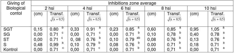
The result of obstacle on the antibiosis soil filtrat
The average inhibition zone from soil filtrat that consists of singular biological
S. griseorubens, the mixed of biological agensia S. griseorubens, G. virens, T. harzianum
and control toward the development of F. oxysporum was different from the 2nd ,4th,
6th, 8th and 10th days after inoculation. The average inhibition zone of soil filtrat
consists of Streptomyces sp, Gliocladium sp, T. harzianum mixture was taller
than soil filtrat that consists of singular S. griseurubens (table 1).
Discussion
The result of squensing DNA Streptomyces sp, that was uploaded from the world
gen bank, had similarity description 98 % with Actinobacterium ZXY004 and with
S . griseorubens strain 2418. Based on the morphology colony character, the size and
the spore shape, the isolat Streptomyces sp had the similarity with strain S. griseorubens
sp. was filament bacterium, small sprouted colony like cotton, and also produces coccus
spore (Brown et al, 2012). Actinobacterium ZXY belongs to Actynomycetes Genus and
another name of Streptomyces griseorubens was Actinomyces griseorubens (Cook and
Baker, 1974).
BLAST analysis result of seqeunsing DNA Gliocladium sp with 18S rRNA
showed the similarity 90 % with Trichoderma sp. Morphology of Gliocladium sp. was
almost the same as T. harzianum. The upload result of BLAST analysis showed the
similarity with the pimary standard used , that was Gliocladium virens. Skreekanth, et al.
Giving of
(2011) also shows the differences on the colony surface on PDA media between
Gliocladium sp. with T. harzianum.
Trichoderma harzianum Isolat from BPPHPTPH Pandaan that had the same
morphology features with isolat T harzianum as is publised by BPTPH Bogor, such
as light green colony to dark green , like wool, produces konodia aseksual with globul
shape and the konodia forms like grape and grows quickly. ( Akladious and Abbas,
2012; Alam et al. 2003; Buchanan and Gibbon, 1974)
Filtrat from the soil observation showed there was antibiosis that can inhibit the
F. oxysporum. Antibiosis on rizofer was antibiosis from G. virens and T.harzianum. This
statement was supported by the observations result of biological agensia population and
the fungus patogen population. While in the 30th days populations of S.griseorubens was
not found and the inhibition zone of soil filtrat consists of S.griseorubens on the 8th, 10th
day was 0 cm .
Biological agensia T. harzianum and G. virens could grow and develop around the
root faster than F. oxysporum, in the 1st day after planting. The competition between
biological agensia fungus and patogen fungus is a nutritious source competition because in
the 3rd day, F. oxysporum grows and develops in the same root of biological agensia
T.harzianum and G. virens. Some researches found that T. harzianum and G. virens are soil
saprofit fungus that can develop quickly. In 48 hours both biological agensia had formed
colony and twists the root and penetrates on the interselluler root (Agrios, 1994;
Allexopoulus 1996; Singh et al., 2002). In vitro observation showed that both biological
agensia grew quickly and can obstruct the development of F. oxysporum, and also forms
colony in the root plant ( Cook and Baker, 1974 ). The competition between Fusarium
patogenik and non patogenik is nutritious competition because both hifa grows on the same
Conclusion
Streptomyces sp. is Streptomyces griseorubens f.sp. capsicum and Gliocladium sp.
is Gliocladium virens. Multi antagonist relationship S. griseorubens f.sp.capsicum, G.virens
and T. harzianum on Fusarium oxysporum F.sp lycopersici in rizofer is : Antagonism of
antibiosis relationship produced by the mixed of biological agensia, S. griseorubens
f.sp.capsicum, G.virens and T.harzianum. Indirect antagonism is the competition and plant
resistance induction of G. virens and T.harzianum.
Reference
Agrios G.N. 1994. Plant Pathology, Second edition. Academic Press. New York, San
Fransisco, London, .
Akladious, S.A., and S. M. Abbas. 2012. Application of Trichoderma harziunum T2 as
a biofertilizer supporting maize growth. African Journal of Biotechnology. 11 (35):
8672-8683.
Alam, M., A.Satar, S. Kumar, A. Samad, and O.P. Dawam. 2003. Streptomyces strain with
potential antimicrobial activity againts phytopatogenik fungi. United States Patent No
US6,558,940 B2.
Alexopoulus, C.J., C,W. Mims, and Blackwell. 1996. Introductory of Mycology, Fourth
edition. John wiley & Sons, Inc. New York Chichester Brisbane Toronto Singapore.
Anitha, A. and M. Rabeeth. 2010 a. Control of fusarium wilt tomato by bioformulation of
Streptomyces griseus in green house condition. African Journal of Basic & Aplaid
Sciences 1 (1-2:) 9 – 14.
Anitha, A. and M. Rabeeth , 2010 b. Degradation of fungal cell walls of phytopatogenic fungi by lytic enzyme of Streptomyces griseus. African Journal of Plant Science. 4 (3):
061-066.
Benitez, T., A.M. Rincon, M.C. Limon, A.C. Codon. 2004. Biocontrol mechanism of strain
4-10.
Brown D.W., R A.E. Butchko, S. E. Baker, and R.H. Proctor. 2012. Phylogenomic and
functional domain analysis of polyketide synthases inFusarium. Fungal Biology
Buchanan, R.E. and N.E. Gibbons. 1974. Bergey's Manual of Determinative Bacteriology.
The Williams & Wilkins comp. Baltimore. 1246 h
Cook, J.R. and K. F. Baker. 1974. Biological Control of Plant Patogens. F.H. Freeman and
company. San Francisco, 348 p.
Chater, KF. and Chandra G. 2006. The evolution of development in Streptomyces analysed
by genome comparisons. Canadian Journal of Botany, Sep :30(5): 651-2.
Doi H., Kinoshita N., Oka T., Hui Z. 2008. Streptomyces strain that decomposes protein
recalcitrant to proteolisis. Microbial Chemistry Research Foundation.
US201230003717AI
Gruber, S. and V. Seiboth. 2012. Self versus non-self: fungal cell wall degradation In
Trichoderma. Microbiology 158:26-34
Kaewchai, S., Soytong, K., and Hyde, K.D. 2009. Mycofungicides and fungal biofertilizers.
Fungal Diversity 38: 25-50.
Olivain C., C. Humbert, J. Nahalkova, J. Fatehi, FL. Haridon, and C. Alobouvete. 2006.
Colonitation of tomato root by phatogenic and non patogenic Fusarium oxysporums
strains inoculated together and separately into the soil. Aplaid and Enviromental
Microbiology. 72 (2): 1523-1531.
Pal, K.K. and B. M. Garderner. 2006. Biological Control of Patogens. The plant health
instructor. DOI: 10.1094/PIH-A-2006-111702.
Sastrahidayat, I.R. , 1994. Guidance Studying Fungi in the Laboratory. Published by UB's
Faculty of Agriculture Publication.
Semangun.2000. Diseases of Horticultural Crops in Indonesia. The fourth edition. Gajah
Mada University Press. Well Bulak Yogyakarta.
Schirmbock, M., M. Lorito, Y. Wang, C.K. Hayes, I.A. Atac, F. Scala, G.e. Harman, and C.P.
antibiotics, moleculer mechanisms involved in antagonistic action of Trichoderma
harzianum agants phytopathogenic fungi. Aplaid and Enviromental Microbiology,
80(12)4364-4370
Singh R.,B.K. Singh, R.S. Upadhyay, B. Rai and Y. S. Lee. 2002 Biological control of
fusarium wilt disease of pigeonpea. Plant Pathology Journal 18(3) : 279-283
Singleton, J.D. Mihail and C.M. Rush. 1993. Methods for research on soilborne
phytopatogenik fungi . APS Press. The American Phytophatological Society. St. Paul
Minesota.
Skreekanth, D., G.k. Sushim, A. Syed, B.M. Khan, and A. Ahmad. 2011. Moleculer and
morphological characterization of taxol-producing endophytic fungus, Gliocladium sp.,
from Taxus baccata. Mycobiology, 39(3): 151–157.
Suryaminarsih and Mulyadi. , 2003. Study of the influence of dose of Trichoderma sp. and
the concentration of leaf fertilizer on the development of Fusarium wilt and yield of
onion. Journal of the Faculty of Biology (in Indonesia). Biology 3: 3.
Suryaminarsih and Mujoko 2012. Compatibility mixture of biological agents Streptomyces sp.
, Gliocladium sp., Trichoderma harzianum in improving the growth of tomato plants
infected with Fusarium wilt disease (In Indonesia). Proceedings of the National Seminar
Perhorti.
Yedidia I, Benhamou N, and Chet I. 1999. Induction of defense responses in cucumber
plants (Cucumis sativus L.) by the biocontrol agent Trichoderma harzianum. Applaid