Association between Serum Vitamin D Level and Clinical Manifestations of Systemic Lupus
Erythematous Patients in Dr. Sardjito General Hospital
Anggi Wahyu Nugroho1, Ayu Paramaiswari2 , Deddy Nur Wachid Achadiono2
1The Specialty Training Program of Internal Medicine, Faculty of Medicine, Public Health and Nursing, Universitas Gadjah Mada/Dr. Sardjito General Hospital
2Division of Rheumatology, Department of Internal Medicine Faculty of Medicine, Public Health, and Nursing Universitas Gadjah Mada/
Dr. Sardjito General Hospital Yogyakarta
Abstract
Background. Vitamin D played a role in the pathogenesis of systemic lupus erythematosus (SLE). Vitamin D made dendritic cells more tolerogenic to autoantigens and inhibited B cells from producing autoantibodies.
Vitamin D deficiency would make dendritic cells intolerant, increased production of interferons, and B cells produce excessive autoantibodies. These excessive autoantibodies and interferons would have caused severe clinical manifestations in SLE patients
Aims. This study was to find out vitamin D deficiency would increase the proportion of severe clinical manifestations in SLE patients in the Dr. Sardjito General Hospital.
Methods. We conducted a cross-sectional study. Data was taken from medical records of SLE patients who went to Dr. Sardjito General Hospital in 2018. The independent variable was serum 25(OH) D levels, which were divided into deficiency (≤12 ng/ml) and not deficiency (>12ng/ml). The dependent variable was the clinical manifestations of SLE patients, which were divided into mild-moderate and severe. Association between two variables was analyzed using Chi-Square.
Results. Vitamin D deficiency was observed in 19(54%) and not deficiency in 16(46%) subjects. SLE patients who underwent vitamin D deficiency more often experienced severe clinical manifestations than those without (52.6% versus 12.5%), prevalence ratio 4.2 CI 1.1-16.5 p=0.015. SLE patients who underwent vitamin D deficiency were more likely to suffer mucocutan, musculoskeletal, and kidney disorders. Also more likely to suffer more than 2 organ disorders than those without (57.9% versus 12.5%), prevalence ratio 4.6 CI 1.2-17.9 p=0.007.
Conclusions. Vitamin D deficiency increased the proportion of severe clinical manifestations in SLE patients at Dr. Sardjito General Hospital. It also increased the proportion of mucocutan disorder, musculoskeletal disorder, and kidney disorder. Also, it increased the proportion of occurrence of more than 2 organ disorders in SLE patients at Dr. Sardjito General Hospital.
Keywords: Vitamin D, SLE, clinical manifestations
Abstrak
Latar Belakang: Vitamin D berperan dalam patogenesis lupus eritematosus sistemik (LES). Vitamin D membuat sel dendritik lebih tolerogenik terhadap autoantigen dan menghambat sel B memproduksi autoantibodi. Defisiensi vitamin D akan membuat sel dendritik tidak tolerogenik, produksi interferon meningkat, dan sel B menghasilkan
Introduction
The role of vitamin D in the immune system was discovered in 1980. Vitamin D receptors are found in the immune system such as dendritic cells, macrophages, and lymphocytes. These cells have the enzyme 1α-hydroxylase and CYP27B1 which can convert vitamin 25(OH)D into its active form.1
Vitamin D acts as an immune modulator, especially in dendritic cells. Dendritic cells present antigens to T cells. Dendritic cells can maintain peripheral tolerance to autoantigens, thereby causing T cell anergy to autoantigens. Vitamin D deficiency can cause autoimmune disorders such as systemic lupus erythematosus (SLE).2
The cause of vitamin D deficiency in SLE patients can be exacerbated by the use of sunscreen, lupus nephritis, the habit of avoiding sunlight, long-term use of glucocorticoids, and the use of chloroquine.1
Clinical manifestations of SLE vary from mild-moderate to severe. Mild clinical manifestations are SLE with musculoskeletal and mucocutaneous disorders. Moderate clinical manifestations are found in one of the following signs, namely mild-moderate nephritis, platelets 20-50 x103/mm3, or serositis. Severe clinical manifestations are found in one of the following disorders, namely pericarditis, pneumonitis, severe nephritis, severe vasculitis, a diffuse rash with ulcers, neurology, and hematology (hemolytic anemia, neutropenia, and thrombocytopenia <20.000/
mm3).3
Miscovick et al (2015), found no relationship between vitamin D levels and clinical manifestations of SLE patients.1 Different result was discovered by Dall’ara et al (2017), found that vitamin D deficiency would make severe clinical manifestations of SLE.4 These result still controversial, so we did this study. The purpose of this study was autoantibodi berlebihan. Autoantibodi dan interferon yang berlebihan ini akan menimbulkan manifestasi klinis pasien LES yang berat.
Tujuan. Tujuan penelitian ini adalah untuk mengetahui apakah defisiensi vitamin D dapat meningkatkan proporsi terjadinya manifestasi klinis berat pada pasien LES di RSUP Dr Sardjito.
Metode. Penelitian ini merupakan penelitian potong lintang. Data diambil dari rekam medis pasien LES yang berobat di RSUP Dr Sardjito tahun 2018. Variabel bebas adalah kadar vitamin D (25(OH)D) serum, yang dibagi menjadi defisiensi (≤12 ng/ml) dan tidak defisiensi (>12ng/ml). Variabel tergantung adalah manifestasi klinis pasien LES, yang dibagi menjadi ringan-sedang dan berat. Hubungan antara kedua variabel dianalisis dengan Chi Square.
Hasil. Subyek yang mengalami defisiensi kadar vitamin D sebanyak 19 (54%) dan yang tidak defisiensi sebanyak 16 (46%). Pasien LES dengan defisiensi vitamin D lebih sering mengalami manifestasi klinis berat dibandingkan dengan yang tidak defisiensi (52,6% dibanding 12,5%) dengan rasio prevalensi 4,2 CI 1,1-16,5 p=0,015. Pada pasien LES dengan defisiensi vitamin D lebih sering mengalami gangguan sistem mukokutan, muskuloskeletal, dan ginjal. Pasien LES dengan defisiensi vitamin D juga lebih sering mengalami gangguan multi organ dibandingkan dengan yang tidak defisiensi (57,9% dibanding 12,5%) dengan rasio prevalensi 4,6 CI 1,2-17,9 p=0,007.
Kesimpulan. Defisiensi vitamin D meningkatkan proporsi terjadinya manifestasi klinis yang berat pada pasien LES di RSUP Dr Sardjito. Defisiensi vitamin D juga meningkatkan proporsi terjadinya gangguan sistem mukokutan, muskuloskeletal, dan ginjal. Selain itu, defisiensi vitamin D juga meningkatkan terjadinya gangguan lebih dari 2 organ pada pasien LES di RSUP Dr Sardjito.
Kata Kunci :Vitamin D, lupus eritematosus sistemik, manifestasi klinis
serositis. Severe manifestations if there was one of the following signs, namely hematological disorders (hemolytic anemia, neutropenia, platelets <20.000/mm3), proteinuria ≥3+, vasculitis, and neuropsychiatry.
After being approved by the Ethics Committee of the Faculty of Medicine, Public Health and Nursing Gadjah Mada University and obtained a research permit from the Director of Dr. Sardjito General Hospital Yogyakarta, we began collecting data from medical records.
The results were tested for normality by the Shapiro-Wilk test. The basic characteristics displayed descriptively included the percentage, median, or mean according to the results of the data normality. The association between vitamin D levels with clinical manifestations of SLE patients was analyzed by Chi-Square and accompanied by p-value and a prevalence ratio with a 95% CI.
Result
Subjects who met the inclusion and exclusion criteria were 35 subjects in total.
The sociodemographic characteristics of the subjects were shown in table 1. The research subjects were 34 (97.1%) women and 1 (2.9%) men. SLE was known to be more frequent in women than men with a ratio of 8-15:1. The median age of subjects was 30 years with a range between 18 to 65 years old. Subjects who were diagnosed at less than 32 years were 25 (71.4%) and those over 32 years were 10 (28.6%). Most SLE diagnosed at the age of 24-32 and 12-17 years. Older age at diagnosis usually has severe clinical manifestations.5 Body mass index (BMI) varies from 16 to 29.7 kg/m2, mean value 20.6 mg/kg2. There were 3 to determine whether vitamin D deficiency
would increase the proportion of severe clinical manifestations in SLE patients at Dr. Sardjito General Hospital. Subanalysis was done to find out whether vitamin D deficiency would increase the proportion of organ system disorder that occurs, and also whether vitamin D deficiency would increase the proportion of more than 2 organs involved in SLE patients at Dr. Sardjito General Hospital.
Method
This research was a cross-sectional study conducted from July 2019 to February 2020.
Data was taken from the medical record of LES patients seeking treatment at Dr. Sardjito General Hospital in 2018.
Inclusion was SLE patients who were diagnosed from ACR 1997 criteria, aged over 18 years old, had data on vitamin D levels, complete blood count, creatinine, and urinalysis results within a maximum examination range of 1 month. Exclusion were pregnant women, cancer patients, tuberculosis, diabetes mellitus, osteoporosis, serum creatinine above 1.3 mg/
dL, obesity, urinary tract infections, using vitamin D supplements, anti-convulsants, anti-fungi, anti-chemotherapy, antiretroviral, isoniazid, and rifampicin in the last 3 months.
The independent variable was serum vitamin D (25(OH) D) levels, which are divided into deficiency (≤12 ng/ml) and not deficiency (> 12 ng/ml). The dependent variable was the clinical manifestations of SLE patients, which are divided into mild-moderate and severe manifestations. Mild-moderate clinical manifestations if there was one of the following criteria, namely mucocutaneous disorders, musculoskeletal, proteinuria <3+
with excess BMI were at risk of experiencing more severe manifestations.6
Table 1. Sociodemographic characteristics of subjects
Characteristics Number
(%) Mean Median
Age (year) 30 (18-65)
Sex
Man 1 (2.9%)
Woman 34 (97.1%) Age at diagnosed
≤32 years
>32 years 25 (71.4%) 10 (28.6%)
Body Mass Index (kg/m3) 21(±2.62) Underweight
Normal Overweight
3 (8.6%) 29 (82.9%)
3(8.6%)
17.3(±1.1) 20.8(±1.5) 27.2(±2.2)
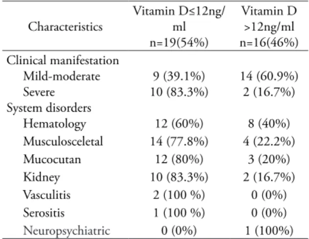
Characteristics of clinical manifestation based on vitamin 25(OH) D levels were shown in table 2. Ten patients (83.2%) with levels of 25(OH) D less than 12 ng/ml had severe clinical manifestations of SLE. Whereas 2 (16.7%) patients with vitamin 25(OH) D levels greater than 12 ng/ml had severe clinical manifestations. Hematologic, musculoskeletal, mucocutaneous, and kidney disorders were more common in patients with vitamin D deficiency compared with those without deficiency. Subjects experiencing vasculitis, serositis, and neuropsychiatric disorders were very few. Vasculitis experienced by 2 subjects, serositis 1 subject, and neuropsychiatric disorder 1 subject.
Characteristics of laboratory results were shown in table 3. Subjects with vitamin D deficiency were 19 (54%) subjects with an average of vitamin D level 7.5 ± 2.4 ng/ml, while those that non-deficiency were 16 (46%) subjects with an average of 18.2 ± 6.5 ng/ml.
The most frequent hematological disorders were anemia 20 (57.1%) and lymphopenia in 12 (34.3%) subjects. This was following the
other study, that anemia and lymphopenia were the most common hematologic disorder in SLE.7 Proteinuria was more common in SLE patients with vitamin D deficiency compared to those without deficiency (69.2% versus 30.8%).
This result was quite interesting because deficiency of vitamin D in SLE patients also occurred in equator areas such as Yogyakarta.
Previous studies mentioned that the prevalence of vitamin D deficiency in SLE patients in China was 84.3%, Australia 27.7%, Hong Kong 27%, Canada 17.9%, United States 20%, and Spain 15%.8 The prevalence of vitamin D deficiency in SLE patients in the Table 3. Characteristics of laboratory result based on vitamin D level
Characteristic Vitamin D≤12ng/ml n=19(54%)
Vitamin D
>12ng/ml n=16(46%) Anemia
Lymphopenia 12 (60%)
7 (58.3%) 8 (40%) 5 (41.7%)
Leucopenia 2 (100%) 0 (0%)
Trombocytopenia 1 (100%) 0 (0%)
Proteinuria 9 (69.2%) 4 (30.8%)
Creatinin (mg/dl)
Vitamin D (ng/mL) 0.73 (±0.24)
7.5 (±2.4) 0.71 (±0.09) 18.2 (±6.5)
Table 2. Characteristics of clinical manifestation based on vitamin D levels
Characteristics Vitamin D≤12ng/
n=19(54%)ml
Vitamin D
>12ng/ml n=16(46%) Clinical manifestation
Mild-moderate Severe System disorders Hematology
9 (39.1%) 10 (83.3%)
12 (60%)
14 (60.9%) 2 (16.7%)
8 (40%) Musculosceletal 14 (77.8%) 4 (22.2%)
Mucocutan 12 (80%) 3 (20%)
Kidney 10 (83.3%) 2 (16.7%)
Vasculitis 2 (100 %) 0 (0%)
Serositis 1 (100 %) 0 (0%)
Neuropsychiatric 0 (0%) 1 (100%)
equator and also four-season countries can be caused by dietary sources of vitamin D, clothing models, sunscreen, medications, or vitamin D antibodies in serum.9
Discussion
The association between vitamin D levels and clinical manifestations of SLE patients are shown in table 4. SLE patients with vitamin D deficiency experienced more severe clinical manifestations compared to those without (52.6% versus 12.5%), with a prevalence ratio of 4.2 CI 1.1-16.5 p-value
= 0.015. These results were in line with the literature that vitamin D deficiency would cause dendritic cells to lose their tolerogenic properties to autoantigens, activation of T helper 1, increased autoantibody production by B lymphocyte, and excessive interferon production, which would cause severe clinical manifestations of SLE.1,2
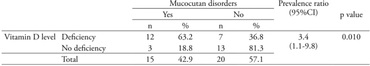
Sub analysis was conducted of the association between vitamin D levels and mucocutaneous disorder in SLE patients shown in table 5. SLE patients with vitamin
D deficiency more often experienced mucocutaneous disorder compared with those without deficiency (63.2% versus 18.8%), with a prevalence ratio of 3.4 CI 1.1 to 9.8 p-value
= 0.010. SLE patients with mucocutaneous disorders would usually be educated to avoid sunlight and use sunscreen. This could cause a decrease in the formation of provitamin D3.
So that the measurement of vitamin 25(OH) D was found to be much lower results.9
Association between vitamin D levels and musculoskeletal disorders in SLE patients was conducted in table 6. SLE patients with vitamin D deficiency more often experienced musculoskeletal disorders compared with those without deficiency (73.7% versus 25%), with a prevalence ratio of 2.9 CI 1.2- 7.2 p-value = 0.005. Immunopathogenesis of musculoskeletal disorders in SLE was caused by increased production of type 1 interferon by plasmacytoid dendritic cells, monocytes, synovial cells, and fibroblasts. Vitamin D deficiency would increase interferon production and musculoskeletal disorders occur.10
Association between vitamin D levels with hematological disorders in SLE patients
Table 4. Association between vitamin D level and clinical manifestations of SLE
Clinical manifestation of SLE Prevalence ratio
(95%CI) p value Severe Mild-moderate
n % n %
Vitamin D level Deficiency 10 52.6 9 47.4 4.2
(1.1-16.5) 0.015
No deficiency 2 12.5 14 87.5
Total 12 34.3 23 65.7
Table 5. Association between vitamin D level and mucocutan disorder in SLE
Mucocutan disorders Prevalence ratio
(95%CI) p value
Yes No
n % n %
Vitamin D level Deficiency 12 63.2 7 36.8 3.4
(1.1-9.8) 0.010
No deficiency 3 18.8 13 81.3
shown in table 7. There was no difference in the proportion of hematological disorders between patients with vitamin D deficiency and no deficiency (63.2% versus 50%) with a ratio of a prevalence of 1.2 CI 0.7-2.3 p-value
= 0.330. Anemia and thrombocytopenia in SLE were due to immunological and non- immunological processes. Immunological causes such as hemolytic anemia and antithrombopoetin antibodies. Non-immunological causes iron deficiency, chronic inflammation, medications, spinal cord suppression, and chronic infections.
The most common causes of anemia in SLE were chronic disease and iron deficiency. It might cause no association was found between vitamin D levels and hematological disorders in this study. 11
Association between vitamin D levels and kidney disorders in SLE patients shown in table 8. SLE patients with vitamin D deficiency
more often experienced kidney disorders than those without deficiencies (52.6% versus 12.5%), with a prevalence ratio of 4.2 CI 1.1 to 16.5 p= 0.015. Vitamin D protected renal podocytes by reducing autophageal activity in SLE patients. Vitamin D deficiency increased podocyte autophagial activity and caused kidney damage.12
We conducted a subanalysis of the association between vitamin D levels and the number of organs affected in SLE (Table 9).
SLE patients with vitamin D deficiency more often experienced more than 2 organs affected compared with those without (57.9% versus 12.5%), with a prevalence ratio of 4.6 CI 1.2- 17.9 p = 0.007.
Multivariate logistic regression analysis was performed to find confounding in this Table 6. Association between vitamin D level and musculoskeletal disorder in SLE
Musculoskeletal disorder
Prevalence ratio
(95%CI) p-value
Yes No
n % n %
Vitamin D
level Deficiency 14 73.7 5 26.3 2.9
(1.2-7.2) 0.005
No deficiency 4 25 12 75
Total 18 51.4 17 48.6
Table 7. Association between vitamin D level and hematological disorder in SLE
Hematological disorders
Prevalence ratio
(95%CI) p-value
Yes No
n % n %
Vitamin D level Deficiency 12 63.2 7 36.8 1.2
(0.7-2.3) 0.330
No deficiency 8 50 8 50
Total 20 57.1 15 42.9
Table 8. Association between vitamin D level and kidney disorders in SLE
Kidney disorders
Prevalence ratio
(95%CI) p-value
Yes No
n % n %
Vitamin D level Deficiency 10 52.6 9 47.4 4.2
(1.1-16.5) 0.015
No deficiency 2 12.5 14 87.5
Total 12 34.3 23 65.7
study. Overweight body mass index (BMI) and age of diagnosed SLE over 32 years were confounding in this study. According to existing research, SLE patients with excess BMI or obesity are associated with poor sleep quality, more comorbidities, and more severe organ damage.6,13
These results are consistent with the research of Saulescu et al (2018), that in systemic lupus erythematosus patients with excess body mass index and / or obesity are associated with poor sleep quality and a lazy lifestyle that can cause various comorbidities in LES patients.
Similar results from the study of Kang et al (2020), in LES patients with obesity will increase the incidence of lupus nephritis and more severe organ damage. This is due to the more comorbids found in LES patients with excessive BMI, such as hypertension, pulmonary hypertension, dyslipidemia, diabetes mellitus, and the risk of atherosclerosis.
Conclusion
Vitamin D deficiency increased the proportion of severe clinical manifestations in
SLE patients at Dr. Sardjito General Hospital.
The deficiency increased the proportion of mucocutan, musculoskeletal, and kidney disorders in SLE patients at Dr. Sardjito General Hospital. Vitamin D deficiency also increased the proportion of occurrence of more than 2 organ disorders in SLE patients at Dr.
Sardjito General Hospital.
References
1. Miskovic, R., Plavsic, A., Raskovic, S., Jovicic, Z., Bolpacic, J. Vitamin D status in patients with systemic lupus erythematosus in Serbia: correlation with disease activity and clinical manifestations. Open access Macedonian journal of medical sciences.
2015; 3(2), 256.
2. Iruretagoyena, M., Hirigoyen, D., Naves, R., Burgos, P. I. Immune response modulation by vitamin D: role in systemic lupus erythematosus. Frontiers in immunology.
2015; 6, 513.
3. Kasjmir, Y.I., Handono, K., Wijaya, L.K., Hamijoyo, L., Labar, Z. Diagnosis dan Table 9. Association between vitamin D level and number of organs affected in SLE
Number of organs affected
Prevalence ratio
(95%CI) p-value
>2 organ system ≤2 organ system
n % n %
Vitamin D level Deficiency 11 57.9 8 42.1 4.6
(1.2-17.9) 0.007
No deficiency 2 12.5 14 87.5
Total 12 34.3 23 65.7
Table 10. Multivariate logistic regression analysis
B S.E. Wald df Sig. Exp(B) 95% C.I. for EXP(B)
Lower Upper
Vitamin D level 2.241 .997 5.053 1 .025 9.403 1.332 66.357
Age diagnosed SLE .492 .995 .244 1 .621 1.635 .232 11.506
Overweight BMI 1.476 1.409 1.098 1 .295 4.377 .277 69.253
Constant -6.069 3.679 2.721 1 .099 .002
Interna Publishing. 2014; 3360-77.
4. Dall’Ara, F., Cutolo, M., Andreoli, L., Tincani, A., Paolino, S. Vitamin D and systemic lupus erythematous: a review of immunological and clinical aspects.
Clinical and Experimental Rheumatology.
2017;36(1), 153-162.
5. Pons-Estel, G. J., Ugarte-Gil, M. F., Alarcón, G. S. Epidemiology of systemic lupus erythematosus. Expert review of clinical immunology. 2017; 13(8), 799-814 6. Saulescu, I., Belinski, D. O., Borangiu, A.,
Groseanu, L., Daia-Iliescu, S., Mazilu, D., Constantinescu, C. 2018. AB0519 Impact of higher body mass index (BMI) in patients with systemic lupus erythematosus. 2018 7. Teke, H. Ü., Cansu, D. Ü.,Korkmaz,
C. Detailed features of hematological involvement and medication-induced cytopenia in systemic lupus erythematosus patients: single center results of 221 patients.
European journal of rheumatology.2017;
4(2), 87.
8. Gaik, O. S., Jen, D. H. Vitamin D status in a monocentric cohort of systemic lupus erythematosus (SLE) patients and correlations with a clinical and immunological profile. Med J Malaysia.
2019; 74(6), 493.
9. Handono, K., Puspitasari, L., Rudijanto, A.,Wahono, S., Kalim, H. Vitamin D serum level and disease activity in patients with systemic lupus erythematosus.
International Journal of Pharmaceutical Science Invention.2013; 2(2), 35-40.
10. Mahmoud, K., Zayat, A., Vital, E. M.
Musculoskeletal manifestations of systemic lupus erythmatosus. Current opinion in rheumatology. 2017; 29(5), 486-492.
11. Teke, H. Ü., Cansu, D. Ü.,Korkmaz, C. Detailed features of hematological involvement and medication-induced cytopenia in systemic lupus erythematosus patients: single center results of 221 patients. European journal of rheumatology.
2017; 4(2), 87.
12. Yu, Q., Qiao, Y., Liu, D., Liu, F., Gao, C., Duan, J., Cui, S. Vitamin D protects podocytes from autoantibodies induced injury in lupus nephritis by reducing aberrant autophagy. Arthritis research &
therapy. 2019; 21(1), 19.
13. Kang, J. H., Lee, S. S., Choi, S. E., Xu, H., Park, D. J. Obesity Increases The Incidence of New Onset Lupus Nephr itis and Organ Damage During Follow Up in Patients with Systemic Lupus Erythematosus.
Sagepub. 2020;0(0)1-9.