Downloaded from www.microbiologyresearch.org by Correspondence
Pierre Flori
pierre.flori@univ-st-etienne.fr
Received 7 November 2003 Accepted 10 February 2004
Comparison between real-time PCR, conventional
PCR and different staining techniques for
diagnosing
Pneumocystis jiroveci pneumonia from
bronchoalveolar lavage specimens
Pierre Flori,
1Bahrie Bellete,
1Fabrice Durand,
1He´le`ne Raberin,
1Ce´line Cazorla,
2Jamal Hafid,
1Fre´de´ric Lucht
2and
Roger Tran Manh Sung
1Laboratory of Parasitology and Mycology, Hoˆpital Nord1and Department of Infectious and Tropical
Diseases, Hoˆpital Bellevue2, University Hospital of Saint Etienne, 42055 Saint Etienne, France
Between January 2002 and July 2003, 173 bronchoalveolar lavage (BAL) specimens from 150 patients (19 HIV-infected and 131 non-HIV-infected patients) were evaluated for identification of Pneumocystis jiroveci(formerly known asPneumocystis cariniif. sp.hominis) using staining techniques, conventional PCR (mtLSUrRNAgene) and real-time PCR (MSGgene). Test results were compared toPneumocystispneumonia (PCP) confirmed by typical clinical findings and response to treatment. Sensitivity and specificity of the techniques were 60 and 100 % for staining (where either one or both techniques were positive), 100 and 87.0 % for conventional PCR and 100 and 84.9 % for real-time PCR, respectively. The use of a concentration of 103copies of DNA per
capillary of BAL as a cut-off (determined by real-time PCR) increased specificity from 84.9 to 98.6 % without reducing the sensitivity of the technique. This technique is rapid (,3 h) and therefore of major interest in differentiating between asymptomatic carriage and PCP. A BAL specimen with
,103copies per capillary ofPneumocystis-specific DNA is more likely to indicate a chronic carrier
state, but in such cases follow-up is required to ensure that the patient is not in the early stage of an active PCP.
INTRODUCTION
Pneumocystis jiroveci
, formerly known as
Pneumocystis carinii
f. sp.
hominis
, is a fungal pathogen that causes
Pneumocystis
pneumonia (PCP), a common and serious opportunistic
infection in immunocompromised patients. Diagnosis of
PCP in the laboratory was, until a few years ago, dependent
on visualization of
Pneumocystis
organisms in stained
pre-parations of appropriate respiratory specimens using the
Giemsa and Gomori
Grocott techniques. Sensitivity of the
staining techniques, although acceptable (70–92 %) for
bronchoalveolar lavage (BAL) specimens (Wakefield
et al.
,
1990; Cregan
et al.
, 1990), was low (35–78 %) for aspirates
and induced sputum samples (Zaman
et al.
, 1988; Wakefield
et al.
, 1991; Lipschik
et al.
, 1992). In the last 10 years, PCR
(Wakefield
et al.
, 1990) has considerably increased sensitivity
of detection of
Pneumocystis
, which is now 86
100 % in
BAL, aspirates and induced sputum specimens (Roux
et al.
,
1994; Cartwright
et al.
, 1994; Caliendo
et al.
, 1998). However,
one problem with this technique is that it is frequently
positive in patients with asymptomatic carriage, with a rate of
2
21 % (Sing
et al.
, 1999, 2000; Nevez
et al.
, 1999;
Helweg-Larsen
et al.
, 2002; Takahashi
et al.
, 2002; Maskell
et al.
,
2003). Real-time PCR, recently described by Larsen
et al
.
(2002), could have a role in distinguishing between
coloniza-tion and clinical disease. The aim of this study was to
compare sensitivity and specificity of different techniques
(standard staining, conventional PCR and real-time PCR)
used in diagnosis of PCP and, as suggested by Larsen
et al
.
(2002), to propose an adapted cut-off value for
differentiat-ing between carriage and PCP usdifferentiat-ing real-time PCR.
METHODS
Patients and clinical samples.Samples for this study came from all patients seen in the University Hospital of Saint Etienne between January 2002 and July 2003 presenting with clinical symptoms of pulmonary infection associated with immunosuppression, justifying a search forPneumocystisin BAL.
From 150 patients with immunosuppression due to different diseases and/or immunosuppressive treatments, 173 BAL samples were ob-tained. Samples comprised 138 specimens retrieved by washing with
50 ml 0.9 % NaCl (BAL-50) and 35 retrieved by washing with 150 ml 0.9 % NaCl (BAL-150). Of these 150 patients, 19 were HIV seropositive and the rest were seronegative.
Sample preparation and classical staining. All BAL samples containing mucous material had twice their volume of 0.9 % NaCl added and they were then mixed vigorously for 5 min. To avoid non-specific inhibition of PCR, no detergents or mucolytic agents were used in this step. Samples were centrifuged at 3000gfor 10 min and pellets were resuspended in 1/10 of the starting volume. A portion of the resuspended pellet (100ìl) was used to prepare smears for Giemsa and
GomoriGrocott staining and smears were examined by two micro-scopists experienced inPneumocystisdiagnosis. Additional samples of 200ìl of the resuspended pellets were stored at 48C until used for DNA
extraction and amplification.
DNA extraction, conventional PCR and real-time PCR.To detect
PneumocystisDNA in BAL samples, 200ìl of each resuspended pellet
was used for DNA extraction with the QIAamp DNA Mini kit (Qiagen) according to the manufacturer’s recommendations.
Conventional PCR.PCR was performed according to Wakefieldet al. (1990). The PCR mixture (50ìl) contained 10 pmol of each primer,
pAZ102E and pAZ102H, derived from the mitochondrial large subunit rRNA (mtLSUrRNA) gene, 200ìM dNTPs (dATP, dCTP, dGTP and
33dUTP), 3 mM MgCl2, 5ìl 103PCR buffer, 1.25 UTaq DNA
polymerase, 1 IU uracyl DNA glycosylase (PCR Core Kit Plus; Roche Molecular Biochemicals) and 10ìl purified DNA. Denaturing,
anneal-ing and extension times were 1 min each, at 95, 58 and 728C, respectively. DNA samples were amplified for 40 cycles. The specific product (364-bp fragment) was separated on a 3 % agarose gel (Metaphor agarose; Tebu) and detected after staining with ethidium bromide under UV illumination.
Real-time PCR with the LightCycler. Real-time PCR was performed with fluorescence resonance energy transfer (FRET) hybridization probes using the LightCycler. PCR was performed with LC FastStart DNA Master hybridization probes (Roche Molecular Biochemicals) in a quantitative touch-down real-time PCR as previously described by Larsenet al. (2002). Briefly, 20ìl of a mixture containing Fast StartTaq
DNA polymerase, dNTPs (with dUTP instead of dTTP), 5 mM MgCl2,
0.5 IU heat-labile uracyl-DNA glycosylase (Roche Molecular Biochem-icals), 10 pg mouse DNA, 5ìl PCR extract, 1ìM of each selected primer
and 0.20ìM of each hybridization probe was used. Internal control was
the mouse galactose-1-phosphate uridyl transferase (GALT) gene (Costaet al., 2001). Specific primers and probes were used for co-amplification of the internal control (Costaet al., 2001) and theMSG
gene ofPneumocystis(Larsenet al., 2002). A double fluorescence reading for each sample was taken at the annealing step: one for LCRed 640 used for thePneumocystisprobe (channel 2) and the other for LCRed 705 used for the internal control probe (channel 3).
Standards and external controls.To obtain standards for real-time PCR, P. jiroveci-positive BAL samples with GomoriGrocott smear were used, and 10-fold dilutions were made to obtain 101104copies
per capillary (Larsenet al., 2002).
Data analysis.In the absence of a sensitive gold standard, diagnosis of PCP is difficult in certain cases. The criteria that we used for confirming an ongoing pneumocystosis were clinical findings of PCP with characteristic X-ray findings (diffuse interstitial infiltrates), dyspnoea with partial pressure of arterial oxygen,70 mmHg and response to anti-Pneumocystistreatment but resistance to other antibiotics. Based on these criteria for diagnosis, sensitivity and specificity of each laboratory technique were estimated.
Statistical assessment of these characteristics was performed using the÷2
test (P,0.05 was considered significant). Statistical assessment of differences of the mean of Pneumocystis DNA concentration [log (copies) per capillary] from PCP or non-PCP cases was performed by unpaired Student’st-test (P,0.05 was considered significant).
RESULTS AND DISCUSSION
Of 150 patients examined, 11 (7.33 %) developed PCP. In the
remaining 139, diagnosis of PCP was not confirmed. Of 11
confirmed cases, seven were HIV seropositive and four were
seronegative.
Comparison of different techniques
Concerning sensitivity and specificity of the diagnostic
techniques, results obtained for the two classical staining
methods (where either one or both were positive) were 60 %
[6/10 (the volume was insufficient for one test)] and 100 %
(139/139), respectively. For conventional PCR, sensitivity
and specificity were 100 % (11/11) and 87 % (121/139) and,
for real-time PCR, 100 % (11/11) and 84.9 % (118/139),
respectively. There was a significant difference (
P
,
0.01) in
both sensitivity and specificity between PCR and staining
techniques; staining gave excellent specificity but lacked
sensitivity, while the two PCR tests were much more sensitive
but could give false positives.
False-positive cases with the molecular techniques
In this study, false-positive cases using the molecular biology
techniques were frequent compared with clinical diagnosis,
and were similar to data published elsewhere (see
Introduc-tion). There were 18/139 (12.9 %) false positives with
con-ventional PCR; these were also positive with real-time PCR,
and there were a further three positives, giving a total of 21/
139 (14.5 %) positives with real-time PCR. With the further
three positives detected by real-time PCR, quantities of DNA
detected were very small (1.3, 1.8 and 3.8 copies per
capillary), indicating a slightly higher sensitivity of
approxi-mately 1 DNA copy per capillary. The coherence in both PCR
techniques confirms the presence of
P. jiroveci-
specific DNA
in asymptomatic patients and their probable carrier state.
False-negative cases with standard staining
techniques
Downloaded from www.microbiologyresearch.org by
number of PCP cases among seronegative patients and, as
described by Ninin
et al
. (1998), deterioration is rapid, and
death results in 35.4 % of cases.
DNA concentration in relation to clinical findings
In this study, one of the objectives was to try to determine a
cut-off value that could discriminate between cases of PCP
and asymptomatic
Pneumocystis
carriers. For this, it is
important to have the same type of specimen for all cases.
In the present study we had 173 BAL specimens divided into
two groups, BAL-50 and BAL-150. No difference in
sensi-tivity or specificity between the two groups regarding
diag-nosis of PCP was demonstrated, and they were therefore
analysed together. However, clinically, BAL-50 was much
better tolerated by patients who were severely hypoxaemic.
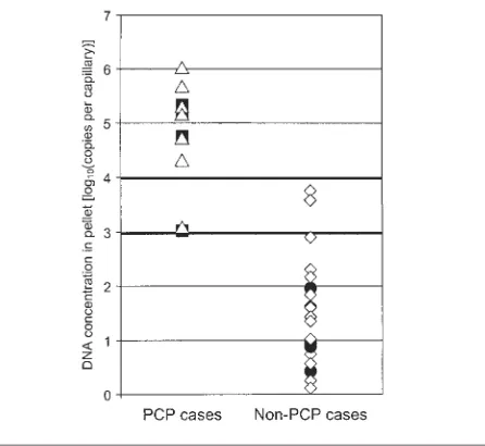
As shown in Fig. 1, concentrations of DNA detected in PCP
patients were very high (
P
,
0.0001). All cases had
.
10
3copies of DNA per capillary, whereas all cases that did not have
PCP but had detectable
Pneumocystis
-specific DNA had
.
10
4copies of DNA per capillary (Table 3). Using 10
3copies of
DNA per capillary of BAL as a cut-off, determined by real-time
PCR, increased specificity from 84.9 (118/139) to 98.6 % (137/
139) without reducing the sensitivity of the technique.
In addition, the limit of detection of the staining techniques
was well below that of real-time PCR. Thus, all BAL
speci-mens that were positive by the staining techniques had DNA
concentrations
.
10
4copies per capillary. This quantitative
difference in sensitivity (a factor of 10
4) has already been
noted in different studies. Leigh
et al
. (1993) defined a factor
between 10
4and 10
6using serial dilution, while Ribes
et al
.
(1997) found a factor of at least 10
2.
As regards a cut-off to differentiate asymptomatic carriage
from PCP, there was an overlap between the two groups (Fig.
1) in spite of a significant difference. That is the reason why a
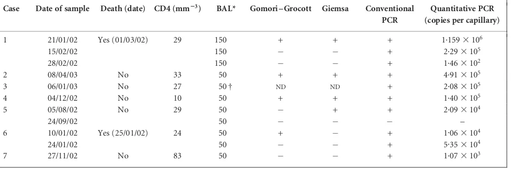
Table 1.Comparison of different diagnostic methods in seven HIV-positive patients who developed pneumocystosis
Dates are given in the form dd/mm/yy.ND, Not done.
Case Date of sample Death (date) CD4 (mm23) BAL* Gomori–Grocott Giemsa Conventional
PCR
Quantitative PCR (copies per capillary)
1 21/01/02 Yes (01/03/02) 29 150 + + + 1.1593106
15/02/02 150 + 2.293105
28/02/02 150 + 1.463102
2 08/04/03 No 33 50 + + + 4.913105
3 06/01/03 No 27 50 † ND ND + 2.083105
4 04/12/02 No 10 50 + + + 1.403105
5 05/08/02 No 29 50 + + 2.093104
24/09/02 50 –
6 10/01/02 Yes (25/01/02) 24 50 + + 1.063104
24/01/02 50 + 5.353104
7 27/11/02 No 83 50 + 1.073103
*50, BAL fluid specimen after washing with 50 ml 0.9 % NaCl; 150, BAL fluid specimen after washing with 150 ml 0.9 % NaCl.
†Volume of sample insufficient (,2 ml) for conventional staining.
Table 2.Comparison of different diagnostic methods in four HIV-negative patients who developed pneumocystosis
See Table 1 for details. CLL, Chronic lymphocytic leukaemia.
Case Date of sample Death (date) Associated disease BAL Gomori–Grocott Giemsa Conventional PCR
Quantitative PCR (copies per capillary)
1 02/04/02 Yes (20/04/02) CLL 150 + 2.383105
15/04/02 150 + 1.113104
2 22/04/02 No CLL 150 + 1.523105
3 14/12/02 Yes (18/12/02) Renal
transplantation, astrocytoma
150 + + + 5.853104
4 27/12/02 No Wegener’s
disease
‘grey zone’ between 10
3and 10
4copies per capillary must be
proposed. This also allows for possible minor
inter-labora-tory differences in performing the techniques. A BAL sample
with
,
10
3copies of DNA per capillary is likely to be a chronic
carrier state but, in such cases, follow-up is necessary, as such
patients could be in the early stage of an active PCP.
To conclude, real-time PCR is a rapid technique (
,
3 h
including extraction, amplification and visualization).
Hav-ing a high sensitivity as with conventional PCR, it has the
added advantage of quantification to determine a cut-off that
permits differentiation between carriage and disease in the
majority of cases.
ACKNOWLEDGEMENTS
We acknowledge the staff of the Laboratory of Parasitology for their skilful technical assistance, and the clinical departments for sending the specimens.
REFERENCES
Caliendo, A. M., Hewitt, P. L., Allega, J. M., Keen, A., Ruoff, K. L. & Ferraro, M. J. (1998).Performance of a PCR assay for detection of
Pneumocystis cariniifrom respiratory specimens.J Clin Microbiol36, 979–982.
Cartwright, C. P., Nelson, N. A. & Gill, V. J. (1994).Development and evaluation of a rapid and simple procedure for detection of Pneumo-cystis cariniiby PCR.J Clin Microbiol32, 1634–1638.
Costa, J. M., Ernault, P., Gautier, E. & Bretagne, S. (2001).Prenatal diagnosis of congenital toxoplasmosis by duplex real-time PCR using fluorescence resonance energy transfer hybridization probes.Prenat Diagn21, 85–88.
Cregan, P., Yamamoto, A., Lum, A., VanDerHeide, T., MacDonald, M. & Pulliam, L. (1990).Comparison of four methods for rapid detection of
Pneumocystis carinii in respiratory specimens. J Clin Microbiol 28, 2432–2436.
Helweg-Larsen, J., Jensen, J. S., Dohn, B., Benfield, T. L. & Lundgren, B. (2002). Detection of Pneumocystis DNA in samples from patients suspected of bacterial pneumonia – a casecontrol study.BMC Infect Dis2, 28. http://www.biomedcentral.com/1471-2334/2/28
Larsen, H. H., Masur, H., Kovacs, J. A. & 7 other authors (2002). Development and evaluation of a quantitative, touch-down, real-time PCR assay for diagnosing Pneumocystis carinii pneumonia. J Clin Microbiol40, 490–494.
Leigh, T. R., Gazzard, B. G., Rowbottom, A. & Collins, J. V. (1993). Quantitative and qualitative comparison of DNA amplification by PCR with immunofluorescence staining for diagnosis ofPneumocystis carinii
pneumonia.J Clin Pathol46, 140–144.
Lipschik, G. Y., Gill, V. J., Lundgren, J. D., Andrawis, V. A., Nelson, N. A., Nielsen, J. O., Ognibene, F. P. & Kovacs, J. A. (1992). Improved diagnosis ofPneumocystis cariniiinfection by polymerase chain reaction on induced sputum and blood.Lancet340, 203–206.
Maskell, N. A., Waine, D. J., Lindley, A., Pepperell, J. C., Wakefield, A. E., Miller, R. F. & Davies, R. J. (2003). Asymptomatic carriage of
Pneumocystis jiroveciin subjects undergoing bronchoscopy: a prospec-tive study.Thorax58, 594–597.
Fig. 1.Quantity of DNA by clinical category. DNA concentrations in PCP cases [n¼11:n, HIV-positive (n¼7);j, HIV-negative (n¼4)] and non-PCP cases in BAL [n¼21:d, HIV-positive (n¼3);e, HIV-negative (n¼18)] are shown. Mean concentrations: log[mean PCP (copies per capillary)]¼4.721.01; log[mean non-PCP(copies per capillary)]¼1.501.01;P,0.0001.
Table 3.Distribution of clinical and biological parameters according to PneumocystisDNA concentration by quantitative real-time PCR in BAL fluid specimens of 150 patients (173 specimens)
Pellet quantitative PCR (copies per capillary)
Number of patients
Clinical symptoms of PCP
Grocott- or Giemsa-positive
Conventional PCR-positive
.104 9 9 6/8* 9 ,104and.103 4 2 0 4 ,103and.102 3 0 0 3 ,102and.101 8 0 0 8 ,101and.100 8 0 0 5
Downloaded from www.microbiologyresearch.org by
Nevez, G., Raccurt, C., Jounieaux, V., Dei-Cas, E. & Mazars, E. (1999). Pneumocystosis versus pulmonaryPneumocystis cariniicolonization in HIV-negative and HIV-positive patients.AIDS13, 535–536.
Ninin, E., Hamidou, M., Germaud, P., Morin, O., Milpied, N. & Raffi, F. (1998).Pneumocystose pulmonaire chez les patients non VIH – e´tude re´trospective de 31 cas.Presse Me´d27, 244–249 (in French).
Ribes, J. A., Limper, A. H., Espy, M. J. & Smith, T. F. (1997).PCR detection ofPneumocystis cariniiin bronchoalveolar lavage specimens: analysis of sensitivity and specificity.J Clin Microbiol35, 830–835.
Roux, P., Lavrard, I., Poirot, J. L., Chouaid, C., Denis, M., Olivier, J. L., Nigou, M. & Miltgen, M. (1994).Usefulness of PCR for detection of
Pneumocystis cariniiDNA.J Clin Microbiol32, 2324–2326.
Sing, A., Roggenkamp, A., Autenrieth, I. B. & Heesemann, J. (1999).
Pneumocystis carinii carriage in immunocompetent patients with primary pulmonary disorders as detected by single or nested PCR.
J Clin Microbiol37, 3409–3410.
Sing, A., Trebesius, K., Roggenkamp, A., Russmann, H., Tybus, K., Pfaff, F., Bogner, J. R., Emminger, C. & Heesemann, J. (2000).
Evaluation of diagnostic value and epidemiological implications of PCR for Pneumocystis carinii in different immunosuppressed and immunocompetent patient groups.J Clin Microbiol38, 1461–1467. Takahashi, T., Goto, M., Endo, T., Nakamura, T., Yusa, N., Sato, N. & Iwamoto, A. (2002).Pneumocystis cariniicarriage in immunocompro-mised patients with and without human immunodeficiency virus infection.J Med Microbiol51, 611–614.
Wakefield, A. E., Pixley, F. J., Banerji, S., Sinclair, K., Milller, R. F., Moxon, E. R. & Hopkin, J. M. (1990).Detection ofPneumocystis carinii
with DNA amplification.Lancet336, 451–453.
Wakefield, A. E., Guiver, L., Miller, R. F. & Hopkin, J. M. (1991).DNA amplification on induced sputum samples for diagnosis ofPneumocystis cariniipneumonia.Lancet337, 1378–1379.
Weig, M., Klinker, H., Bogner, B. H., Meier, A. & Gross, U. (1997). Usefulness of PCR for diagnosis ofPneumocystis cariniipneumonia in different patient groups.J Clin Microbiol35, 1445–1449.