Tetsuya Takakuwa,1 Tadashi Hongyo,2 Mukh Syaifudin,2 Hiroyuki Kanno,1 Fumio Matsuzuka,3
Isamu Narabayashi,4 Taisei Nomura2 and Katsuyuki Aozasa1, 5
1Departments of Pathology, 2Radiation Biology, Osaka University Medical School, 2-2 Yamada-oka,
Suita, Osaka 565-0871, 3Section of Surgery, Kuma Hospital, Kobe 650-0011 and 4Department of
Radiol-ogy, Osaka Medical College, Takatsuki, Osaka 569-8686
Patho-epidemiological studies showed that thyroid lymphoma (TL) arises in inflammatory lesions of chronic lymphocytic thyroiditis (CLTH). Replication error (RER) is found in inflammatory lesions and associated cancer, suggesting that chronic inflammation could be a risk factor for neo-plastic development through causing RER. To clarify whether RER is involved in the pathogenesis of TL, we examined the microsatellite instability (MSI) in 9 cases with CLTH and 19 with TL, including 10 diffuse large B-cell lymphoma (DLBL), 4 follicle center cell lymphoma, 3 marginal zone B-cell lymphoma of extranodal (MALT) type, and 2 lymphoplasmacytic type. Sixteen distinct microsatellite repeats were analyzed. Mutations of p53 and k-ras genes were also examined. When alterations at 2 or more microsatellite loci were judged as positive, only 5 DLBL cases exhibited MSI. The frequency of MSI in DLBL was significantly higher than that in other types of TL and CLTH (P<<<<0.05). Four of 19 cases (21.1%) showed point mutation of the k-ras gene. The k-ras
mutations occurred in the cases with DLBL with RER, and four of five cases with RER had a k-ras
mutation, indicating a close association between RER and k-ras mutation. p53 mutations were not found in the CLTH. Two of 19 TL cases showed mutations of p53 gene. There was no significant association between RER and p53 mutation. These findings indicate that genomic instability con-tributes to the progression of TL from low grade to high grade, but not to the development of low grade lymphoma in CLTH lesions.
Key words: Chronic lymphocytic thyroiditis — Malignant lymphoma — Microsatellite instability — p53 — k-ras
Thyroid lymphoma (TL) is a minor constituent of non-Hodgkin’s lymphoma, accounting for 2.5% of all cases of extranodal lymphomas in the series of Freeman and asso-ciates from North America1) and 2.2% in our series from
Japan.2) Several studies have suggested that TL originates
from active lymphoid cells in autoimmune lymphocytic thyroiditis, i.e., Hashimoto’s thyroiditis or chronic lym-phocytic thyroiditis3) (CLTH). Follow-up studies
con-firmed an important role of CLTH in the development of TL.4) An immunophenotypic study revealed that TL are
exclusively of B-cell origin.5)
From patho-epidemiological studies on malignant lymphomas, we proposed that malig-nant lymphoma, exclusively of B-cell type, develops in chronic inflammation.6) Malignant lymphomas of the
pleu-ral cavity, urinary bladder, stomach, and thyroid develop-ing in patients sufferdevelop-ing from chronic pyothorax,7) chronic
urocystitis,8) follicular gastritis caused by Helicobacter pylori infection,9) and CLTH6) are included in this concept.
Reactive oxygen species released from polymorphonuclear leukocytes and macrophages cause DNA damage in the inflammatory lesions,10) and thus might contribute to
tumorigenesis. Indeed, chronic inflammation appears to enhance tumorigenesis in lung, bowel, and skin.
Replication error (RER), as revealed by widespread microsatellite instability (MSI), is a manifestation of genomic instability caused by a defect in DNA mismatch repair function, and facilitates the fixation of genetic dam-age.11)
Microsatellites are nucleotide repeat sequences which occur scattered throughout the genome. MSI has been detected in cancers associated with the hereditary nonpolyposis colorectal cancer syndrome, as well as in a variety of sporadic cancers, including gastric, endometrial, and colorectal cancers.11) MSI was reported in colonic
mucosa with ulcerative colitis and associated carcinomas or in the parenchymal cells of pancreas affected by pan-creatitis, suggesting that impaired DNA repair increases risk for the development of neoplasias in these patients.12, 13) Although MSI is uncommon in ordinary
lym-phomas,14) relatively frequent RER has been reported in
gastric lymphoma or AIDS-related lymphoma. These find-ings suggest that infection and chronic inflammation could be risk factors for neoplastic development through causing RER.15 – 17)
In this study, we examined the MSI in TL and CLTH to clarify whether RER is involved in the pathogenesis of
TL. Mutations of p53 and k-ras genes were also examined by polymerase chain reaction-single strand conformation polymorphism (PCR-SSCP) followed by direct sequenc-ing, because p53 and k-ras genes play both direct and indirect roles in maintaining genetic integrity, and muta-tions of these genes are commonly found in human malig-nancies.
MATERIALS AND METHODS
Cases Thyroid specimens were collected from 19 patients with TL and 9 with CLTH, who were admitted to the Kuma Hospital (Kobe) during the period 1995–98. All but 4 cases of TL were female. Age of patients on admission ranged from 45 to 70 (median 66) years in TL and 27 to 80 (median 67) years in CLTH. All patients underwent surgery, including total, partial thyroidectomy, or open biopsy. All of the histologic specimens were fixed in 10% formalin and routinely processed for paraffin-embedding or snap-frozen at −150°C and stored at −180°C until use. Criteria for the diagnosis of CLTH included increased con-sistency of the thyroid-gland, occasional hypothyroidism, high level of thyroid-stimulating hormone, low 123I-uptake,
and the presence of antimicrosomal and/or antithyroglobu-lin antibodies in the serum. Histologic findings of CLTH included lymphocytic infiltration, usually forming lym-phoid follicles with germinal centers, varying degrees of fibrosis and oxyphilic change or squamous metaplasia in epithelial cells of the thyroid follicles. TL were classified according to the revised European-American classification for lymphoid neoplasms (REAL). Immunohistochemical study on paraffin sections from TL was carried out using the avidin-biotin-peroxidase complex method:
mono-clonal antibodies used as the primary antibody included L26 (CD20), CD3, UCHL-1 (CD45RO) (DAKO, Glost-rup, Denmark), MB-1 and MT-1 (CD43) (Bioscience, Emmenbrucke, Switzerland).
Selection of microsatellite loci Sixteen distinct microsat-ellite repeats were analyzed by PCR in all cases (Table I). D3S1261, D3S1265, and c-myc have previously been shown to have a high frequency of instability in gastric lymphoma,16, 17) as does D9S171 in non-Hodgkin’s
lym-phoma of B-cell lineage.18) BAT25 and BAT26, a
mononu-cleotide repeat, were reported to show shortened alleles in human tumors with RER.19)
Three tumor suppressor loci (p53×2, DCC) were also selected. One of each primer pair was fluorescence-labeled with XRITC, HEX, NED or FITC dye.
Microsatellite analysis DNA was extracted from fresh-frozen thyroid tissues, as well as from peripheral blood leukocytes of the same patient, using the phenol-chloro-form extraction method. A 15 µl aliquot of reaction mix-ture containing 100 ng of genomic DNA, 0.2 µM of each primer of the appropriate pair, 0.25 µM of each deoxynu-cleotide triphosphate, 1× PCR buffer, and 0.6 U of Ampli Taq Gold DNA polymerase (Applied Biosystems, Foster City, CA) was used for PCR. PCR conditions were as fol-lows: 95°C for 10 min followed by 37 cycles (95°C for 30 s, 50°C for BAT25 and 26, 58°C for D3S643, 55°C for other markers, and 72°C for 30 s), and a final elongation at 72°C for 10 min. After amplification, reaction products (3 µl) were denatured and separated on 6% polyacryl-amide gels containing 7 M urea. The gels were placed in an FMBIO-II (Takara, Kusatsu) and analyzed. The elec-trophoretic patterns of peripheral blood leukocytes and thyroid tissue from the same patient were then compared
Table I. Microsatellite Markers Used for MSI Analysis
Locus Repeat Location Dye Size (bp) Source
D3S643 CA 3p21.3 XRITC 103 Takara
D3S1261 CA 3p12-14 XRITC 217 Takara
D3S1265 CA 3q27 XRITC 126–150 Takara
D6S309 CA 6p24-25 NED 307–333 Applied Biosystems
D9S171 CA 9p21 NED 164–188 Applied Biosystems
D11S1314 CA 11q13 HEX 97–123 Applied Biosystems
D14S74 CA 14p23 HEX 301–325 Applied Biosystems
D15S978 CA 15q11-13 HEX 187–215 Applied Biosystems
D22S539 CA 22q11 NED 203–221 Applied Biosystems
DCC TA 18q21 FITC 150–210 Takara
MYC CA 8q24 XRITC 113 Takara
P53(1) CA 17p13 FITC 103–135 Takara
P53(2) AAAAT 17p13 FITC 140–175 Takara
(AT)TSHR AT 14q31 FITC 292–328 Takara
BAT25 Poly(A) tracts 4q12 XRITC 125 Takara
to detect different alleles caused by expansion or deletion of repeat tracts.
p53 and k-ras mutations Partially intron-based PCR primers of p53 gene exons 5–8 and k-ras gene exons 1–2 are shown in Table II. PCR amplification and nonradioac-tive (cold) SSCP analysis were carried out to detect muta-tions as described previously.20) The aberrant SSCP bands
were extracted from the gel and reamplified by PCR for 20 or 25 cycles to enrich the mutated alleles. Sequencing was performed by the dideoxy chain termination method using the Ampli Taq FS cycle-sequencing kit (Applied Biosystems). Sequencing primers were the same as those used for PCR. Cycle sequencing was performed following the usual protocol, i.e., 30 cycles of denaturation (95°C, 30 s), annealing (52°C, 30 s), and extension (72°C, 4 min) followed by 20°C after the final cycle. After ethanol pre-cipitation, the samples were analyzed with a Genetic Ana-lyzer (ABI PRISM 310′, Applied Biosystems). PCR-SSCP analysis and sequencing of mutated bands were repeated three times for each sample to rule out the possibility of contamination and PCR fidelity artifacts.
Statistical method Fisher’s exact test was used to evalu-ate the significance of differences in the frequencies of MSI and mutations in p53 and k-ras genes between CLTH and TL.
RESULTS
Histologic and immunohistologic findings Lymphoma cells in all cases showed a B-cell phenotype, i.e., CD20+
and/or MB-1+, CD3−, CD45RO−, CD43−. The TL of this
series were classified as diffuse large B-cell lymphoma (DLBL) in 10 cases, follicle center cell lymphoma in 4, marginal zone B-cell lymphoma of extranodal (MALT) type in 3, and lymphoplasmacytic type in 2.
Microsatellite alterations and RER The cases showing alterations at ≥2 microsatellite loci were judged as posi-tive for MSI. Single-locus microsatellite changes in nontu-mor DNA are reported to be detectable at low frequency (1 to 4×10⁻³ per cell generation) in the absence of
detect-able defects in DNA mismatch repair,21) so such occasional
microsatellite alterations were considered to be a
back-Table II. Oligonucleotide Primers Used for PCR Reactions
k-ras
Exon 1 5′-CAT GTT CTA ATA TAG TCA CA-3′ 5′-CTC TAT TGT TGG ATC ATA TTC GTC C-3′ Exon 2 5′-ACT GTG TTT CTC CCT TCT CA-3′
5′-CAC AAA GAA AGC CCT CCC CA-3′
p53
Exon 5 5′-GTA CTC CCC TGC CCT CAA CA-3′ 5′-CTC ACC ATC GCT ATC TGA GCA-3′ Exon 6 5′-TTG CTC TTA GGT CTG GCC CC-3′
5′-CAG ACC TCA GGC GGC TCA TA-3' Exon 7 5′-TAG GTT GGC TCT GAC TGT ACC-3′
5′-TGA CCT GGA GTC TTC CAG TGT-3′ Exon 8 5′-AGT GGT AAT CTA CTG GGA CGG-3′
5′-ACC TCG CTT AGT GCT CCC TG-3′
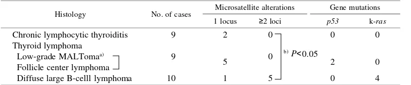
Table III. Microsatellite Alterations and Gene Mutations
Histology No. of cases Microsatellite alterations Gene mutations 1 locus ≥2 loci p53 k-ras
Chronic lymphocytic thyroiditis 9 2 0 0 0
Thyroid lymphoma
Low-grade MALTomaa) 9
5 0 2 0
Follicle center lymphoma
Diffuse large B-celll lymphoma 10 1 5 0 4
a) Marginal zone lymphoma and lymphoplasmacytic lymphoma. b) Fisher’s exact test.
ground and not significant. RER is usually characterized by alterations in multiple loci,22) and this threshold would
significantly lower the probability of regarding back-ground mutations as positive.
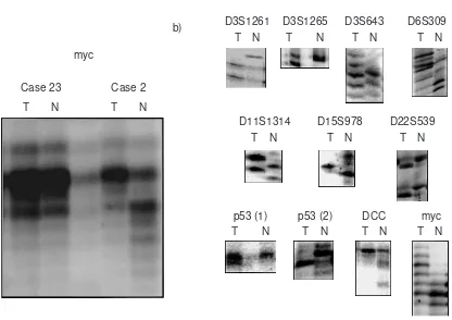
The results are summarized in Table III and representa-tive cases are illustrated in Fig. 1. Six cases of TL and 2
cases of CLTH showed alterations at a single microsatel-lite locus. Only 5 cases of DLBL exhibited alterations at
≥2 loci, i.e., 2 loci in 2 cases, 3 loci in 2 cases, and 8 loci in one case. The frequency of MSI in DLBL was signifi-cantly higher than that in other types of TL and CLTH (P<0.05). One DLBL case showed alterations at D3S1261
a)
myc
Case 23
b)
T N
Case 2
T N
T N
D3S1261
T N
D3S1265
T N
D3S643
T N
D11S1314
T N
p53 (1)
T N
p53 (2)
T N
DCC
T N
myc
T N
D15S978
T N
D22S539
T N
D6S309
Fig. 1. Representative examples of microsatellite analysis in thyroid lymphomas (T) and peripheral blood cells (N) from the same patients. Sixteen distinct microsatellite repeats were analyzed as described in “Materials and Methods.” a) Case 23 showed no microsat-ellite alteration, whereas Case 2 showed microsatmicrosat-ellite alteration at the myc locus. b) D22S539 locus from marginal zone B-cell lym-phoma of extranodal (MALT) type. Remaining figures were from cases of DLBL with RER. No cases showed alterations at D9S171, D14S74, BAT25 and BAT26 loci.
Table IV. Summary of Cases with Mutations of p53 and k-ras Gene
Case Age Sex Histology RER p53 k-ras exon 1
a)
Exon Codon Nucleotide Codon Nucleotide
1 60 F DLBL 8/16 — — — 12 GGT→GCT
2 61 F DLBL 3/16 — — — 12 GGT→TGT
3 67 M DLBL 2/16 — — — 13 GGC→GAC
4 80 F DLBL 3/16 — — — 13 GGC→GAC
5 54 F FCL 1/16 6 190 CCT→CTT — —
6 73 F LP 1/16 8 272 GTT→GGT — —
a) No mutations were found in exon 2.
and D3S1265, and three cases at the c-myc locus. No cases exhibited shortened alleles at BAT25 and BAT26, which are indicators of RER in many human tumors.19)
p53 and k-ras mutations Detected mutations of p53 and k-ras genes are summarized in Table IV. Four of 19 cases (21.1%) had mutations at exon 1 of the k-ras gene: two were missense mutations in codon 13 and two were muta-tions in codon 12, one of which was a missense change and the other resulted in substitution of glycine (GGT) with cysteine (TGT). Two of the four substitutions were G:C→A:T, one was G:C→C:G, and one was G:C→T:A. CpG pairs were involved in none of the cases. None of the cases showed point mutations in exon 2 of the k-ras gene. It is noteworthy that the k-ras mutations occurred in the cases with DLBL with RER, and four of five cases with RER had a k-ras mutation. There is a significant associa-tion between RER and k-ras mutation (P<0.05).
p53 mutations were not found in the CLTH. Two of 19 TL cases (10.5%) showed mutations of the p53 gene: one was a missense change in codon 190 at exon 6, and the other was a substitution mutation of valine (GTT) with glycine (TGT) in codon 272 at exon 8. One substitution was C:G→T:A and the other was T:A→G:C. CpG sites were not involved. No significant association between RER and p53 mutation was found.
DISCUSSION
CLTH is an organ-specific autoimmune disease and pre-disposes to TL, with a delay of over 10 years until devel-opment of TL.4) In this situation, MSI was suggested to
occur in the thyroid lesions, because oxidative stress is responsible for as many as 10000 DNA-damaging events/ cell/day under normal cellular conditions.23) However,
none of the present CLTH and TL of low grade cases showed MSI. Chronic inflammation in CLTH might inten-sify oxidative stress and induce DNA damage, but might be compensated by enhanced capacity of DNA repair.
Gastric lymphoma is now classified as MALT lym-phoma,24)
and is considered to develop in H. pylori -induced chronic gastritis.9) Chong et al.16) reported that
66.7% of low-grade gastric MALT lymphomas showed RER. However, Xu et al.25) have recently reported that
none of 33 cases of primary gastric lymphoma of the MALT type exhibited RER, after examining 9 microsatel-lite loci including the same 7 microsatelmicrosatel-lite loci as selected by Chong et al.16) These discrepancies might derive from
the different criteria used to define RER: only one locus was enough for some studies and more than 2 loci in another. We regarded the cases showing alterations at ≥2 microsatellite loci as positive for MSI, and none of the 9 cases of primary TL of low grade or follicle center cell lymphoma exhibited RER, including alterations at D3S1261, D3S1265 and c-myc loci, which have been
reported to have a high frequency of instability.16) Thus,
genomic instability might not contribute significantly to the molecular pathogenesis of low-grade MALT lym-phoma developing in chronic inflammation.
In this study, 50% of DLBL exhibited RER, i.e., alter-ations at ≥2 loci. The frequency of MSI in DLBL was sig-nificantly higher than that in other types of TL and CLTH (P<0.05), suggesting that increase in the genomic instabil-ity contributes to the progression from low grade to high grade lymphoma of the thyroid. In gastric lymphoma, accelerated genomic instability in DLBL compared to low grade lymphomas was reported,16, 17, 25)
although the differ-ence in MSI frequency was not statistically significant in the study by Xu et al.25) Frequency of MSI in DLBL
aris-ing in the wall of chronic pyothorax, a type of lymphoma developing in chronic inflammation, was found to be about 30% (data not published). In nodal lymphomas, Gamberi et al.14) found that MSI was not responsible
for the clinical progression and histologic transformation, which are common in the late phase of nodal lymphomas. These findings might suggest different pathogenetic mech-anisms for progression of nodal and extranodal lympho-mas.
The k-ras gene-encoded protein, referred to as p21, functions as a G protein, which plays a role in signal transduction from growth factor receptors on the cell membrane. Point mutations in k-ras gene result in forma-tion of proteins that can not be inactivated, thus leading to autonomous cell growth and proliferation. It was reported that leukocyte-derived potent oxidants could cause k-ras
oncogene activation, which is believed to play a critical role in the pathogenesis of many human malignancies.26)
In our studies, a significant association between RER and k-ras mutation (P<0.05) was found; four of five DLBL with RER had k-ras mutation. MSI might lead to accumu-lation of genetic aberrations such as k-ras mutations, caus-ing progression of low grade to high grade lymphoma. Alternatively, MSI may be present per se in some cases with DLBL. The association of RER with k-ras mutation was also reported in pancreatic cancer, non-small cell lung cancer, and a type of sarcoma in mice.13, 27)
p53 is a well-known tumor suppressor gene, and has an anti-oncogenic role by causing cells with damaged DNA to arrest at the G1 phase of the cell cycle or stimulating expression of the bax gene, the protein product of which promotes apoptosis. p53 mutation was involved in the development of splenic marginal zone lymphoma,28) or
associated with progression of MALT lymphoma.29)
In our cases, p53 mutations were found in only 2 of 9 cases with low-grade MALT lymphoma, but in none of DLBL. These findings suggest that genomic instability does not always occur in concert with p53 inactivation.
change DNA sequences as follows30); G:C→A:T,
A:T→G:C, G:C→T:A, A:T→T:A. Indeed, in our cases, such changes were found in 3 of 4 cases with k-ras muta-tion, and in 1 of 2 cases with p53 mutations.
In conclusion, the present findings on TL indicate that the genomic instability contributes to the molecular patho-genesis of the progression from low grade to high grade lymphoma.
ACKNOWLEDGMENTS
This study was supported by grants from the Vehicle Racing Commemorative Foundation and the Ministry of Education, Sci-ence, Sports and Culture, Japan (08457061, 08670202, 08770126, 09670184, 09770148, 10151225).
(Received October 20, 1999/Revised December 7, 1999/ Accepted December 10, 1999)
REFERENCES
1) Freeman, C., Berg, J. W. and Cutler, S. J. Occurrence and prognosis of extranodal lymphomas. Cancer, 29, 252–260 (1972).
2) Aozasa, K., Tsujimoto, M., Sakurai, M., Honda, M., Yamashita, K., Hanada, M. and Sugimoto, A. Non-Hodgkin’s lymphomas in Osaka, Japan. Eur. J. Cancer Clin. Oncol., 21, 487–492 (1985).
3) Volpe, R. Thyroiditis: current views of pathogenesis. Med. Clin. North Am., 59, 1163–1175 (1975).
4) Kato, I., Tajima, K., Suchi, T., Aozasa, K., Matsuzuka, F., Kuma, K. and Tominaga, S. Chronic thyroiditis as a risk factor of B-cell lymphoma in the thyroid gland. Jpn. J. Cancer Res. (Gann), 76, 1085–1090 (1985).
5) Aozasa, K., Ueda, T., Katagiri, S., Matsuzuka, F., Kuma, K. and Yonezawa, T. Immunologic and immunohistologic analysis of 27 cases with thyroid lymphomas. Cancer, 60, 969–973 (1987).
6) Aozasa, K. Malignant lymphoma of the mucosa-associated lymphoid tissue. Am. J. Surg. Pathol., 16, 90–92 (1992). 7) Aozasa, K., Ohsawa, M. and Kanno, H.
Pyothorax-associ-ated lymphoma: a distinct type of lymphoma strongly asso-ciated with Epstein-Barr virus. Adv. Anat. Pathol., 4, 58– 63 (1997).
8) Ohsawa, M., Aozasa, K., Horiuchi, K. and Kanamaru, A. Malignant lymphoma of bladder. Report of three cases and review of the literature. Cancer, 72, 1969–1974 (1993). 9) Parsonnet, J., Hansen, S., Rodriguez, L., Gelb, A. B.,
Warnke, R. A., Jellum, E., Orentreich, N., Vogelman, J. H. and Friedman, G. D. Helicobacter pylori infection and gas-tric lymphoma. N. Engl. J. Med., 330, 1267–1271 (1994). 10) Cerutti, P. A. and Trump, B. F. Inflammation and oxidative
stress in carcinogenesis. Cancer Cells, 3, 1–7 (1991). 11) Eshleman, J. R. and Markowitz, S. D. Microsatellite
insta-bility in inherited and sporadic neoplasms. Curr. Opin. Oncol., 7, 83–89 (1995).
12) Suzuki, H., Harpaz, N., Tarmin, L., Yin, J., Jiang, H. Y., Bell, J. D., Hontanosas, M., Groisman, G. M., Abraham, J. M. and Meltzer, S. J. Microsatellite instability in ulcerative colitis-associated colorectal dysplasias and cancers. Cancer Res., 54, 4841–4844 (1994).
13) Brentnall, T. A., Chen, R., Lee, J. G., Kimmey, M. B., Bronner, M. P., Haggitt, R. C., Kowdley, K. V., Hecker, L. M. and Byrd, D. R. Microsatellite instability and K-ras mutations associated with pancreatic adenocarcinoma and pancreatitis. Cancer Res., 55, 4264–4267 (1995).
14) Gamberi, B., Gaidano, G., Parsa, N., Carbone, A., Roncella, S., Knowles, D. M., Louie, D. C., Shibata, D., Chaganti, R. S. and Dalla-Favera, R. Microsatellite instability is rare in B-cell non-Hodgkin’s lymphomas. Blood, 89, 975–979 (1997).
15) Bedi, G. C., Westra, W. H., Farzadegan, H., Pitha, P. M. and Sidransky, D. Microsatellite instability in primary neo-plasms from HIV+ patients. Nat. Med., 1, 65–68 (1995). 16) Chong, J. M., Fukayama, M., Hayashi, Y., Hishima, T.,
Funata, N., Koike, M., Matsuya, S., Konishi, M. and Miyaki, M. Microsatellite instability and loss of hetero-zygosity in gastric lymphoma. Lab. Invest., 77, 639–645 (1997).
17) Peng, H., Chen, G., Du, M., Singh, N., Isaacson, P. G. and Pan, L. Replication error phenotype and p53 gene mutation in lymphomas of mucosa-associated lymphoid tissue. Am. J. Pathol., 148, 643–648 (1996).
18) Fernandez, P. J., Santos, J., Perez, D. C. I., Melendez, B., Martinez, B., Robledo, M., Rivas, C. and Benitez, J. Fre-quent allelic losses of 9p21 markers and low incidence of mutations at p16 (CDKN2) gene in non-Hodgkin lympho-mas of B-cell lineage. Cancer Genet. Cytogenet., 98, 63– 68 (1997).
19) Zhou, X. P., Hoang, J. M., Li, Y. J., Seruca, R., Carneiro, F., Sobrinho, S. M., Lothe, R. A., Gleeson, C. M., Russell, S. E., Muzeau, F., Flejou, J. F., Hoang, X. K., Lidereau, R., Thomas, G. and Hamelin, R. Determination of the replica-tion error phenotype in human tumors without the require-ment for matching normal DNA by analysis of mononucleotide repeat microsatellites. Genes Chromosom. Cancer, 21, 101–107 (1998).
20) Hongyo, T., Buzard, G. S., Calvert, R. J. and Weghorst, C. M. ‘Cold SSCP’: a simple, rapid and non-radioactive method for optimized single-strand conformation polymor-phism analyses. Nucleic Acids Res., 21, 3637–3642 (1993). 21) Weber, J. L. and Wong, C. Mutation of human short
tan-dem repeats. Hum. Mol. Genet., 2, 1123–1128 (1993). 22) Aaltonen, L. A., Peltomaki, P., Leach, F. S., Sistonen, P.,
Pylkkanen, L., Mecklin, J. P., Jarvinen, H., Powell, S. M., Jen, J., Hamilton, S. R., Peterson, G. M., Kinzler, K. W., Vogelstein, B. and de la Chapelle, A. Clues to the patho-genesis of familial colorectal cancer. Science, 260, 812– 816 (1993).
8-hydroxy-2′-deoxyguanosine in rat organ DNA and urine. Proc. Natl. Acad. Sci. USA, 87, 4533–4537 (1990). 24) Isaacson, P. G. and Spencer, J. Malignant lymphoma of
mucosa-associated lymphoid tissue. Histopathology, 11, 445–462 (1987).
25) Xu, W. S., Chan, A. C., Liang, R. and Srivastava, G. No evidence of replication error phenotype in primary gastric lymphoma of mucosa-associated lymphoid tissue. Int. J. Cancer, 76, 635–638 (1998).
26) Jackson, J. H., Vollenweider, M., Hill, J., Rodriguez, H., Schwabacher, A. W., Mitra, G. and Kuo, C. Y. Stimulated human leukocytes cause activating mutations in the K-ras protooncogene. Oncogene, 14, 2803–2808 (1997). 27) Niwa, O., Kamiya, K., Furihata, C., Nitta, Y., Wang, Z.,
Fan, Y. J., Ninomiya, Y., Kotomura, N., Numoto, M. and Kominami, R. Association of minisatellite instability with
c-myc amplification and K-ras mutation in methylcholan-threne-induced mouse sarcomas. Cancer Res., 55, 5670– 5676 (1995).
28) Baldini, L., Fracchiolla, N. S., Cro, L. M., Trecca, D., Romitti, L., Polli, E., Maiolo, A. T. and Neri, A. Frequent p53 gene involvement in splenic B-cell leukemia/ lymphomas of possible marginal zone origin. Blood, 84, 270–278 (1994).
29) Du, M., Peng, H., Singh, N., Isaacson, P. G. and Pan, L. The accumulation of p53 abnormalities is associated with progression of mucosa-associated lymphoid tissue lym-phoma. Blood, 86, 4587–4593 (1995).