A CASE REPORT
ABSTRACT
Address for corespondance :
dr. Raymond Pranata, Faculty of Medicine, Universitas Pelita Harapan, Tangerang, Banten, Indonesia
Email :raymond_pranata@hotmail.com
How to cite this article :
PREmATuRE VEnTRiCulAR COmPlEx-induCEd CARdiOmyOPAThy:
A CASE REPORT And REViEw Of liTERATuRE
PREmATuRE VEnTRiCulAR COmPlEx-induCEd CARdiOmyOPAThy: A CASE REPORT And REViEw Of liTERATuRE
Raymond Pranata1, Emir Yonas2, Veresa Chintya3
1Assistant Physician, Siloam General Hospital - Faculty of Medicine, Universitas Pelita Harapan, Tangerang, Banten, Indonesia 2Faculty of Medicine, YARSI University, Jakarta, Indonesia
3General Practitioner, Sanjiwani General Hospital, Gianyar, Bali, Indonesia
introduction: Frequent premature ventricular complexes (PVCs) have theability to cause or contribute to cardiomyo-pathy and heart failure symptoms in long-term.
Case illustration: A 46 years old male presented with recurrent palpitations, the latest since 6 hours before admission. PMH of hypertension, diabetes and heart disease were denied. BP: 120/80 mmHg, HR: 138 bpm, RR 22x/minute. ECG: LV focus multifocal PVC bigeminy. lab: within normal limits. CxR: cardiomegaly. Echocardiography: MR and LVH. The patient was administered diltiazem IV in the ED. Bisoprolol was given during discharge.
Discussion: PVC-induced cardiomyopathy’s mechanisms are not entirely clear; a more likely explanation is abnormal ventricular activation resulting in mechanical dyssynchrony, >10.000 PVCs/day has high risk and this patient’s PVC burden should be calculated. It is a diagnosis of exclusion andmade after exploring possible causes and excluding them.Pharmacotherapy to suppress PVCs includes β-Blockers, CCB, and other antiarrhythmic drugs. Results of the use of β-Blockers and CCB are modest with reported efficacy rates in the 20% range.In patients with PVC-induced cardiomyopathy, successful elimination of PVCs with ablation frequently restores ventricular function acutely and has aprocedural success rate of PVC elimination of 84%. Unfortunately, this patient is not a suitable candidate for catheter ablation. Hence, β-Blockers was chosen for long-term therapy.
Conclusion: Frequent PVCs with high burden has potential to cause adverse cardiovascular events and should be treated to prevent its deleterious consequences. Suppression of PVCs using either antiarrhythmic pharmacological agents or emerging catheter ablation techniques appears to reverse the LV dysfunction.
Keywords: PVC, Cardiomyopathy, burden
ABSTRAK
Pendahuluan: Kompleks ventrikular prematur (KVP) memiliki kemampuan untuk menyebabkan kardio miopati atau gejala gagal jantung dalam jangka panjang.
Ilustrasi Kasus: Seorang laki-laki berusia 46 tahun datang dengan keluhan berdebar-debar sejak 6 jam sebelum masuk rumah sakit. Riwayat penyakit hipertensi, diabe-tes, dan gagal jantung disangkal. Tekanan darah 120/80 mmHg, detak jantung 138 kali/menit, laju nafas 22x/ menit. EKG menunjukan KVP bigemini multifocal. Pe-meriksaan laboratorium dalam batas normal. Ro thoraks: kardiomegali. Ekokardiografi: MR dan LVH. Pasien diberikan diltiazem IV saat di IGD. Pasien diberikan bisoprolol saat pulang.
diskusi: Mekanisme mengapa kardiomiopati yang disebabkan oleh VKP masih belum sepenuhnya jelas; penjelasan yang paling memungkinkan adalah aktivasi abnormal pada ventrikel yang menyebabkan fungsi me-kanis menjadi tidak sinkron, VKP >10.000/hari memiliki risiko yang tinggi. Hal tersebut merupakan diagnosis eksklusi setelah menyingkirkan kemungkinan penyebab lain. Farmako terapi untuk mengurangi VKP adalah dengan penyekat beta, penyekatk anal kalsium, dan obat antiaritmik lainnya. Pengunaan penyekat beta dan ka-nal kalsium memiliki efikasi hanya sekitar 20%. Ablasi
dengan angka keberhasilan 84%, namun, pasien ini bukan merupakan kandidat yang cocok untuk ablasi kateter. Oleh karena itu penyekat beta merupakan pilihan untuk terapi jangka panjang.
Kesimpulan: Kontraksi ventrikular prematur yang sering berpotensi untuk menyebabkan adverse cardiovascular events dan harus ditangani untuk mencegah komplikasi. Mengurangi VKP dengan obat anti aritmik atau ablasi kateter dapat mengembailkan fungsi ventrikel.
ABSTRAK
Pendahuluan: Kompleks ventrikular prematur (KVP) memiliki kemampuan untuk menyebabkan kardio miopati atau gejala gagal jantung dalam jangka panjang.
ilustrasi Kasus: Seorang laki-laki berusia 46 tahun datang dengan keluhan berdebar-debar sejak 6 jam sebelum masuk rumah sakit. Riwayat penyakit hipertensi, diabetes, dan gagal jantung disangkal. Tekanan darah 120/80 mmHg, detak jantung 138 kali/menit, laju nafas 22x/menit. EKG menunjukan KVP bigemini multifocal. Pemeriksaan laboratorium dalam batas normal. Ro thoraks: kardiomegali. Ekokardiografi: MR dan LVH. Pasien diberikan diltiazem IV saat di IGD. Pasien diberikan bisoprolol saat pulang.
diskusi: Mekanisme mengapa kardiomiopati yang disebabkan oleh VKP masih belum sepenuhnya jelas; penjelasan yang paling memungkinkan adalah aktivasi abnormal pada ventrikel yang menyebabkan fungsi mekanis menjadi tidak sinkron, VKP >10.000/hari memiliki risiko yang tinggi. Hal tersebut merupakan diagnosis eksklusi setelah menyingkirkan kemungkinan penyebab lain. Farmako terapi untuk mengurangi VKP adalah dengan penyekat beta, penyekatk anal kalsium, dan obat antiaritmik lainnya. Pengunaan penyekat beta dan kanal kalsium memiliki efikasi hanya sekitar 20%. Ablasi kateter yang berhasil dapat memperbaiki fungsi ventrikel dengan angka keberhasilan 84%, namun, pasien ini bukan merupakan kandidat yang cocok untuk ablasi kateter. Oleh karena itu penyekat beta merupakan pilihan untuk terapi jangka panjang.
Kesimpulan: Kontraksi ventrikular prematur yang sering berpotensi untuk menyebabkan
adverse cardiovascular events dan harus ditangani untuk mencegah komplikasi. Mengurangi
VKP dengan obat anti aritmik atau ablasi kateter dapat mengembailkan fungsi ventrikel.
Kata kunci: VKP, kardiomiopati, beban
inTROduCTiOn
Premature ventricular complex (PVCs)
without underlying cardiac disease were
longed thought to be benign, it is true in
most cases. However, recent evidence
showed that frequent PVCs have theability
to cause or contribute to cardiomyopathy
and heart failure symptoms in long
term.1Premature ventricular contractions-induced cardiomyopathy is reversible
cardiomyopathy due to PVCs.2 In this paper we will present a patient without other
cardiovascular risk factors developed a left
ventricular hypertrophy accompanied with
frequent PVCs.
CASE illuSTRATiOn
A 46 years old male presented with
recurrent palpitations, with the latest was
since 6 hours before admission. He
frequently complained of palpitations for
most days of the week, however, he felt that
the symptoms were more severe at that time.
He did not experience chest pain, shortness
of breath, nausea, vomiting or sweating. He
dyspne a, orthopnea, syncope or edema. Past
medical history of hypertension, diabetes
and heart disease were denied. The patient
did not smoke and avoid caffeine. On
presentation, to emergency department the
patient was fully conscious with blood
pressure of 120/70 mmHg, heart rate of 138
bpm, and respiratory rate of 22x/minute.
Cardiac examination revealed a
grade II/VI systolic murmur at the apex
radiating to axilla suggestive of mitral
regurgitation with apex displaced downward
and leftward. There was no hepatomegaly,
edema or enlarged thyroid glands.
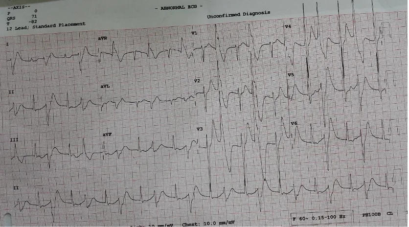
Electrocardiography showed frequent
multifocal PVC (bigeminy) originating from
left ventricular focus(fig 1). Complete
blood counts, glucose, electrolytes, renal and
liver function werewithin normal limits.
Chest X-Ray showed cardiomegaly(fig 2).
The patient was admitted and given
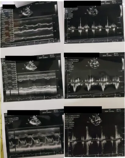
intravenous diltiazem. Echocardiography
showed mitral regurgitation (MR) and left
ventricular hypertrophy (LVH)(fig 3). The
patient was diagnosed with
tachycardia-induced cardiomyopathy with differential
diagnosis of primary valvular heart disease.
The patient was given bisoprolol and
discharged from the hospital.
diSCuSSiOn
Premature ventricular complex-induced
cardiomyopathy’s mechanisms are not
entirely clear; a more likely explanation is
abnormal ventricular activation resulting in
mechanical dyssynchrony, >10.000
PVCs/day has high risk and this patient’s
PVC burden was >10.000 PVCs/day.3It is a diagnosis of exclusion and made after
exploring possible causes and excluding
them.This patient denies previous history
and other risk factors for cardiovascular
disease. Blood pressure, glucose,and
electrolytes were also within normal limits.
Electrocardiography, chest x-ray, and
echocardiography in this patient showed
LVH with MR. Frequent palpitation and
evidence of frequent PVCs without other
cardiovascular risk factors or history may
explain the cause of LVH. Mitral
regurgitation may be due to LVH or primary
valvular disease, hence, a differential
diagnosis.
Further investigations should be
performed, including 24-hour Holter
monitor to quantify PVC burden and can
detect any related arrhythmias. In those with
exercise-inducedPVCs, treadmill test may
be ordered. Testing for sleep apnea is
appropriate if there is high nocturnal PVCs
burden. Typically, electrophysiological
cardiomyopathy’s mechanisms are not
PVCs/day has high risk and this patient’s
helpful to detect potential causes of PVCs
such as arrhythmogenic right ventricular
dysplasia and infiltrative
cardiomyopathies.2,4
In healthy people with PVC, adjusted
hazard ratio for cardiac death is 3,98 & 0,95
in male and female, respectively.5Indications to initiate therapy for patients with PVCs
include bothersome symptoms, the presence
of complications (cardiomyopathy with
decreased EF or ventricular dilatation and
PVC induced tachycardia), PVCs triggering
malignant ventricular arrhythmias, and
PVCs limiting optimal biventricular
pacing.3Management in asymptomatic patients with normal LV function and a very
frequent PVCs (>20%) is still controversial. Whether it should be managed with
medication or ablation to prevent future risk
of cardiomyopathyor follow-up assessment
of left ventricular function is still
undetermined.3No treatment other than reassurance needed in patients without
inherited arrhythmia syndromes who have
asymptomatic or mildly symptomatic PVCs.
In those with asymptomatic PVCs and a
normal LV function without accompanying
structural heart disease,consider follow-up
in those with PVC burden >10,000 in 24
hours or in patients with PVC burden
<10,000 in 24 hours if symptoms worsen.
Medications are reported to suppress PVCs
in 10%-40% of patients.Pharmacotherapy to suppress PVCs includes β-Blockers (1st line), CCB, and other antiarrhythmic drugs.6,7 Results of the use of β-Blockers and CCB
are modest with reported efficacy rates in
the 20% range. In CHF-STAT trial,
amiodarone decreased hourly PVCs
(44 ± 145 vs 254 ± 370, P < .001) with 69%
of patients experiencing an 80% decrease in
PVC burden at 3 months. However,
long-term use is limited by its adverse effect
profile.
Catheter ablation has emerged as a
relatively safe and effective option to
pharmacotherapy for PVC elimination. It is
recommended in right ventricular outflow
tract (RVOT) PVC or VT with symptoms,
failed pharmacotherapy or decline in LV
function. It is also indicated in PVCs that
trigger recurrent ventricular fibrillation
leading to implantable cardioverter
defibrillation (ICD) shock or lead to
electrical storms.2,6,7This patient fulfilled neither of those criteria. In patients with
PVC-induced cardiomyopathy, successful
elimination of PVCs with ablation
frequently restores ventricular function and
has anacute procedural success rate of PVC
elimination of 84% (80%-90%) of patients
patients.2 Predictors of success were RVOT PVC location and monomorphic as opposed
to multiple PVC morphologies.
Unfortunately, both were not present in this
patient, the likelihood of success and clinical
improvement vs the potential risk should be
weighted. Factors to evaluate include PVC
frequency, anticipated PVC location, the
number of PVC morphologies,
pharmaceutical alternatives, and patient age
and comorbidities.We chose
pharmacotherapy over ablation withβ
-Blockers(1st line medication) was chosen as the initial therapy.
COnCluSiOn
Frequent PVCs with high burden has
potential to cause adverse cardiovascular
events and should be treated to prevent its
deleterious consequences. Suppression of
PVCs using either antiarrhythmic
pharmacological agents or emerging catheter
ablation techniques appears to reverse the
LV dysfunction.
REfEREnCES
1. Baman TS, Lange DC, Ilg KJ, Gupta
SK, Liu TY, Alguire C, et al.
Relationship between burden of
premature ventricular complexes and
left ventricular function. Heart
Rhythm 2010;7:865-9.
2. Luebbert J, Auberson D, Marchlinski
F. Premature Ventricular Complexes
in Apparently Normal Hearts. Card
ElectrophysiolClin 2016;8(3):503-14
3. Latchamsetty R, Bogun F. Premature
Ventricular Complex-induced
Cardiomyopathy. Rev EspCardiol
2016;69:365-9. DOI:
10.1016/j.rec.2015.12.015
4. Cantillon DJ. Evaluation and
management of premature
ventricular complexes. Cleve Clin J
Med 2013;80(6):377-87.
5. Hirose H, et al. Cardiac mortality of
premature ventricular complexes in
healthy people in Japan. J Cardiol
consensus on ventricular arrhythmias.
Heart Rhythm 2014;11(10):e166-96.
doi: 10.1016/j.hrthm.2014.07.024
7. Priori SG, et al. 2015 ESC
Guidelines for the management of
patients with ventricular arrhythmias
and the prevention of sudden cardiac
β
Management of Patients with
Ventricular Arrhythmias and the
Prevention of Sudden Cardiac Death
of the European Society of
Cardiology (ESC).Eur Heart J
2015;36(41):2793-867. doi:
Fig 1. Patient’s ECG
Fig 2. Patient’s Chest X-Ray