ISSN 2331-1851 ©2013 World Science Research Journals
Full Length Research Paper
Profile of Retinoblastoma in East Java, Indonesia
Hendrian D. Soebagjo
1*,
Reni Prastyani
1, Hidayat Sujuti
2, Diana Lyrawati
3and
Sutiman B. Sumitro
41
Department of Ophthalmology, Faculty of Medicine, University of Airlangga, Dr.Soetomo Hospital Surabaya, Surabaya, East Java, Indonesia.
2
Department of Biochemistry-Molecular Biology, Faculty of Medicine, Brawijaya University – Department of Ophthalmology, Dr. Saiful Anwar Hospital, Malang, East Java, Indonesia.
3
Departement of Pharmacy, Faculty of Medicine, Brawijaya University – Dr. Saiful Anwar Hospital, Malang, East Java, Indonesia.
4
Departement of Biology, Faculty of Mathematic and Natural Sciences, University of Brawijaya, Malang, East Java, Indonesia.
Accepted 24 September, 2013
This study showed the profile of retinoblastoma in East Java, Indonesia and correlations between important factors for retinoblastoma based on the stage of malignancy and pedigree. This is a retrospective study based on hospital medical record. Fourty four patients were diagnosed of retinoblastoma by histopathology examination. The classification of pedigree was determined by unilaterality/bilaterality and family history of retinoblastoma. IRSS (International Retinoblastoma Staging System) classification was used to determine the stage of retinoblastoma, that is, intraocular, extraocular and metastasis stage. The mean age of patients was 44.64 months and most patients were in the age group of 60-119 months (22.72%). The study showed 38 (86.36%) cases of unilateral retinoblastoma and 6 (13.64%) cases of bilateral retinoblastoma. Retinoblastoma surface in patients mostly at stage III-a with the total of 27 (61.36%) cases and stage I or II each 6 (13.64%) cases. Pedigree classification showed 1 (2.27%) familial hereditary retinoblastoma patient, 6 (13.64%) sporadic hereditary retinoblastoma patients, and 37 (84.09%) patients of non-hereditary retinoblastoma. Spearman correlation test between age and IRSS show a significant results at stage III-a (rs = 0,785, p=0.002). Most patients with unilateral retinoblastoma (84.09%) were non-hereditary retinoblastoma survivors and patients with bilateral retinoblastoma (15.91%) were hereditary retinoblastoma which include familial and sporadic retinoblastoma.
Key words: Retinoblastoma, lateralization, staging IRSS, pedigree.
INTRODUCTION
Retinoblastoma is an embryonal tumor originating from retinal cells, which account for the most frequent intraocular malignant tumor among children with incidence rate of 1 in every 15,000–20,000 live birth and occurrence rate up to 9,000 cases each year (Linn, 2005; Kivela and Paulino, 1999; Chintagumpala et al., 2007; Dimaras et al., 2012). As reported, it occurs 1 in every 18,000–34,000 live birth in developing countries of Africa
Based on its etiology or hereditary pattern, which can be investigated through pedigree system, retinoblastoma is classified into 3 types; that is, familial hereditary retinoblastoma; sporadic hereditary retinoblastoma; and non–hereditary retinoblastoma (Bunin and Orjuela, 2007).
In familial hereditary retinoblastoma, the entire patient cells contained mutated RB1 gene, and this process act as “the first hit” in retinoblastoma development. Next mutation occurs if the process of copying the RB1 gene ensues, termed as “the second hit”. Familial hereditary retinoblastoma may reach 10% of total retinoblastoma patients (Bunin and Orjuela, 2007; Knudson, 2001).
Sporadic hereditary retinoblastoma occurs in 30% of children with retinoblastoma. Patients will suffer from malignancy in both eyes, although there were no family history of retinoblastoma. Mutation of the RB1 gene is purely a de novo (Bunin and Orjuela, 2007; Abramson et al., 1998).
Most of retinoblastoma in developing country are a non–hereditary type. These type of retinoblastoma manifest in one eye as a result of two RB1 somatic genes mutation in a single cell, not long after conception (Bunin and Orjuela, 2007; Abramson et al., 1998).
Most cases of familial or sporadic hereditary retinoblastoma are bilateral, only 10–15% are unilateral. In contrast, all non–hereditary retinoblastoma cases are unilateral. Therefore, bilateral retinoblastoma cases can be interpreted as hereditary retinoblastoma (familial and sporadic hereditary) and unilateral cases as non– hereditary retinoblastoma (Bunin and Orjuela, 2007; Richter et al., 2003; Akhriwu and Alex, 2012; Abramson et al., 1998).
Data from several countries showed that diagnosis of retinoblastoma is usually made at the age of 18 months, with bilateral cases averagely diagnosed at 12 months old and unilateral cases diagnosed at 23 months to 29– 30 months old. Retinoblastoma diagnosed in children aged > 5 years old is very rare (Draper et al., 1992).
In this study, we sought to identify the profile of retinoblastoma patients in East Java Indonesia and evaluate the relationship between age and pedigree pattern of retinoblastoma. This study intend to show the older age at diagnosis, the higher stage on, and to prove that patients with unilateral retinoblastoma are classified as non–hereditary retinoblastoma, and bilateral retinoblastoma are classified as hereditary retinoblastoma, which include familial and sporadic hereditary retinoblastoma.
*Corresponding author. E-mail: [email protected]. Tel: +62811347609.
Abbreviations: IRSS, International retinoblastoma staging system; RB1, retinoblastoma 1.
METHODS
This study was carried out to evaluate the medical records of all retinoblastoma patients that were hospitalized at Dr. Soetomo General Hospital Surabaya between January 2010 to December 2012.
Hereditary classification was determined by lateralization factor and family history using pedigree of retinoblastoma. Lateralization obtained from clinical symptoms, and family history were taken from medical record evaluation. IRSS classification was used to determine staging, which consist of intraocular, extraocular, and metastatic stage. Data was statistically analyzed using Spearmann correlation test.
RESULTS
There were 44 patients histopathologically diagnosed with retinoblastoma enrolled at Dr. Soetomo general hospital, Surabaya from January 2010 to December 2012. Pedigree classified patients to the status of the hereditary. Of all patients, 1 (2.27%) was familial hereditary retinoblastoma, 6 (13.64%) were sporadic hereditary retinoblastoma, and 37 (84.09%) which is the highest were non– hereditary retinoblastoma (Table 1).
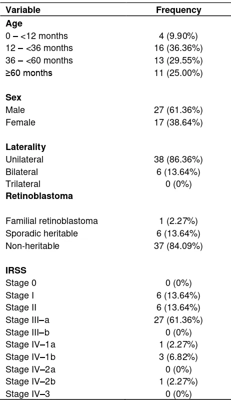
Patient’s age and sex were analyzed. Patients age ranged from 3 to 154 months old, with the mean age of 44.64 months. Twenty seven (61.36%) were male, and 38.64% were female. No significant relationship was found between age and heredity, but there was significant relationship between age and staging (IRSS). Stage III–a was the most dominant among others and range of age were 60–119 months group as seen in Figure 1 and Table 1. Based on the aforementioned age group, 61.36% patients were at stage III–a.
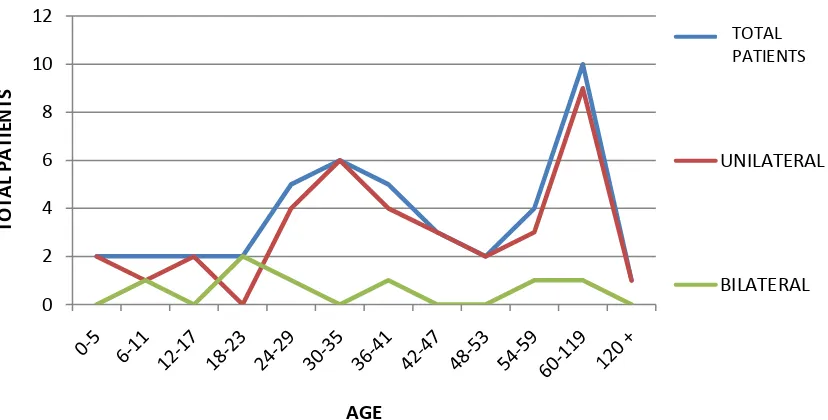
Spearman correlation test analysis showed positive significant correlation between age and IRSS staging for stage III–a (r=0.785, p=0.002), and not significant for other stages. There were no data for stage III–b, IV–2a and IV–3, therefore, they were not analyzed (Table 2). Significant distributions of laterality and age are shown in Figure 2. Unilateral retinoblastoma was the majority type in 60 – 119 months age group, as well as in total patients. From the total 35 unilateral patients, 9 (25.7%) were in group 60–119 months.
Table 1. Profile of retinoblastoma patients.
Familial retinoblastoma 1 (2.27%) Sporadic heritable 6 (13.64%)
Non-heritable 37 (84.09%)
retinoblastoma and International Staging of Retinoblastoma (rs=0.866, p=0.333).
DISCUSSION
In this study, we analyzed 44 new cases of retinoblastoma in Dr. Soetomo general hospital Surabaya with median age of 44.64 months old. Retinoblastoma incidence in foreign countries occurs earlier. Khan et al. (2013) in Pakistan studied 52 cases with median age of 42.5 months. In contrast, Antoneli et al. (2003) studied 83 cases in Sao Paolo, Brazil with median age 32.9 months and Essuman et al. (2010) in Ghana studied 23 cases with median age of 36.3 months. The median age in Nigeria was 29 months, studied by Owoeye et al. (2005).
In Netherland, median age of screened children diagnosed for familial hereditary retinoblastoma was 17.2 months. Our study showed that median age of diagnosis in our patients was older (44.64 months). Such later age of first diagnosis may be due to less advance early diagnosis and screening methods especially in developing countries. Most of our patients usually arrive in terminal state. Moreover, late diagnoses could be due to other problems such as, poor affordability and accessibility to available medical facilities. All of these contributes to high mortality rate.
The late median age correlated with heredity, shown in quantity distribution of familial retinoblastoma patients, where we found just a few in this study. It is associated with the severity and staging during first diagnosis and then advanced because of hereditary. Mostly patients were first diagnosed in proptotic and bilateral state. Survival rate for this group was very low and have poor outcome. Thus, familial retinoblastoma patients is uncommon since they cannot survive to produce offsprings (Khan et al., 2013; Moll et al., 2000).
Figure 1 showed that stage III–a mainly occurred at age 60 – 119 months, whereas lower stage occurred at younger age and only few children experienced late stage. This study showed that extraocular retinoblastoma usually happen in children of older age and most of them came in late stage. This statement supported by the minimum data for metastatic stage/stage IV may be as a result of fact that the patient had expires due to malignancy. Similar results have been reported in other developing countries. In India, stage III retinoblastoma is mostly suffered by children of median age 46.8 months. This was projected due to lack of early detection, and so the patients came in late stage (Chong et al., 2006; Pratt et al., 1997; Radhakrishnan et al., 2012).
Retinoblastoma stages in our study were found mostly at stage III–a, which were suffered by 26 patients (61.36%) out of total 44 patients. Meanwhile 12 patients (27.28%) suffered at stage I and II retinoblastoma, and the rest have already experienced stage IV by which the cancer have spread to orbital tissue, bone or CNS. More than half of the participants of our study suffered from stage III–a retinoblastoma, that is, extraocular retinoblastoma stage, where cancer cells invaded periocular tissue without invasion to periauricular and extension to cervical lymph node. Previous study also lead to similar result, a study of the relationship between retinoblastoma grading and retinoblastoma stage found that stage III with metastasis was the most commonly found stage (Murphree and Chantada, 2007; Radhakrishnan et al., 2012; Soebagjo et al., 2011).
Figure 1. Relationship of retinoblastoma patients with IRSS staging.
Table 2. Correlations of age and IRSS.
Stadium IRSS Correlations coef. p value*
Stage I 0.552 0.063
Stage II 0.552 0.063
Stage III–a 0.785 0.002*
Stage III–b 0 0
Stage IV–1a 0.136 0.673
Stage IV–1b 0.034 0.917
Stage IV–2a 0 0
Stage IV–2b 0.045 0.889
Stage IV–3 0 0
*significant if p<0.05.
(2006) found unilateral retinoblastoma to be 60–70% compared to 30–40% cases of bilateral retinoblastoma. Same result was revealed by Essuman et al. (2010) in Ghana where 82.6% were unilateral cases and 17.4% were bilateral. Lateralization of retinoblastoma is one of the factor that correlates with heredity. Familial retinoblastoma occurs in genetic fashion, and this type of retinoblastoma always occur bilaterally. Retinoblastoma is established on a child if RB1 gene in his/her system is found, and this gene is inherited. According to Knudson theory, the existent of RB1 gene in the system is believed
to be the “first hit”. Mutation of gene copy that occurs some times after delivery in retinal cells on both eyes became the “second hit”. Genetic factors has no role in sporadic heritable retinoblastoma that manifests in both eyes as well as in the non–heritable retinoblastoma that manifests in one eye (Knudson, 2001; Abramson et al., 1998; Grossniklaus, 2006).
A corellation was found between lateralization of retinoblastoma and hereditary status. A significant result was obtained from correlation test. Unilateral retinoblastoma was 84.09% compared to 15.91%
0 2 4 6 8 10 12
0-5 6-11 12-17 18-23 24-29 30-35 36-41 42-47 48-53 54-59 60-119 120 +
TOT
A
L PA
TIE
N
TS
AGE
I
II
IIIA
IIIB
IV-1A
IV-IB
IV-2A
IV-2B
IV-3
TOTAL PENDERITA
Figure 2. Relationship of retinoblastoma patients with laterality. unilateral retinoblastoma patients were from non– heritable group. This indicates that lateralization can be used as an indicator of retinoblastoma classification according to heredity. This classification is important to determine therapy and there is need to take preventive steps (Kivela and Paulino, 1999; Dimaras et al., 2012).
CONCLUSION
The largest group of retinoblastoma patients age were 60-119 months. Most patients were at stage III–a, which is an indicator of advanced stage. Lateralization is correlated with heredity in retinoblastoma. Of all patients, 1 (2.27%) was familial retinoblastoma, 6 (13.64%) were sporadic hereditary retinoblastoma, and the majority, non–hereditary retinoblastoma, were 37 (84.09%). Detection of retinoblastoma in children at East Java, Indonesia, should be done as early as possible.
REFERENCES
Abramson DH, Mendelsohn ME, Servodidio CA, Tretter T, and Gombos DS (1998). Familial retinoblastoma: where and when? Acta Ophthalmol. Scand., 76(3): 334-338.
Ajaiyeoba IA, Pindiga HU, Akang EE (1992). Tumours of the eye and orbit in Ibadan. East Afric. Med. J., 64: 487-9.
Akhriwu WO, Alex PI (2012). Epidemiology of Retinoblastoma. Chapter 5. Retinoblastoma: An Update on Clinical, Genetic Counseling, Epidemiology and Molecular Tumor Biology, Prof. Govindasamy Kumaramanickavel (Ed.), ISBN: 978-953-51-0435-3, InTech.
Antoneli CBG, Steinhorst F, Ribeiro KCB, Novaes PERS, Chojniak MM, Arias V, Camargo B (2003). Extraocular Retinoblastoma: A 13-Year Experience, American Cancer Society. Cancer, 98: 1292-1298
Berge EO, Knappskog S, Geisler S, Staalesen V, Pacal M., Puntervol P, Lillehaug JR, Lønning PE (2010). Identification and characterization of retinoblastoma gene mutations disturbing apoptosis in human breast cancers. Mole. Cancer, 9:173.
Bunin GR, Orjuela M (2007). Geographic and environmental factors. Retinoblastoma. Chapter 67. Section 6. In: Singh AD, Demato BE, Pe’er J., Clinical Ophthalmic Oncology. Philadelphia: Elsevier Sauders, 410-416.
Chintagumpala M, Chevez-barrios P, Paysse EA, Plon SP, Hurwitz R (2007). Retinoblastoma: review of current management. The Oncologist, 12: 1237-1246. Chong EM, Coffee RE, Chintagumpala M (2006).
Extensively necrotic retinoblastoma is associated with high-risk prognostic factors. Arch. Pathol. Lab. Med., 130(11): 1669.
Dimaras H, Kimani K, Dimba EAO, Gronsdahl P, White A, Chan HSL, Gallie BL (2012). Retinoblastoma. Seminar. Lancet, 379: 1436-1446.
66: 211-219.
Essuman V, Amponsah CTNTIM, Akafo S, Renner L, Edusei L (2010). Presentation of Retinoblastoma at A Paediatric Eye Clinic in Ghana, Ghana. Med. J., 44(1): 10-15.
Grossniklaus HE (2006). Ophthalmic Pathology and Intraocular Tumor. American Academy Of Ophthalmology. Basic Clin. Sci. Course, 4: 263-267. Khan AA, Bukhari MH, Mehboob R (2013). Association of
retinoblastoma with clinical and histopathological risk factors. Nat. Sci., 5(4): 437-444.
Kivela T, Paulino AC (1999). Trilateral retinoblastoma: is the location of the intracranial tumour important?. Cancer, 86: 135-141.
Knudson AG (2001). Two genetic hits (more or less) to cancer. Nat. Rev., 1: 157-170.
Linn MA (2005). Intraocular retinoblastoma: The case for a new group classification. Ophthalmol. Clin. North. Am., 18: 453-458.
Moll AC, Imhof SM, Schouten-Van Meeteren AVM, Boers M (2000). At what age could screening for familial retinoblastoma be stopped? A register based study 1945–98. Br.J. Ophthalmol.,84: 1170-1172.
Murphree AL, Chantada G (2007). Staging and Grouping of Retinoblastoma in: Singh A.D., Demato B.E., Pe’er J., Clin. Ophthalmic Oncol., 69: 422-427.
Owoeye JF, Enoch AOA, Dupe SAP (2006). Retinoblastoma a clinico pathological study in Ilorin, Nigeria. Afr. J.Health Sci., 13: 117-123.
Pratt CH, Fontanesi J, Lu X, Parham DM, Elfervig J, David M (1997). Proposal for a New Staging Schema for Intraocular Retinoblastoma Based on a Analysis of 103 Globes, 2(1): 1-5.
Radhakrishnan R, Kumar A, Malhotra, Bakhshi S (2012). Role of PET/CT in Staging and Evaluation of Treatment Response After 3 Cycles of Chemotherapy in LocallyAdvanced Retinoblastoma: A Prospective Study Venkatraman. J. Nucl. Med., 53: 191-198.
Richter S, Vandezande K, Chen N, Zhang K, Sutherland J, Anderson J, Han L, Panton R, Branco P, and Gallie B (2003). Sensitive and efficient detection of RB1 gene mutations enhances care for families with retinoblastoma. Am. J. Hum. Genet., 72: 253-269. Sitorus RS, Gumay S, Valk PVD, (2009). The apoptosis
paradox in retinoblastoma. Natural compounds and their role in apoptotic cell signaling pathways. Ann. N.Y. Acad. Sci., 1171: 77-86.