Article
65
Introduction
Uniquely in humans, the male germ line plays a sentinel role in both the aetiology of infertility and the generation of mutations responsible for genetic disease. Human semen samples are characterized by their poor quality exhibiting frequent morphological defects (World Health Organization, 1999), impaired motility (Aitkenet al., 1982), poor chromatin compaction (Sakkaset al., 1999), a high incidence of DNA fragmentation (Kaoet al., 1998; Irvine et al., 2000) and an impaired capacity for oocyte recognition and fusion (Liu and Baker, 1994; Aitken 1986). As a result of these abnormalities, defective sperm function is recognized as the most common defined cause of human infertility (Hull et al., 1985). In addition, the male germ line has been identified as a major source of disease-causing mutations in humans, including
dominant genetic conditions such as achondroplasia or Apert’s syndrome (Crow, 1997) as well as childhood cancers (Jiet al., 1997) and birth defects (Sawyer and Aitken, 2001).
Causes of impaired germ cell
function
The impoverished state of the male germ line in humans is undoubtedly due to a multiplicity of genetic and environmental factors. One key contributor is the recent lack of selection pressure for high fertility genes in developed countries. Across the world today, there is a high correlation between fertility, measured as the number of children born per woman, and infant mortality rate (Montgomery, 2003). When infant mortality rates are high (around one in five births), the average family size approximates to seven, as typified by several
Oxidative stress in the male germ line and its
role in the aetiology of male infertility and
genetic disease
John Aitken is currently Director of the ARC Centre of Excellence in Biotechnology and Development and Professor of Biological Sciences at the University of Newcastle, NSW. He moved to the University of Newcastle in 1998 from the University of Edinburgh where he held an Honorary Professorship in the Faculty of Medicine and a special appointment with the MRC Reproductive Biology Unit. His research interests are focused on the differentiation of male and female germ cells and the practical application of that knowledge in a clinical and biotechnology context.
R John Aitken1, Mark A Baker, Dennis Sawyer
ARC Centre of Excellence in Biotechnology and Development, Discipline of Biological Sciences, School of Environmental and Life Sciences, University of Newcastle, NSW 2308, Australia
1Correspondence: Tel: +61 2 49216143; Fax: +61 2 49216923; e-mail: [email protected]
Professor John Aitken
Abstract
The human male is characterized by extremely poor semen quality as reflected in the number, morphology and motility of the spermatozoa and a high incidence of nuclear and mitochondrial DNA damage. As a consequence of these factors, defective sperm function is thought to be a major contributor to the aetiology of human infertility, as well as childhood diseases including dominant genetic mutations such as achondroplasia and cancer. Factors associated with the origin of poor semen quality include: (i) a lack of selection pressure for high fecundity genes in developed countries, (ii) an evolutionary lineage associated with the deterioration of several male fertility genes in humans and their close ancestors, (iii) genetic factors including, but not limited to, Y-chromosome deletions (iv) paternal age and (v) environmental factors. A model is proposed whereby factors such as ageing or environmental toxicants initiate DNA strand breakage in the spermatozoa of affected males, eventually leading to a mutation in the embryo. This hypothesis stresses the importance of discovering the identity of those environmental factors that are capable of damaging DNA integrity in the male germ line. Such information could make an important contribution to understanding of the origins of both male infertility and a variety of pathological conditions that affect humans, including cancer and dominant genetic disease.
66
African countries where a high fertility/high mortality pattern of demographic change is evident. However, in areas of the developed world where perinatal mortality rates have fallen to less than 1% of births, the average woman has only one or two children. In such societies, the highly fecund have no selective advantage over the subfertile and, as a consequence, there is no mechanism to preserve high fecundity genes in the population. Under these circumstances, baseline fertility rates are certain to decline.
Another factor to consider in the origins of human infertility is its hominid ancestry. The only other mammal to have semen quality as poor as man is the gorilla (Seuanez, 1980). Linked with this observation is the fact that many genes that appear to be central to the reproductive performance of male mammals have been lost from humans and their close evolutionary ancestors. Examples of genes associated with male reproduction that have become degraded during hominid evolution include fertilin alpha (Juryet al., 1998) and a sperm-specific, endozepine-like peptide discovered by Ivell et al. (2000).
Genetic and environmental factors also make powerful contributions to the aetiology of male infertility. The role of Y-chromosome deletions in the causation of azoospermia and severe oligozoospermia has been a major advance in understanding of defective spermatogenesis and will be considered later in this article. Analysis of environmental influences on male reproduction has also generated a great deal of discussion, much of which has focused on the impact of environmental endocrine disruptors. There can be no doubt that exposure of the developing fetus to xenobiotics that perturb steroid hormone balance in the fetal testes can disrupt normal testicular development. An excellent review of this field has recently been published, to which interested readers are referred (Sharpe, 2003).
Semen quality is also influenced by paternal age. Perhaps
surprisingly, it is not the fertilizing ability of the spermatozoa that deteriorates with a man’s age (Nieshlaget al., 1982), it is his ability to generate normal offspring free of genetic mutations (Crow, 1997). Since spermatogonial stem cells replicate throughout life, the older the man, the more rounds of replication his stem cells will have undergone, before finally differentiating into spermatozoa. As a consequence, the gametes of older men can be expected to exhibit a proportionately higher incidence of mutations due to replication errors. Such errors have, until recently, been held to account for the paternal origin of dominant genetic diseases, such as achondroplasia, the incidence of which rises exponentially with paternal age. Paradoxically, a recent analysis of achondroplasia mutations in the germ line of ageing males revealed that the number of mutant spermatozoa is much lower than would be anticipated from the incidence of this disease in their children (Hurst and Ellegran, 2002). One possible explanation for this discrepancy is set out below.
DNA damage in spermatozoa
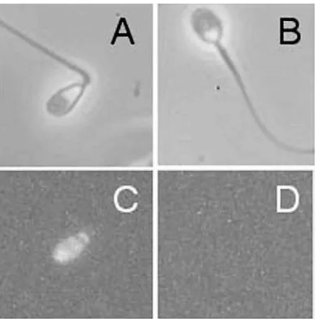
Human spermatozoa are unusual in that they frequently possess high levels of nuclear DNA damage that can be detected with a variety of methods, including modified nick translation methods, TUNEL (terminal deoxynucleotidyl transferase mediated dUTP nick end labelling), sperm chromatin stability assay (SCSA) and Comet analyses (Sunet al., 1997; Irvineet al., 2000; Evensonet al., 2002). These techniques detect either existing DNA fragmentation (TUNEL) (Figure 1) or chromatin instability leading to DNA fragmentation on exposure to extremely high (Comet) or low (SCSA) pH values. The incidence of DNA damage detected with such assays is positively correlated with age (Morris et al., 2002) and negatively correlated with normal embryonic development, whether conventional IVF or ICSI (intracytoplasmic sperm injection) is used as the fertilization technique (Sunet al., 1997; Twigget al., 1998 Morriset al., 2002).
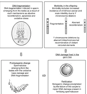
Since spermatozoa have no capacity for DNA repair (Drost and Lee, 1995), it is the oocyte that shoulders the responsibility for repairing the genetic damage brought into the zygote by the fertilizing spermatozoon (Matsuda and Tobari, 1989). If DNA damage is not repaired at this stage, checkpoint controls will be exerted at each cleavage division and cells still found to contain damaged genes, will be deleted by apoptosis. Such a mechanism could readily explain the observed association between DNA damage in human spermatozoa and high rates of fragmentation in embryos generated by assisted conception therapy (Morriset al., 2002; Tomsuet al., 2002). However, if the mutations introduced by such mechanisms are sufficiently subtle, they will escape recognition and have the potential to interfere with the health and wellbeing of the offspring. Such a mechanism could readily account for the elevated incidence of dominant genetic mutations, such as achondroplasia, in the offspring of ageing fathers (Crow, 1997). As indicated above, such mutations are not frequently induced in the germ line of older men, as was previously suspected (Tiemann-Boeggeet al., 2002). An alternative hypothesis is presented in Figure 2, wherein the significantly elevated levels of DNA damage observed in the spermatozoa of ageing males are held to constitute a pro-mutational change that then becomes fixed as a mutation in the embryo as a result of defective repair in the oocyte or early embryo (Figure 2).
67
DNA damage and Y-chromosome
deletion
A corollary of this hypothesis is that any mutational change created in the fertilized zygote will be transferred to the germ line of the affected embryo. Such a mechanism may underlie the involvement of genetic factors in the aetiology of poor semen quality. Specifically, deletions in the Yq11 region of the Y-chromosome have been clearly associated with male infertility (Pryoret al., 1977). The incidence of such deletions in the infertile male population varies with the infertility phenotype, but recent studies suggest that around 14% of subjects with severe oligozoospermia/azoospermia are so affected (Roberts, 1998). Current evidence suggests that gene-specific deletions are probably rare events in such patients and that in general, only large Y deletions that remove several genes are associated with the infertile phenotype (Krausz and McElreavey, 1999). Since the Y-chromosome deletions causing infertility cannot be inherited, such damage can either originate de novoin the father’s spermatozoa, or in the embryo as a consequence of DNA fragmentation in the father’s germ line. Which of these alternative mechanisms is responsible for the creation of Y-chromosome deletions is still uncertain, but they are not mutually exclusive.
It is of interest, however, that most deletions on the Y chromosome are flanked by repeat sequences that could act as substrates for homologous recombination (Kampet al., 2000; Kuroda-Kawaguchiet al., 2001; Reppinget al., 2002). It could therefore be suggested that primary DNA damage in the male germ line involves double strand breaks, and that in attempting
to repair such damage, repeat sequences from two distant regions of the Y chromosome are united in such a way that the intervening DNA becomes deleted (Figure 2).
The fact that patients exhibiting severe disruptions of their semen profile show a 20-fold increase in somatic chromosomal mutations, including rearrangements and translocations (Vincentet al., 2002), is additional support for the contention that male infertility is associated with widespread DNA fragmentation in the father’s germ line. Furthermore, since DNA fragmentation in spermatozoa is compatible with fertilization following ICSI, it is possible that infertility, as well as other genetically determined conditions, might ultimately arise in children conceived using this technique, or their descendents (Twigget al., 1998).
If DNA fragmentation in the male germ line is such a consistent feature of male infertility, it will be important to determine the cause of such genetic damage. It is unlikely that significant DNA fragmentation occurs in spermatogonia, since this stem cell population exhibits an extremely stable genetic structure (Hill et al., 2003). Later in spermatogenesis, it is possible that double strand breaks are created as a result of one or more of three fundamental mechanisms: (i) aberrant recombination during meiosis; (ii) defects in the testicular microenvironment that initiate apoptosis in the germ line; or (iii) chemically induced DNA fragmentation.
A well-researched example of an environmental factor that can induce DNA fragmentation in human spermatozoa is smoking. In heavy smokers, the oxidative stress created by their habit
68
appears to induce significantly elevated levels of DNA fragmentation and oxidative base damage in their spermatozoa (Fragaet al., 1996). In addition, the xenobiotics in cigarette smoke can trigger Fas-mediated apoptosis in the male germ line and, where this process is incomplete, generate spermatozoa with nuclear DNA fragmentation that is, presumably, endonuclease-driven (Sawyer and Aitken, 2001). Whatever the causes of the DNA fragmentation induced in the germ line by heavy smoking, the consequence of such environmentally triggered DNA damage can be seen in the increased risk of childhood cancer recorded in their children (Jiet al., 1997).
The oxidative stress hypothesis
Following on from the above example, a unifying hypothesis for the aetiology of both genetic damage and infertility in the male germ line, would be oxidative stress. According to this hypothesis, oxidative stress in the male germ line leads to the induction of damage to the sperm plasma membrane and a loss of genetic integrity in the nucleus and mitochondria. When oxidative stress is at its most severe, the result is infertility due to an inability of the spermatozoa to engage in the membrane fusion events associated with the acrosome reaction and sperm–oocyte fusion. Under these conditions, the fusogenicity of the plasma membrane is lost as a consequence of peroxidative damage to the unsaturated fatty acids that dominate this cellular structure (Jones et al., 1979; Aitken, 1999).Oxidative stress in the male germ line is also associated with DNA damage in the sperm nucleus and mitochondria (Aitken, 1999). When the stress levels are high, this DNA damage is of little clinical consequence because, for the reasons given above, the spermatozoa are incapable of fertilization. In a sense, the negative impact of oxidative stress on the fertilizing potential of human spermatozoa amounts to a safety strategy, ensuring that fertilization is not achieved when DNA damage in the male germ line is excessive. However at lower levels of oxidative stress, DNA damage may be induced in the nuclear and mitochondrial genomes under conditions where the spermatozoa retain their capacity for fertilization (Aitken, 1999). This is significant because it means that spermatozoa are potentially capable of passing damaged genes onto the embryo that could ultimately impact upon the health and wellbeing of the offspring (Aitken and Marshall Graves, 2002).
Origins of oxidative stress –
xenobiotics and cytoplasmic
retention
If pro-oxidant exposure is a contributory factor in the aetiology of male infertility and genetic disease, then discovering the source of this oxidative stress becomes an important objective for biomedical research. Both intrinsic and extrinsic factors appear to be involved. Extrinsic, environmental chemicals capable of inducing oxidative stress include polycyclic aromatic hydrocarbons, transition metals, anaesthetic gases, smoking, solvents, pesticides and ionizing radiation (Fragaet al., 1996; Sawyer and Aitken, 2001). Intrinsic factors contributing to oxidative stress in spermatozoa appear to
involve high levels of redox activity, detectable with chemiluminescent probes such as luminol, as well as with morphological defects in the spermatozoa associated with the retention of excess residual cytoplasm (Aitken et al., 1997; Maket al., 2000).
During spermiogenesis, spermatozoa undergo a unique phenotypic change involving removal of most of their cytoplasm. In man, this change is effected with varying degrees of efficiency with the result that retention of excess residual cytoplasm is a common feature of defective human spermatozoa (Gomezet al., 1996; Gil-Guzmanet al., 2001). The suggestion that oxidative stress in spermatozoa might be associated with cytoplasmic retention stems from a series of independent studies revealing that poor semen quality is statistically associated with high cellular contents of key cytoplasmic enzymes such as creatine kinase (Huszaret al., 1988), lactic acid dehydrogenase (Casano et al., 1991), superoxide dismutase (Aitken et al., 1996) and glucose-6-phosphate dehydrogenase (Gomezet al., 1996). The presence of these cytosolic enzymes correlates with the amount of residual cytoplasm retained in the midpiece of human spermatozoa (Gomezet al., 1996). The loss of sperm motility and fertilizing potential associated with varicocoeles and idiopathic male infertility is also associated with the retention of excess residual cytoplasm by the spermatozoa (Ziniet al., 1998, 1999, 2000). Furthermore recent studies of IVF patients have also demonstrated a strong negative correlation between fertilization rate and the presence of residual cytoplasm in the sperm midpiece (Keatinget al., 1997).
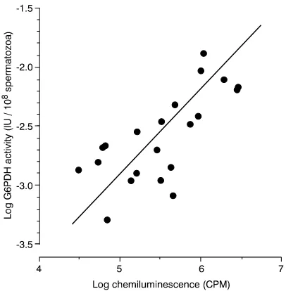
The mechanism by which the presence of excess residual cytoplasm disrupts human sperm function is thought to involve the induction of oxidative stress (Gomez et al., 1996). Specifically, the presence of excess glucose-6-phosphate dehydrogenase (G-6-PDH) is held to enhance the cytoplasmic generation of NADPH that, in turn, fuels the production of free radicals by a proposed sperm NADPH oxidase (Aitkenet al., 1997), a candidate for which is NOX5 (Banfiet al., 2001). In support of this proposal there is a very clear relationship between free radical generation by mammalian spermatozoa and the cellular content of G-6-PDH (Figure 3). According to this model, the increased concentration of cytoplasmic enzymes and enhanced ROS generation observed in cases of male infertility are simply a passive consequence of cytoplasmic retention and enhanced substrate generation. Whatever its cause, the ability of human spermatozoa to generate ROS is negatively correlated with semen quality and the fertilizing capacity of human spermatozoa in vivoand in vitro(Aitken, 1999; Gomezet al., 1998).
69
Summary
In summary, it is now clear that defective sperm function is common causative factor in the aetiology of human infertility. In this context the term ‘sperm function’ refers not only to the fertilizing ability of the cell, but also its ability to support normal embryonic development. Concern over the latter stems from an accumulating body of data indicating that human spermatozoa frequently exhibit high levels of DNA damage. The latter is, in turn, associated with abnormal embryonic development and morbidity in the offspring including cancer, infertility and diseases caused by dominant genetic mutations. Although the origins of defective fertilizing potential and DNA damage are still poorly understood, it is possible that they are linked by their dependence on oxidative stress. The latter is known to cause a loss of fertilizing potential as a consequence of peroxidative damage to the sperm plasma membrane. Reactive oxygen metabolites are also known to induce DNA damage in spermatozoa whether oxidants are added directly to the cell or their production is induced by the addition of substrate nicotinamide adenine dinucleotide phosphate (NADPH) (Aitken et al., 1998). The origins of oxidative stress may in turn, involve defects in spermiogenesis resulting in the retention of excess residual cytoplasm by the spermatozoa. Analysis of the genetic and environmental factors regulating cytoplasmic extrusion in the male germ line may therefore be of strategic relevance to a variety of male-mediated pathological conditions.
References
Aitken RJ (ed.) 1986 The zona-free hamster oocyte penetration test and the diagnosis of male fertility. World Health Organization Symposium. International Journal of Andrology Supplement6, 1–199.
Aitken RJ 1999 The human spermatozoon: a cell in crisis? The Amoroso Lecture. Journal of Reproduction and Fertility115, 1–7.
Aitken RJ, Marshall-Graves JA 2002 The future of sex. Nature415, 963.
Aitken RJ, Best FSM, Richardson DWet al.1982 An analysis of sperm function in cases of unexplained infertility: conventional criteria, movement characteristics and fertilizing capacity.
Fertility and Sterility38, 212–221.
Aitken RJ, Buckingham DW, Carreras A, Irvine DS 1996 Superoxide dismutase in human sperm suspensions: relationships with cellular composition, oxidative stress and sperm function. Free Radical Biology and Medicine21, 495–504.
Aitken RJ, Fisher H, Fulton Net al.1997 Reactive oxygen species generation by human spermatozoa is induced by exogenous NADPH and inhibited by the flavoprotein inhibitors diphenylene iodonium and quinacrine. Molecular Reproduction and Development47, 468–482.
Aitken RJ, Gordon E, Harkiss Det al.1998 Relative impact of oxidative stress on the functional competence and genomic integrity of human spermatozoa. Biology of Reproduction59, 1037–1046.
Banfi B, Molnar G, Maturana Aet al.2001 A Ca2+-activated NADPH oxidase in testes, spleen, and lymph nodes. Journal of Biological Chemistry276, 375594–375601.
Casano R, Orlando C, Serio M, Forti G 1991 LDH and LDH-X activity in sperm from normospermic and oligozoospermic men.
International Journal of Andrology14, 257–263.
Crow JF 1997 The high spontaneous mutation rate: is it a health risk? Proceedings of the National Academy of Sciences94, 8380–8386.
Drost JB, Lee WR 1995 Biological basis of germline mutation: comparisons of spontaneous germline mutation rates among drosophila, mouse, and human. Environmental and Molecular Mutagenesis Supplement26, 48–64.
Evenson DP, Larson KL, Jost LK 2002 Sperm chromatin structure assay: its clinical use for detecting sperm DNA fragmentation in male infertility and comparisons with other techniques. Journal of Andrology23, 25–43.
Fraga CG, Motchnik PA, Wyrobek AJet al.1996 Smoking and low antioxidant levels increase oxidative damage to DNA. Mutation Research351, 199–203.
Gil-Guzman E, Ollero M, Lopez MCet al.2001 Differential production of reactive oxygen species by subsets of human spermatozoa at different stages of maturation. Human Reproduction16, 1922–1930.
Gomez E, Buckingham DW, Brindle Jet al.1996 Development of an image analysis system to monitor the retention of residual cytoplasm by human spermatozoa: correlation with biochemical markers of the cytoplasmic space, oxidative stress and sperm function. Journal of Andrology17, 276–287.
Gomez E, Irvine DS, Aitken RJ 1998 Evaluation of a spectrophotometric assay for the measurement of
malondialdehyde and 4-hydroxyalkenals in human spermatozoa: relationships with semen quality and sperm function.
International Journal of Andrology21, 81–94.
Hill KA, Wang J, Farwell KD, Sommer SS 2003 Spontaneous tandem-base mutations (TBM) show dramatic tissue, age, pattern and spectrum specificity. Mutation Research534, 173–186. Hull MGR, Glazener CMA, Kelly NJet al.1985 Population study of
causes, treatment and outcome of infertility. British Medical Journal291,1693–1697.
Hurst LD, Ellegran H 2002 Mystery of the mutagenic male. Nature
420, 365–366.
Huszar G, Vigue L, Corrales M 1988 Sperm creatine phosphokinase quality in normospermic, variablespermic and oligospermic men.
Biology of Reproduction38, 1061–1066.
Irvine DS, Twigg J, Gordon Eet al.2000 DNA integrity in human spermatozoa: relationship with semen quality. Journal of Andrology21, 33–44.
Ivell R, Pusch W, Balvers Met al.2000 Progressive inactivation of the haploid expressed gene for the sperm-specific endozepine-like peptide (ELP) through primate evolution. Gene255, 335–345. Ji BT, Shu XO, Linet MSet al.1997 Paternal cigarette smoking and
the risk of childhood cancer among offspring of nonsmoking mothers. Journal of the National Cancer Institute89, 238–244. Figure 3.Close correlation between the G-6-PDH
70
Jones R, Mann T, Sherins RJ 1979 Peroxidative breakdown of phospholipids in human spermatozoa: spermicidal effects of fatty acid peroxides and protective action of seminal plasma. Fertility and Sterility31, 531–537.
Jury JA, Frayne J, Hall L 1998 Sequence analysis of a variety of primate fertilin alpha genes: evidence for non-functional genes in the gorilla and man. Molecular Reproduction and Development
51, 92–97.
Kamp C, Hirschmann P, Voss Het al.2000 Two long homologous retroviral sequence blocks in proximal Yq11 cause AZFa microdeletions as a result of intrachromosomal recombination events. Human Molecular Genetics9,2563–2572.
Kao SH, Chao HT, Wei YH 1998 Multiple deletions of mitochondrial DNA are associated with the decline of motility and fertility of human spermatozoa. Molecular Human Reproduction4,
657–666.
Keating J, Grundy CE, Fivey PSet al.1997 Investigation of the association between the presence of cytoplasmic residues on the human sperm midpiece and defective sperm function. Journal of Reproduction and Fertility110, 71–77.
Krausz C, McElreavey K 1999 Y chromosome and male infertility.
Frontiers in Bioscience4, 1–8.
Kuroda-Kawaguchi T, Skaletsky H, Brown LGet al.2001 The AZFc region of the Y chromosome features massive palindromes and uniform recurrent deletions in infertile men. Nature Genetics29, 279–286.
Liu DY, Baker HWG 1994 A new test for the assessment of sperm–zona pellucida penetration: relationship with results of other sperm tests and fertilization in vitro. Human Reproduction
9, 489–496.
Mak V, Jarvi K, Buckspan Met al.2000 Smoking is associated with the retention of cytoplasm by human spermatozoa. Urology56, 463–466.
Matsuda Y, Tobari I 1989 Repair capacity of fertilized mouse eggs for X-ray damage induced in sperm and mature oocytes. Mutation Research210, 35–47.
Montgomery K 2003 The demographic transition.
http://www.uwmc.uwc.edu/geography/ Demotrans/demtran.htm. Morris ID, Ilott S, Dixon L, Brison DR 2002 The spectrum of DNA
damage in human sperm assessed by single cell gel
electrophoresis (Comet assay) and its relationship to fertilization and embryo development. Human Reproduction17, 990–998. Nieschlag E, Lammers U, Freischem CWet al.1982 Reproductive
functions in young fathers and grandfathers. Journal of Clinical Endocrinology and Metabolism55, 676–681.
Pryor JL, Kent-First M, Muallem Aet al.1977 Microdeletions in the Y chromosome of infertile men. New England Journal of Medicine336, 534–539.
Repping S, Skaletsky H, Lange Jet al.2002 Recombination between palindromes P5 and P1 on the human Y chromosome causes massive deletions and spermatogenic failure. American Journal of Human Genetics71, 906–922.
Roberts KP 1998 Y chromosome deletions and male infertility: state of the art and clinical implications. Journal of Andrology19, 255–259.
Sakkas D, Mariethoz E, Manicardi Get al.1999 Origin of DNA damage in ejaculated human spermatozoa. Reviews of Reproduction4, 31–37.
Sawyer DE, Aitken RJ 2001 Male mediated developmental defects and childhood disease. Reproductive Medicine Reviews8, 107–126.
Seuanez HN 1980 Chromosomes and spermatozoa of the African great apes. Journal of Reproduction and Fertility Supplement28,
91–104.
Sharpe RM 2003 The oestrogen hypothesis – where do we stand now? International Journal of Andrology26, 2–15.
Sun JG, Jurisicova A, Casper RF 1997 Detection of deoxyribonucleic acid fragmentation in human sperm: correlation with fertilization in vitro. Biology of Reproduction56, 602–607.
Tiemann-Boege I, Navidi W, Grewal Ret al.2002 The observed
human sperm mutation frequency cannot explain the
achondroplasia paternal age effect. Proceedings of the National Academy of Sciences of the USA99, 14952–14957.
Tomsu M, Sharma V, Miller D 2002 Embryo quality and IVF treatment outcomes may correlate with different sperm comet assay parameters. Human Reproduction17, 1856–1862. Twigg JP, Irvine DS, Aitken RJ 1998 Oxidative damage to DNA in
human spermatozoa does not preclude pronucleus formation at ICSI. Human Reproduction13,1864–1871.
Vincent M-C, Daudin M, De Mas Pet al.2002 Cytogenetic investigations of infertile men with low sperm counts: a 25 year experience. Journal of Andrology23, 18–22.
World Health Organization 1999 Laboratory Manual for the Examination of Human Semen and Sperm-Cervical Mucus Interaction, 4th edn. Cambridge University Press, Cambridge. Zini A, O’Bryan MK, Israel L, Schlegel PN 1998 Human sperm
NADH and NADPH diaphorase cytochemistry: correlation with sperm motility. Urology51, 464–468.
Zini A, Buckspan M, Jamal M, Jarvi K 1999 Effect of
varicocelectomy on the abnormal retention of residual cytoplasm by human spermatozoa. Human Reproduction14, 1791–1793. Zini A, Defreitas G, Freeman Met al.2000 Varicocele is associated
with abnormal retention of cytoplasmic droplets by human spermatozoa. Fertility and Sterility74, 461–464.
Paper based on a contribution presented at the First Meeting of the Mediterranean Congress on Reproductive Medicine in Taormina, Sicily, November 2002.