ISBN: 978-979-95093-6-9
Bogor, desember 2010
Seminar Nasional Sains III
13 november 2010
Sains Sebagai Landasan Inovasi Teknologi
dalam Pertanian dan Industri
Prosiding
-200.00 400.00 600.00 800.00 1,000.00 1,200.00 1,400.00 1,600.00 0.25 0.3 0.35 0.4 0.45 0.5 0.55 0.6 0.65 0.7 λ11 K , K 11, K 12, K 21, K 22 ( % ) K K11 K12 K21 K22ISBN: 978-979-95093-6-9
Seminar Nasional Sains III
13 November 2010
Sains Sebagai Landasan Inovasi Teknologi
dalam Pertanian dan Industri
Prosiding
Dewan Editor
Ence Darmo Jaya Supena
Endar Hasafah Nugrahani
Hamim
Hasim
Indahwati
Kiagus Dahlan
Fakultas MIPA – Institut Pertanian Bogor
bekerja sama dengan
MIPAnet
__________________________________________________________________
Copyright© 2010Fakultas Matematika dan Ilmu Pengetahuan Alam (FMIPA), Institut Pertanian Bogor (IPB) Prosiding Seminar Nasional Sains III ”Sains Sebagai Landasan Inovasi Teknologi dalam Pertanian dan Industri” di Bogor pada tanggal 13 November 2010
Penerbit : FMIPA-IPB, Jalan Meranti Kampus IPB Dramaga, Bogor 16680 Telp/Fax: 0251-8625481/8625708
http://fmipa.ipb.ac.id
Terbit 30 Desember 2010 ix + 427 halaman
KATA PENGANTAR
Ketahanan pangan dan kemandirian energi merupakan isu sentral nasional dan dunia untuk mengimbangi terus bertambahnya jumlah penduduk, semakin menyempitnya lahan yang disertai tidak terlalu signifikannya peningkatan produktivitas pertanian, ditambah lagi dengan masalah global menurunnya kualitas lingkungan. Untuk mengatasi permasalahan-permasalahan ini tentunya dibutuhkan inovasi-inovasi. Inovasi akan menjadi lebih bermakna dan berhasil guna bila berlandaskan kepada sains dan teknologi. Banyak perguruan tinggi dan lembaga litbang departemen atau bahkan divisi litbang di perusahaan terus melakukan penelitian dan pengembangan yang didasarkan pada pemanfaatan dan pengembangan sains dan teknologi untuk mengembangkan dan menghasilkan inovasi-inovasi dalam upaya untuk meningkatkan produktivitas serta meningkatkan nilai tambah. Seminar Nasional Sains III (2010) yang diselenggarakan atas kerjasama FMIPA-IPB dan MIPAnet, diharapkan menjadi sarana dan upaya untuk menjalin komunikasi antar pelaku dan institusi yang terlibat untuk mengoptimumkan pemanfaatan sains sebagai landasan dalam mengembangkan dan menghasilkan inovasi-inovasi dalam upaya menjawab tantangan ketahanan pangan dan kemandirian energi. MIPAnet adalah Jaringan Kerjasama Nasional Lembaga Pendidikan Tinggi Bidang MIPA yang didirikan pada tanggal 23 Oktober 2000.
Makalah-makalah hasil penelitian dipresentasikan pada empat kelas paralel yaitu
Biological Science, Biochemistry, Chemistry, serta Physics & Mathematical Science. Selain itu beberapa makalah juga ditampilkan pada sesi Poster. Makalah-makalah tersebut sebagian besar merupakan isi dari prosiding ini. Seminar dihadiri oleh peneliti dari balitbang-balitbang terkait dan dosen-dosen perguruan tinggi, mahasiswa pascasarjana serta guru-guru SMA.
Ucapan terima kasih disampaikan kepada FMIPA-IPB dan MIPAnet yang telah mendukung penuh kegiatan Seminar Nasional Sains III ini. Juga kepada Panitia Seminar, para mahasiswa, dan semua pihak yang telah mensukseskan acara seminar ini. Kami juga sangat berterima kasih kepada semua pemakalah atas kerjasamanya, sehingga memungkinkan prosiding ini terbit. Semoga prosiding ini bermanfaat bagi semua pihak.
Bogor, Desember 2010
Dekan FMIPA-IPB,
DAFTAR
ISI
No.
Penulis
Judul
Hal
Biological
Science
1
1 Turati, Miftahudin, Ida
Hanarida
Penapisan Galur‐galur Padi Toleran Cekaman
Aluminium pada Populasi RIL F7 Hasil Persilangan
antara Padi Var IR64 dan Hawara Bunar
2
2 Dedi Suryadi, Miftahudin, Ida
Hanarida
Penapisan Galur‐galur Padi Toleran Cekaman Besi
pada Populasi RIL F7 Hasil Persilangan antara Padi
Var IR64 dan Hawara Bunar
12
3 Riana Murti Handayani, Gayuh
Rahayu, Jonner Situmorang
Interaksi Kultur Tunas in vitro Aquilaria spp. dengan
Hifomiset (Acremonium spp.)
19
4 Ahmad Basri, Hamim,
Nampiah Sukarno
Teknik Perkecambahan dan Respon Beberapa Aksesi
Jarak Pagar terhadap Aplikasi Pupuk Hayati Selama
Pemantapan Bibit
28
5 Martha Sari, Hamim Jarak Pagar (Jatropha curcas L.) sebagai Sumber
Senyawa Metabolit Sekunder Antimikrob Alternatif
36
6 Jeni, Hamim, Aris
Tjahjoleksono, Ida Hanarida
Soemantri
Viabilitas dan Efektifitas Pupuk Hayati dari Beberapa
Teknik Pengeringan dan Lama Penyimpanan
50
7 Risa Swandari Wijihastuti,
Tatik Chikmawati, Miftahudin
Optimasi Lingkungan Tumbuh Mikroalga dari Kawah
Ratu Sukabumi yang Berpotensi sebagai Sumber
Biodiesel
61
8 Suprihatin, Muhammad Romli,
Andes Ismayana
Kajian Produksi Mikroalga dengan Media Limbah Cair
Rumah Pemotongan Hewan
68
9 Yahmi Ira Setyaningrum,
Hamim, Dorly
Respon Morfologi Buah dan Kemunculan Getah
Kuning terhadap Aplikasi Kalsium secara Eksternal
pada Buah Manggis (Garcinia mangostana)
80
10 Ari Fina Bintarti, Iman
Rusmana, Dave B. Nedwell
Aktivitas Oksidasi Metan dan Akumulasi Ammonium
Isolat Bakteri Metanotrof Asal Sawah
89
11 Anthoni Agustien Produksi Protease Serin dari Sel Amobil Brevibacillus
agri A‐03 dengan Matriks Alginat
99
12 Rahmat Hidayat, Usamah
Afiff, Fachriyan Hasmi
Pasaribu
Pemeriksaan Serologik Brucellosis dan Mikrobiologik
Susu di Peternakan Sapi Perah Kabupaten Bogor dan
Sukabumi
No.
Penulis
Judul
Hal
13 Tania June Perubahan Iklim: Observasi Fungsi Supply dan
Demand terhadap CO2 pada Tanaman dan
Implikasinya
118
14 Baba Barus, U. Sudadi, B.
Tahjono, L.O.S. Iman
Pengembangan Geoindikator untuk Penataan Ruang 133
15 Wien Kusharyoto, Martha Sari Ekspresi Fragmen Antibodi Fab yang Spesifik
terhadap Virus Dengue DEN‐2 di Escherichia coli
145
Biochemistry
153
1 Dyah Iswantini, Latifah K
Darusman, Lany Yulinda
Daya Inhibisi Ekstrak Pegagan, Kumis kucing,
Sambiloto dan Tempuyung terhadap Aktivitas ACE
secara In vitro
154
2 Christofferus SY, Dyah
Iswantini
Daya Inhibisi Ekstrak Rimpang Jahe Merah dan Kulit
Kayu Manis terhadap Aktivitas Enzim
Siklooksigenase‐2 dan Enzim Xantin Oksidase secara
In vitro
163
3 Anggi Susanti, Dyah Iswantini Kinetika Inhibisi Ekstrak Tempuyung (Sonchus
arvensis L.) terhadap Enzim Xantin Oksidase secara In
Vitro
172
4 Dyah Iswantini, Deden
Saprudin, R Aghyar Rudita
Pengaruh Ekstrak Bangle (Zingiber cassumunar
Roxb.) terhadap Aktivitas Enzim Kolesterol Oksidase
secara In vitro
181
5 Rini Madyastuti Purwono,
Bayu Febram Prasetyo, Ietje
Wientarsih
Aktivitas Diuretik Fraksi Etil Asetat Ekstrak Etanol
Daun Alpukat (Persea americana mill.) pada Tikus
Sprague‐Dawley
190
6 Eti Rohaeti, Irmanida
Batubara, Anastasia Lieke
LDN, Latifah K Darusman
Potensi Ekstrak Rhizophora sp sebagai Inhibitor
Tirosinase
196
7 Popi Asri Kurniatin, Laksmi
Ambarsari, Juliana
Komposisi dan Aktivitas Bioflokulan dari
Flavobacterium sp.
202
Chemistry
212
1 Muhammad Bachri Amran Metoda Analisis Ion Besi Berbasis Cyclic‐Flow
Injection Analysis (cy‐FIA) sebagai Suatu Usaha
Menuju Analisis Kimia Ramah Lingkungan (Green
Analytical Chemistry)
No.
Penulis
Judul
Hal
2 Purwantiningsih Sugita, Tuti
Wukirsari, Tetty Kemala, Bayu
Dwi Aryanto
Perilaku Disolusi Mikrokapsul Ketoprofen‐Alginat
Berdasarkan Ragam Konsentrasi Surfaktan
221
3 Purwantiningsih Sugita, Yunia
Anggi Setyani, Tuti Wukirsari,
Bambang Srijanto
Dissolution Behavior of Ketoprofen Double Coated by
Chitosan‐Gum Guar with Alginat‐CaCl2
230
4 Dwi Wahyono,
Purwantiningsih Sugita,
Laksmi Ambarsari
Sintesis Nanopartikel Kitosan dengan Metode
Ultrasonikasi dan Sentrifugasi serta Karakterisasinya
241
5 Siti Latifah, Purwantiningsih
Sugita, Bambang Srijanto
Stabilitas Mikrokapsul Ketoprofen Tersalut Kitosan‐
Alginat
248
6 Salih Muharam,
Purwantiningsih Sugita, Armi
Wulanawati
Adsorption of Au (III) onto Chitosan Glutaraldehyde
Cross‐linked in Cyanide Solution
260
7 Wulandari Kencana Wardani,
Purwantiningsih Sugita,
Bambang Srijanto
Sintesis dan Karakterisasi Glukosamina Hidroklorida
Berbasis Kitosan
271
8 Setyoningsih, Akhiruddin M,
Deden S
Kajian Penggunaan Asam Oleat dan Teknik
Hidrotermal pada Sintesis Nanokristal Magnetit
282
9 Sugiarti, S.; Abidin, Z.;
Shofwatunnisaa ; Widyastana,
P.; Hediana, N
Sintesis Nanokomposit Beberapa Material Clay/TiO2
dari Bahan Dasar Kaolin Indonesia
288
10 Sugiarti, S.; Abidin, Z.; Henmi,
T
Zeolit/TiO2 Nanokomposit sebagai Fotokatalis pada
Penguraian Biru Metilena
298
11 Syafii, F; Sugiarti, S; Charlena Modifikasi Zeolit Melalui Interaksi dengan Fe(OH)3
untuk Meningkatkan Kapasitas Tukar Anion
307
Physics
316
1 Wiwis S., Agus Rubiyanto Pengembangan Metode Penyetabil Sumber Cahaya
Laser He‐Ne dengan Menggunakan Plat λ/4
317
2 Harmadi, Gatut Yudoyono,
Mitrayana, Agus Rubiyanto,
Suhariningsih
Pola Spekel Akusto‐Optik untuk Pendeteksian
Getaran (Vibrasi) Akustik pada Objek yang Bergetar
322
No.
Penulis
Judul
Hal
3 Stepanus Sahala S. Alat Peraga Fisika Menggunakan Interfacing Sensor
Cahaya dengan Stopwatch pada Percobaan Gerak
Jatuh Bebas dalam Pembelajaran Fisika
331
4 Akhiruddin Maddu, Deni
Christopel Pane, Setyanto Tri
Wahyudi
Pengaruh Konsentrasi Dopan HCl pada Polianilin
terhadap Karakteristik Sensor Gas Amonia (NH3)
341
5 M.N. Indro, R. Permatasari, A.
Insani
Pembuatan Nano Alloy MgNi dengan Teknik Ball
Milling
349
6 Rani Chahyani, Zahroul
Athiyah, Kiagus Dahlan
Sintesis dan Karakterisasi Membran Polisulfon
Didadah Karbon Aktif untuk Filtrasi Air
354
7 Abdul Djamil Husin, M.
Misbakhusshudur, Irzaman,
Jajang Juansah, Sobri Effendy
Pemanfaatan dan Kajian Termal Tungku Sekam untuk
Penyulingan Minyak Atsiri dari Daun Cengkeh
sebagai Pengembangan Produk dan Energi Alternatif
Terbarukan
364
8 S.U. Dewi, K. Dahlan, R.S.
Rahayu, B.M. Bachtiar
Pengujian Biphasic Calcium Phosphate (BCP) dalam
Sel Fibroblas
373
Mathematical
Science
381
1 Tri Handhika, Murni Kajian Stabilitas Model Tingkat Bunga
Rendleman‐Bartter
382
2 Agus Santoso Randomisasi Pemilihan Butir Awal pada Algoritma
Computerized Adaptive Test sebagai Upaya
Mengurangi Item Exposure
391
3 Endar H. Nugrahani Pengaruh Parameter Tingkat Produktivitas Manusia
pada Model Pertumbuhan Ekonomi Regional
401
4 Mohammad Masjkur Perbandingan Metode Peragam Papadakis
Rancangan Nearest Neighbour
410
5 Mohammad Masjkur Perbandingan Rancangan Spasial Nearest Neighbour
dan Rancangan Acak Kelompok Percobaan
Pemupukan Padi Sawah
C
C
H
H
E
E
M
M
I
I
S
S
T
T
R
R
Y
Y
DISSOLUTION BEHAVIOR OF KETOPROFEN Chemistry
DISSOLUTION BEHAVIOR OF KETOPROFEN DOUBLE COTED BY
CHITOSAN-GUM GUAR WITH ALGINAT-CaCl
2Purwantiningsih Sugita1, Yunia Anggi Setyani1, Tuti Wukirsari1, and Bambang Srijanto2
1Department of Chemistry, Bogor Agricultural University, 2Agency Assessment and Application of Technology, Jakarta
Abstract
High dose ketoprofen administration as anti-inflammatory drug can cause gastrointestinal bleeding. To minimize this drawback, we have successfully double coated guar gum-modified chitosan microcapsules containing ketoprofen with alginate. The results showed that this double coating had improved the microcapsule’s stability in the gastric acid medium. The ketoprofen releasing kinetic models, both at gastric and intestinal pH, are dominated by the Korsmeyer-Peppas model suggesting that this action is following the diffusion mechanism. This kinetic model is the closest approximation to the real releasing condition.
Keywords: chitosan, ketoprofen, microcapsule, dissolution
1. INTRODUCTION
Ketoprofen is one of the non-steroidal anti-inflammatory drugs (NSAID) which is able to suppress pain by inhibiting the prostaglandin synthesis. Its solubility in water is low and high dose administration (> 300 mg) can cause gastrointestinal bleeding [1]. Thus a specific drug delivery system, which is able to minimize those drawbacks, is highly needed.
Controlled release system method is able to minimize the negative side effect to the digestive system and overcome the short elimination time [2]. One of this is the microencapsulation. This method is able to maintain the therapeutic dose and activity of a continually administered drug. Some coating materials have already been studied for this application, alginate, chitosan [3], pectin [4], gelatine [5], polyacrylate, hydroxypropylmethylcellulose, and ethylcellulose [6] are just a few examples. Besides that, chitosan-carboxymethyl cellulose (CMC) has already been developed [7]. The difference between chitosan and CMC solubility is the main problem of this system. This difference made the resulted gel not homogenous. Sugita et al. [8] used chitosan-guar
gum to coat ketoprofen because this modified chitosan is able to form homogenous gel. However, guar gum was not strong enough to protect the microcapsules in the gastric environment and destroyed at the 90th minute [8]. Thus another kind of polymer which is
Prosiding Seminar Nasional Sains III: Bogor, 13 November 2010
DISSOLUTION BEHAVIOR OF KETOPROFEN Chemistry
strong enough to protect the microcapsules from the gastric acid is needed. In this study, alginate was applied as the external coating material for the chitosan-guar gum microcapsule. This choice is made based on alginate’s properties that is indestructible and able to form gel at gastric pH (< 3) [9], and also can undergo a spontaneous reaction with chitosan [10].
Chitosan application as drug coating material has been extensively studied, for example as ketoprofen [11] and propanolol hydrochloride [6] coating material. Beside its application as single material, chitosan has also been combined with other polymers in its application, for example chitosan-CMC for indometasin coating material and alginate-chitosan for insulin hormone [12]. Meanwhile study regarding combination between chitosan and guar gum has been conducted by Sugita et al. (2007) which resulting the
uniform sized microcapsules (0.4-5 μm). However, double coating of chitosan-guar gum microcapsule with alginate has never been done before.
2. MATERIAL AND METHODS 2.1. Material and Instrument
Chitosan used in this study was obtained from Bratachem with moisture content, ash content, deacetylation degree, and molecular weight specifications of 7.5%, 0.11%,
74.41%, and (2.43894–6.31553)106 g/mol respectively. The other materials used were
glutaraldehyde, guar gum, chloride buffer solution (KCl-HCl)-water pH 1.2, phosphate buffer solution (KH2PO4-NaOH)-water pH 7.4, Tween-80, CaCl2, alginate, and ketoprofen
active compound which was obtained from PT Kalbe Farma.
Instruments used in this study were glass wares, hot plate, J.P. SELECTA oven, magnetic stirrer, Bruker Tensor 37 FTIR spectrophotometer, Ostwald-Cannon-Fenske viscometer, diffusion cell instrument with water bath, aerator, UV-1700 PharmaSpec spectrophotometer, SEM JEOL JSM-5310LV, sieve shaker, Hansen paddle dissolution assay, and Minitab Release 14 software.
2.2. Method
2.2.1. Microcapsule Preparation [10,13]
As much as 228.6 ml of 2.5% (w/v) chitosan solution in 1% (v/v) acetic acid solution mixed with 38.1 ml of 0.35% guar gum solution with stirring. After that, 7.62 ml of 3.75% glutaraldehyde was added to the mixture and stirred until homogenous.
As much as 250 ml 0.8% (w/v) ketoprofen solution in 96% ethanol was mixed with chitosan-guar gum so the weight ratio of chitosan-ketoprofen becomes 2:1. After that, 5 ml of 2% Tween-80 was added to the mixture and stirred at room temperature (Sugita et
Prosiding Seminar Nasional Sains III: Bogor, 13 November 2010
DISSOLUTION BEHAVIOR OF KETOPROFEN Chemistry
al., 2006). The microcapsule was made by adding the chitosan-guar gum mixture to
alginate solutions of different concentration (1, 2, and 3% (w/v)) dropwise via a syringe.
Then the microcapsules was filtered and washed with CaCl2 solutions with different
concentration of 0.05, 0.10, and 0.15 M. The chitosan-guar gum and alginate coated ketoprofen microcapsules then dried by oven at 40 °C for 3 hours.
2.2.2. Encapsulation efficiency [14]
NaOH was used to extract ketoprofen from the microcapsule. As much as 50 mg microcapsules of each variant was digested, then extracted with 80 ml of 0.1 M NaOH for 5 minutes. The extract was filtered and diluted by NaOH to 100 ml volume. The extracted ketoprofen concentration was measured by UV spectrophotometer at 257 nm. The obtained absorbance were used to calculate the ketopfofen concentration via a standard curve.
2.2.3. In vitro dissolution test [15]
The dissolution test was conducted using the type 2 dissolution device (paddle method). The microcapsule was weighted (500 mg) and placed at the dissolution chamber. The test was conducted on gastric medium (pH 1.2) for 3 hours and intestinal medium (pH 7.4) for 6 hours at 37 ± 0.5 °C with paddling speed 150 rpm. Fifteen milliliters of aliquots were sampled every 15 minutes from the gastric and intestinal medium. After each time an aliquot was taken, the removed volume was replaced with the new medium solution with the same volume and temperature. The dissolution medium volume was 500 ml. Aliquot’s ketoprofen concentration was measured at 258.6 nm (for dissolution at pH 1.2) and 260 nm (for dissolution at pH 7.4). The dissolution kinetic was studied by plotting the ketoprofen release percentage versus dissolution time and then determines the reaction order and the ketoprofen release model.
2.2.4. Microcapsule characterization
The microcapsule’s morphology characterization was conducted to the empty, filled, and acid and base dissolute ketoprofen microcapsule using scanning electron microscope (SEM). While the particle size measurement was conducted using sieve shaker and photo-stereo microscope.
3. RESULTS
Alginate concentration variation effect the resulting gel strength. Microcapsule gel made using 1% (w/v) alginate is the most fragile. Besides that, this microcapsule gel also has the biggest size compared to the gel made using 2 and 3% (w/v) alginate. After
Prosiding Seminar Nasional Sains III: Bogor, 13 November 2010
DISSOLUTION BEHAVIOR OF KETOPROFEN Chemistry
washed by CaCl2, the gel becomes harder and clustered during drying. The dried
microcapsules containing ketoprofen are yellow while the empty ones are paler.
Microcapsules resulted from all formula could not through the 35 mesh sieve, in other words the size are ≥ 500 μm and they looks more bulky and filled than the blank (microcapsules without ketoprofen). These results are in agreement with the photo-stereo microscope observation of which results are listed in Table 1.
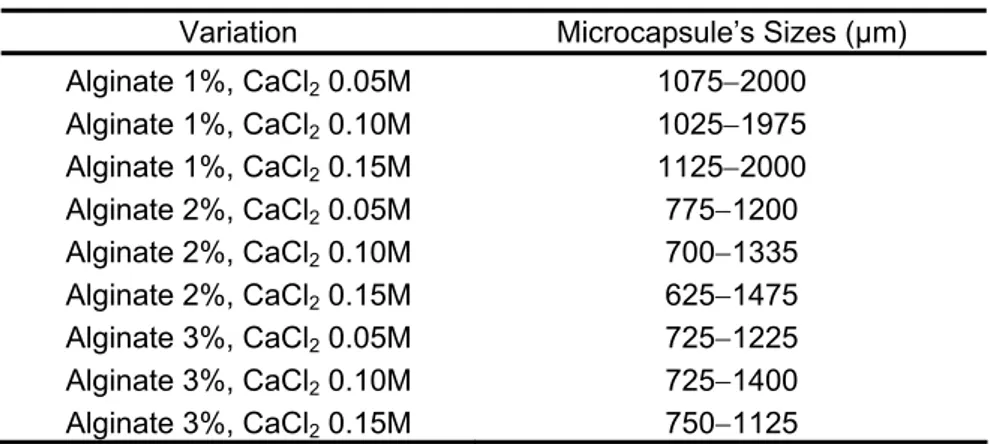
Table 1 Microcapsule’s sizes
Variation Microcapsule’s Sizes (μm) Alginate 1%, CaCl2 0.05M 1075−2000 Alginate 1%, CaCl2 0.10M 1025−1975 Alginate 1%, CaCl2 0.15M 1125−2000 Alginate 2%, CaCl2 0.05M 775−1200 Alginate 2%, CaCl2 0.10M 700−1335 Alginate 2%, CaCl2 0.15M 625−1475 Alginate 3%, CaCl2 0.05M 725−1225 Alginate 3%, CaCl2 0.10M 725−1400 Alginate 3%, CaCl2 0.15M 750−1125
3.1. Microcapsule Moisture Content
The obtained microcapsules had different moisture contents ranging from 13.91 to 25.84% (Table 2). Table 2 shows that the increasing CaCl2 concentration will increase the
moisture content of the microcapsules made using 1 and 2% (w/v) alginate. Besides that, the increase in alginate concentration is also tending to increase moisture content.
Table 2 Chitosan-alginate ketoprofen microcapsule’s moisture contents
Treatment Alginate (% [w/v]) CaCl2 (M) Moisture Content (%) 1 0.05 14.48 1 0.10 14.73 1 0.15 23.36 2 0.05 16.24 2 0.10 18.14 2 0.15 25.84 3 0.05 24.79 3 0.10 17.43 3 0.15 13.91 3.2. Ketoprofen Release
The maximum ketoprofen release percentage in gastric medium was very low, ranging from 3.18-12.97%. It was also not released completely in intestinal medium until
the 360th minute. However, the maximum ketoprofen release percentages in intestinal
medium were still higher compared to the gastric medium release, i.e. 18.34-54.97%.
Prosiding Seminar Nasional Sains III: Bogor, 13 November 2010
DISSOLUTION BEHAVIOR OF KETOPROFEN Chemistry
Fehling tests to the 360th minute’s aliquots taken from the intestinal medium dissolution
test showed negative results.
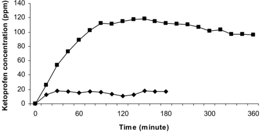
The best microcapsules were obtained from the H formula with 3% (w/v) alginate
and 0.15 M CaCl2. Figure 1 shows that ketoprofen releases at gastric medium were
controlled. While at the intestinal medium, ketoprofen concentration reached its maximum
value at the 90th minutes and became relatively steady afterwards. Ketoprofen
concentrations at the equilibrium condition were 110-120 mg/l.
0 20 40 60 80 100 120 140 0 60 120 180 300 360 Tim e (minute) K et o p ro fen co n cen tr at io n ( p p m )
Figure 1 Acidic (♦) and basic (■) dissolution behavior of the best produced microcapsules (3% (w/v) alginate and 0.15 M CaCl2).
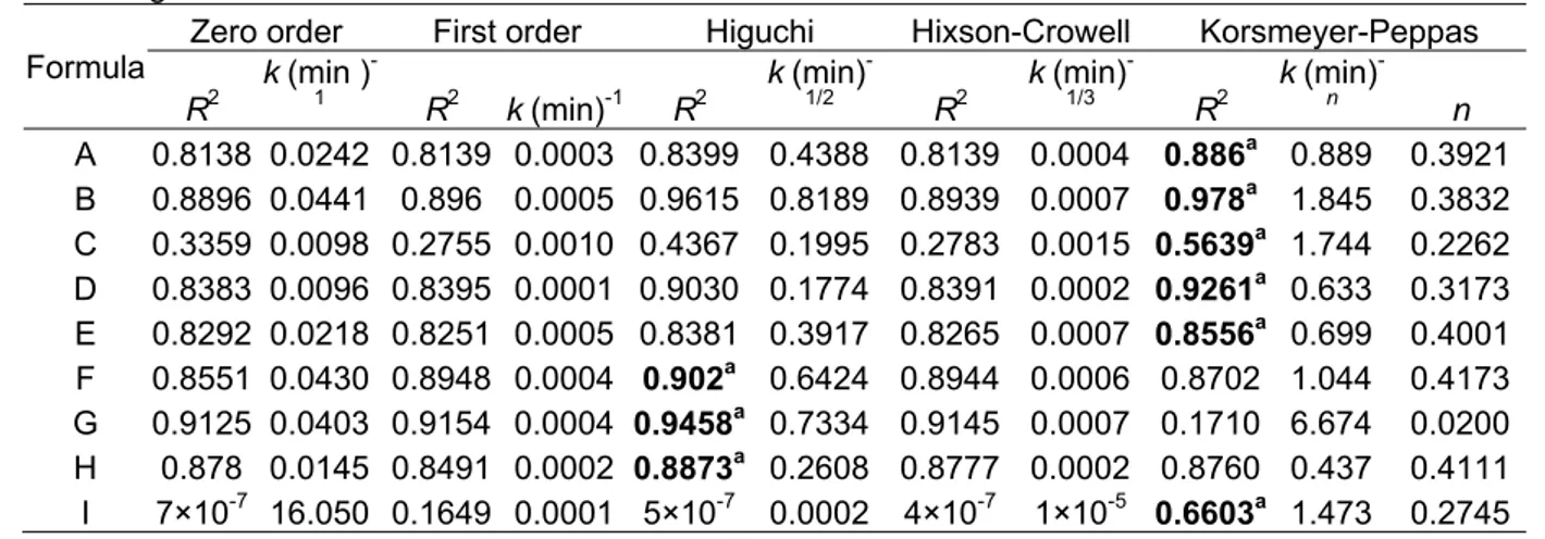
According to Table 3, ketoprofen releases were dominated by the Korsmeyer-Peppas kinetic model both in artificial gastric and intestinal medium. However, the F, G, and H formula were following the Higuchi kinetic model in the gastric medium dissolution. These models were assigned by calculating their determination coefficient towards each formula. Comparison between dissolutions in artificial gastric and intestinal medium shows that the rate constant (k) in gastric medium is tends to be lower than in base medium. The
same thing was also happening to the maximum rate of release calculated by the Korsmeyer-Peppas and Higuchi kinetic models (Figure 2). Figure 2 also implied that the kinetic models had provided the best approximation to the real value.
Prosiding Seminar Nasional Sains III: Bogor, 13 November 2010
DISSOLUTION BEHAVIOR OF KETOPROFEN Chemistry
Figure 2 Maximum ketoprofen release percentages in acidic (a) and basic (b) medium based on model calculations and experimental results.
Table 3 Dissolution kinetic determination in artificial gastric and intestinal medium
a The highest determination coefficient
Captions: a A = alginate 1%, CaCl
2 0.05 M F = alginate 2%, CaCl2 0.15 M
B = alginate 1%, CaCl2 0.1 M G = alginate 3%, CaCl2 0.05 M
C = alginate 1%, CaCl2 0.15 M H = alginate 3%, CaCl2 0.1 M
D = alginate 2%, CaCl2 0.05 M I = alginate 3%, CaCl2 0.15 M
E = alginate 2%, CaCl2 0.10 M
Artificial gastric medium
Zero order First order Higuchi Hixson-Crowell Korsmeyer-Peppas Formula R2 k (min ) -1 R2 k (min)-1 R2 k (min) -1/2 R2 k (min) -1/3 R2 k (min) -n n A 0.8138 0.0242 0.8139 0.0003 0.8399 0.4388 0.8139 0.0004 0.886a 0.889 0.3921 B 0.8896 0.0441 0.896 0.0005 0.9615 0.8189 0.8939 0.0007 0.978a 1.845 0.3832 C 0.3359 0.0098 0.2755 0.0010 0.4367 0.1995 0.2783 0.0015 0.5639a 1.744 0.2262 D 0.8383 0.0096 0.8395 0.0001 0.9030 0.1774 0.8391 0.0002 0.9261a 0.633 0.3173 E 0.8292 0.0218 0.8251 0.0005 0.8381 0.3917 0.8265 0.0007 0.8556a 0.699 0.4001 F 0.8551 0.0430 0.8948 0.0004 0.902a 0.6424 0.8944 0.0006 0.8702 1.044 0.4173 G 0.9125 0.0403 0.9154 0.0004 0.9458a 0.7334 0.9145 0.0007 0.1710 6.674 0.0200 H 0.878 0.0145 0.8491 0.0002 0.8873a 0.2608 0.8777 0.0002 0.8760 0.437 0.4111 I 7×10⁻⁷ 16.050 0.1649 0.0001 5×10⁻⁷ 0.0002 4×10⁻⁷ 1×10⁻⁵ 0.6603a 1.473 0.2745
Artificial intestinal medium
Zero order First order Higuchi Hixson-Crowell Korsmeyer-Peppas Formula R2 k (min ) -1 R2 k (min)-1 R2 k (min) -1/2 R2 k (min) -1/3 R2 k (min) -n N A 0.0781 0.0148 0.0527 0.0001 0.2938 0.5697 0.0676 0.0002 0.8184a 1.1393 0.4726 B 0.0991 0.0295 0.0676 0.0003 0.3347 1.0785 0.0777 0.0005 0.8504a 1.1534 0.5796 C 0.0693 0.0145 0.0527 0.0001 0.0693 0.8718 0.058 0.0002 0.8173a 1.1429 0.4788 D 0.1918 0.0211 0.1784 0.0002 0.3042 0.0044 0.1829 0.0003 0.8742a 1.1048 0.4744 E 0.0923 0.0309 0.0584 0.0003 0.3225 1.1485 0.0691 0.0005 0.8500a 1.1553 0.5908 F 0.2798 0.0713 0.1632 0.0008 0.4358 1.4738 0.1796 0.0011 0.8980a 1.1397 0.6644 G 0.1692 0.0461 0.1283 0.0005 0.4509 0.7057 0.1419 0.0008 0.8768a 1.1475 0.6237 H 0.1791 0.0223 0.2202 0.0003 0.6424 1.6924 0.1587 0.0003 0.9198a 1.1153 0.4971 I 0.3512 0.0628 0.3353 0.0008 0.0178 0.1345 0.0002 1×10⁻⁵ 0.9198a 1.1120 0.6284 0 2 4 6 8 10 12 14 16 A B C D E F G H I Formula %release 0 10 20 30 40 50 60 1 2 3 4 5 6 7 8 9 Formula %release
Research results Release model calculation
Research results Release model calculation
Prosiding Seminar Nasional Sains III: Bogor, 13 November 2010
DISSOLUTION BEHAVIOR OF KETOPROFEN Chemistry
Equation for zero order Q = kt; first order ln [A]t = ln [A]o −kt; Higuchi Q= kt12; Hixson-Crowell
; Korsmeyer-Peppas
n kt
Q= Qo13 −Qt13 =kt. (Q = persen release, k = rate constant, and t =
time of release)
3.3. Microcapsule Morphology
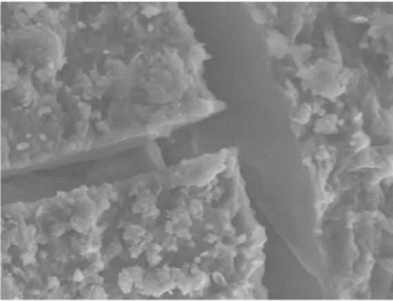
SEM image of microcapsules obtained from the best formula, 3% (w/v) alginate and 0.10 M CaCl2, clearly shows that the surfaces were very tight with a few shallow cracks
(Figure 3). Besides that, there were also fragments of crushed layer in the surface.
Figure 3 Ketoprofen microcapsule’s surface SEM image (3% alginate and 0.15 M CaCl2)
at 1500 times magnification.
After gastric and intestinal dissolution, the microcapsule’s surface looks remain unchanged but with bigger size. However, the surface swelling in gastric medium is bigger than in intestinal medium. During the dissolution test, the gastric medium color turned to yellowish. But none of these were happened in the intestinal medium.
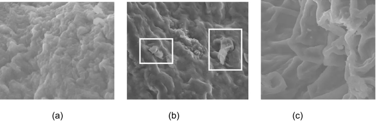
Observation to the SEM images taken from the microcapsule surface that had been spent 180 minutes in the gastric dissolution test shows that the surface had not significantly changed compared to the initial image (Figure 4a and b). Beside that, the surface contains fragments after gastric dissolution. Figure 4c has a different morphology compared to the initial image.
Prosiding Seminar Nasional Sains III: Bogor, 13 November 2010
DISSOLUTION BEHAVIOR OF KETOPROFEN Chemistry
(a) (b) (c)
Figure 4 SEM images taken from ketoprofen microcapsule before dissolution (a), after acidic dissolution (b), and after basic dissolution (c) at 1000 times magnification (inset: crushed alginate layer fragments).
4. DISCUSSION
Ketoprofen microencapsulation with guar gum-modified chitosan and alginate double coating has been successfully conducted by means of a chemical method. The first layer coated the ketoprofen is the glutaraldehyde linked chitosan with guar gum as the interpenetrating agent. Due to its low solubility in chitosan solution, the ketoprofen was dissolved in ethanol prior to mixing with chitosan solution.
The second coating layer was introduced via soaking in alginate solution. Swelling occurred to microcapsule gel due to high free water content around the gel in 1% alginate solution. While in 2 and 3 % alginate solution, the free water contents around the gel were lower. The smaller gel size in the 1% alginate solution was also due to the stronger cross-linking between the negative charged alginate and the positive charged chitosan [10].
The washing step with CaCl2 solution was intended to strengthen the outmost layer
of alginate [16] by means of cross-linking formation at the alginate’s guluronate residue [17] thus made the dried microcapsule tend to clustered. Besides that, during the drying process, intermolecular hydrogen bonds between alginates from different microcapsules were formed [16]. Alginate at the external surface of the microcapsules also made the surface more hygroscopic. Alginate is a hydrocolloid which contain large amount of hydroxyl groups.
4.1. Ketoprofen Release from the Microcapsule
Compared to Sugita et al. [8], double coating with alginate as the additional layer
has been proven here to be able to minimize ketoprofen release in gastric acidic environment. This fact reinforce Silva et al. [16] who stated that double coating is able to
enhance microcapsule’s stability in gastric environment. Tan et al. [18] stated that the
increasing alginate concentration caused the resulted microcapsule’s surface to have too
Prosiding Seminar Nasional Sains III: Bogor, 13 November 2010
DISSOLUTION BEHAVIOR OF KETOPROFEN Chemistry
few pores thus made ketoprofen difficult to get through it. This statement was proved by the fact that the k value we obtained from the dissolution at the intestinal medium is higher
than the value obtained from the gastric dissolution. However, the strong cross-linking between guar gum-modified chitosan and glutaraldehyde also caused the ketoprofen won’t be released completely before the 360th minute.
Because ketoprofen can initiate gastric bleeding and its absorption process is happening in the intestine, the microcapsules are considered good if only a little ketoprofen released in the gastric and more in the intestine. Besides that, another parameter which needs to be met by a good microcapsule is the low moisture content and high encapsulation efficiency. According to the scoring result, the best microcapsule was obtained from the H formula with 3% alginate and 0.15 M CaCl2.
4.2. Microcapsule Morphology
In gastric medium, the medium solution will penetrate the microcapsule’s surfaces thus swelled and even dissolved the chitosan. This also observed by the color change of the medium solution to yellowish after the gastric dissolution. However, this could not be happened in the intestinal medium due to the characteristic of chitosan which is insoluble in alkali environment.
The resemblances between SEM images of the microcapsule’s surface before and after gastric dissolution test occurred because the outermost microcapsule’s layer, i.e. Ca2+ cross linked alginate, was not totally affected by the gastric environment. Alginate
layer was swelled because it was pushed by the swelled inner chitosan-guar gum matrix. However, some alginate layer, which is too thin, was unable to hold this force and then crushed. The fragments of this destroyed alginate layer was scattered around the microcapsule’s external surface.
The differences between the SEM images taken before and after the intestinal dissolution reinforced Ivanova et al. (2000) [19], who stated that intestinal buffer solution is
able to destroy cross-linking bonds between alginate and Ca2+, thus alginate will be
dissolved. This means that the SEM image after intestinal dissolution is showing the surface of chitosan-guar gum matrix. Because alginate layer has been destroyed, ketoprofen will be released more easily from the matrix when it made contact with the intestinal medium. This release is also accommodated by large amount of channels which enlarged the microcapsule’s surface area that in contact with the medium.
5. CONCLUSIONS
Alginate double coating application to chitosan-guar gum microcapsule has been proved able to enhance microcapsule’s stability in gastric acidic medium. The best Prosiding Seminar Nasional Sains III: Bogor, 13 November 2010
DISSOLUTION BEHAVIOR OF KETOPROFEN Chemistry
produced microcapsule was made from 3% (w/v) alginate and 0.15 M CaCl2. Ketoprofen
releases both in acidic gastric pH and basic intestine pH were dominated by Korsmeyer-Peppas kinetic model. This assigned model is the best approximation to the real condition in this study.
ACKNOLEDGEMENTS
This work was supported by Higher Education Directorate General, Indonesia. The author would also like to thank Professor Suminar S Achmadi as a head of Organic Chemistry Laboratory for her support.
REFERENCES
[1] American Medical Association. Drug Evaluations. 8thEdition. (1991)
[2] Sutriyo, J. Djajadisastra, A. Novitasari, Mikroenkapsulasi propanolol hidro-klorida dengan penyalut etil selulosa menggunakan metode penguapan pelarut, Majalah Ilmu Kefarmasian1 (2004) 93-99.
[3] Q. Ul-Ain, S. Sharma, G.K. Khuller, S.K. Garg, Alginate-based oral drug delivery system for tuberculosis pharmacokinetics and therapeutics effects, J. Antimicrob. Chemother. 51 (2003) 931-938.
[4] W. Ouyang et al, Artificial cell microcapsule for oral delivery of live bacterial cell for
therapy: Design, preparation, and in-vitro characterization, J. Pharm. Pharmaceut Sci. 7 (2004) 315-324.
[5] P.T. Tayade, R.D. Kale, Encapsulation of drug-insoluble drug by cross-linking technique: effect of process and formulation variables on encapsulation efficiency, particle, size, and in vitro dissolution rate, AAPS Pharm. Sci. 6 (2006) 12th article. [6] Sutriyo, J. Djajadisastra, R. Indah, Perbandingan pelepasan propanolol hidroklorida
dari matriks kitosan, etil selulosa (EC) dan hidroksi propil metil selulosa (HPMC), Majalah Ilmu Kefarmasian,2 (2005) 145-153.
[7] W. Tiyaboonchai, G.C. Ritthidej, Development of indomethacin sustained release microcapsules using chitosan-carboxymethylcellulose complex coarcevation, Songklanakarin J. Sci. Technol. 25 (2003) 245-254.
[8] P. Sugita, F. Amelia, B. Srijanto, B. Arifin, T. Wukirsari, Perilaku disolusi ketoprofen tersalut gel kitosan-gom guar. JSChem in press (2007)
[9] K. Parfitt, editor. Martindale: The Complete Drug Reference, second volume, thirty-second ed., Pharmaceutical Pr, London, 1999.
[10] A.C. Friedli, I.R. Schlager, Demonstrating encapsulation and release: a new take on alginate complexation and the nylon rope trick, J. Chem. Educ. 82 (2005) 1017-1020. [11] T. Yamada, H. Onishi, Y. Machida, In vitro and in vivo evaluation of sustained release
chitosan-coated ketoprofen microparticles, Yakugaku Zasshi 121 (2001) 239-245. [12] S.A. Timmy, S.P. Victor, C.P. Sharma, V. Kumari, Betacyclodextrin complexes insulin
loaded alginate microsphere oral delivery system, Trend Biomater. Artif. Organs 15 (2002) 48-53.
Prosiding Seminar Nasional Sains III: Bogor, 13 November 2010
DISSOLUTION BEHAVIOR OF KETOPROFEN Chemistry
[13] P. Sugita, A. Sjahriza, S.I. Lestari, Sintesis dan optimalisasi gel kitosan-gom guar, J Nature 9 (2006) 32-36.
[14] H. Varma, Comparative studies on the dissolutiom profile of flubriprofen from coated and uncoated alginate microspheres, Pharmacol. 2 (2007) 187-202.
[15] Departemen Kesehatan RI, Farmakope Indonesia, fourth ed., Depkes, Jakarta, 1995. [16] C.M. Silva, A.J. Riberio, M. Figueiredo, D. Ferreira, F. Veiga, Microencapsulation of
hemoglobin in chitosan-coated alginate microspheres prepared by emulsification/ internal gelation, AAPS J.7 (2006) E903-E912.
[17] M. George, T.E. Abraham, Polyionic hydrocolloids for the intestinal delivery of protein drugs: Alginate and chitosan-A review, J. Controlled Release 114 (2006) 1-14.
[18] J.Y. Tan, Release behaviour of Ketoprofen from chitosan/alginate microcapsules, J. Bioact. and Compatible Polym. 18 (2003) 207.
[19] E. Ivanova, V. Chipeva, I. Ivanova, X. Dousset, D. Poncelet, Encapsulation of lactic acid bacteria in calcium alginate beads for bacteriocin production, J. Culture Collect. 3 (2000) 53-58.
Prosiding Seminar Nasional Sains III: Bogor, 13 November 2010