Jurnal Kedokteran Dan Kesehatan Volume 2 Nomor 4, Oktober 2015 538
Melittin Treatment for Biofilm of MRSA (Methicillin Resistant Staphylococcus aureus)
Dwi Marlina
ABSTRACT
Background:According to the Centers for Disease Control and Prevention (CDC) there were estimated to be 94,360 MRSA invasive infections in the US with approximately18,650 deaths annually since 2005.Other organizations estimate the true numbers to be over one million infected in the US with MRSA and over 100,000 deaths from 2005-2010.These infections attack all ages, from the elderly to the young, mainly because their immune system is suppressed. In acute care hospitals, MRSA colonization is a common cause of nosocomial infection and increased hospital costs (Huh, Kim, &Chae, 2012). MRSA universally attach to surfaces and produce extracellular polysaccharides, resulting in the formation of abiofilm. Biofilms pose a serious problem for public health because of the increased resistance of biofilm-associated organisms to antimicrobial agents and the potential for these organisms to cause infections in patients with indwelling medical devices.An appreciation of the role of biofilms in infection should enhance the clinical decision-making process.Currently, biofilm is considered as a major mediator of infection with an estimated 80% incidence of infection associated with the formation of biofilms. Biofilm resistance is usually multifactorial, which makes it difficult to eradicate biofilms. Therefore, this study will focus on creatingMethicilin ResistantStaphylococcus aureus biofilms in culture and on testing the effects on that biofilm of the antibacterial peptide: Melittin.
Method:This study is an experiment and the strain of Methicillin Resistant Staphylococcus aureus(MRSA) was WBG 8287 which was donated by France O Brien who is from the medical research facility of royal Perth hospital.
Result:The biofilm formation produces large amounts of non-cellular material with very few visible cells. The thickness of film shows that MRSA produced biofilm well. Melittin able to treat MRSAwith 60 minutes having stable color
Conclusion:The resultsshowed that the procedures used were capable of inducing this MRSA strain to form a biofilm and melittin able to treat MRSA.
Suggestion:However, it is still not a perfect trial. Therefore, for the foreseeable future will be carried out the modified experiment and the experiment should be done with the variation of treatment and increasing the time of treatment. It aims to get the best results in the healing process of the disease which is caused by MRSA.
Key words:MRSA, biofilm, Melittin.
Introduction
Staphylococcus aureusis a major human pathogen and has many virulence factors that allow the infection of humans and animals, from superficial lesions to life-threatening systemic conditions such as endocarditis, osteomyelitis, pneumonia, meningitis and sepsis (Sorum et al., 2013).Staphylococcus aureusis a gram-positive bacterium, with a cell wall that contains two major components;peptidoglycan and teichoic acid. The speciesis found on the surface of the skin and upper respiratory tract, (Iwatsuki, et al., 2006). These organisms evolved methicillin resistance by acquiring the mecA gene and are
known widely as methicillin resistant
Staphylococcus aureus (MRSA). MecA expression results in the production of a penicillin-binding protein (PBP2a), which has reduced affinity for β-lactam antibiotics and confers resistance to all β-lactams, including the extended spectrum β-lactams, in practical use as antimicrobial agents (Lulitanond et al., 2013). According to the Centers for Disease Control and Prevention (CDC) there were estimated to be 94,360 MRSA invasive infections in the US with approximately18,650 deaths annually since 2005.Other organizations estimate the true numbers to be over one million infected in the US with MRSA and over 100,000 deaths from
Jurnal Kedokteran Dan Kesehatan Volume 2 Nomor 4, Oktober 2015 538 2010.85% of all invasive MRSA infections
were from healthcare facilities with patients with two-thirds showing symptoms of infections after their stay and one-third while in the facility.14% of all infections occurred in the community with no exposure to healthcare facilities and this number is continuing to grow. Increasing antibiotic resistance is defined by an increase in the number of hospitalizations in in the last 15 years, with almost 5% of hospitalized patients acquiring an infection (Klevens - 2007). These infections attack all ages, from the elderly to the young, mainly because their immune system is suppressed. In acute care hospitals, MRSA colonization is a common cause of nosocomial infection and increased hospital costs (Huh, Kim, &Chae, 2012). Biofilm resistance is usually multifactorial, which makes it difficult to eradicate biofilms. Biofilms pose a serious problem for public health because of the increased resistance of biofilm-associated organisms
to antimicrobial agents and the potential for these organisms to cause infections in patients with indwelling medical devices.
An appreciation of the role of biofilms in infection should enhance the clinical decision-making process.Therefore, this study will focus on creatingmethicillin resistantStaphylococcus aureus biofilms in culture and on testing the effects on that biofilm of the antibacterial peptide: Melittin.
Method
Preparation of MRSA
1 colony of MRSA was suspended in 200µL Bacteriological Peptone. The concentration of bacteria was estimated to be 5 × 10⁷ bacteria per mL (calculation: 1 colony has previously been shown to contain approximately 107 cells (J. Ravensdale, personal). One colony was suspended in 200µL Bacteriological Peptone to produce a suspension of 5 × 10⁷ cells/mL.
Preparation of MRSA Biofilm
30 µL of human serum was spread on the surface of 12 electron microscope stubs which were incubated at 37oC for 30 minutes. After 30 minutes, all stubs were rinsed with 50 µL water. Then 2 mL of Bacteriological Peptone and 10 µL of an MRSA suspension (5 × 10⁷ bacteria per mL) were added to the well
containing the stub. Cultures were shaken for 48 hours at 70 rpm and 37oC.
Treatment of Biofilm MRSA with Melittin and Melittin Fragment
The biofilm in each well was rinsed with Bacteriological Peptone, 100 µL Melittin (5 µg / mL) was placed on the stubs, which were incubated for 15, 30, or 60 minutes.
Fixing of MRSA Biofilm
Stubs were rinsed with HpH2O, PBS, or
Bacteriological peptone and then 50 µL Glutaraldehyde (2.5%) was placed onto the biofilm for 3 hours. Bacteria on the stubs were then dehydrated by sequential treatment withethanol at 70%, 90% and 100% for 10 minutes each, dried in desiccator until overnight.
Measurement of MRSA Viability
200µL of bacteriological peptone containing 1%was placed into the wells of the culture plate containing biofilm cultures.
Process of bacteria shooting
All images were taken by scanning electron microscope. Zeiss Neon EsB focussed ion beam scanning electron microscope (FIBSEM) located within the Centre for Materials Research, at Curtin University. The bacteria were coated in a way, evaporative deposition of platinum at 3 or 10 nm thickness.
Result
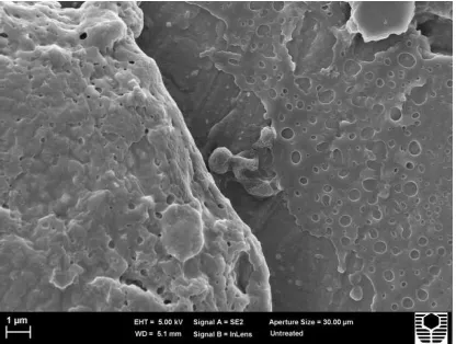
There are three types of biofilm formed in this experiment. In the first type of biofilm shown in figure (1, 2, & 3) shows that the film is produced thick enough. Figure 3 is taken from a low magnification, showing the number of cells that are not too much but movies are pretty much. This is reinforced, when the pictures are taken from different sides (shown by figure 2) looks very thick blobs but no visible cell there. This phenomenon raises a question in this trial, whether the bacteria is below the blob or no bacteria at all there.Biofilm on the second and third types is shown in figure 4 and 5. Figure 4 is a biofilm formed by use of the Bacteriological peptone medium were added ethanol. The result shows that a lot of cells are formed but a little film. With a low magnification, the cell looks
Jurnal Kedokteran Dan Kesehatan Volume 2 Nomor 4, Oktober 2015 538 almost cover the entire surface of the stub.
Figure 5 shows the presence of more cells of figure 4 it is clear that even looks like a blob of biofilm. This type of biofilm using Bacteriological peptone medium were added sucrose. But the figure 5, if carefully observed seem obvious presence of other bacteria in the biofilm (looks different forms of type MRSA cells, foreign cells and elongated oval).
Fig.1. In this figure, from the high magnification shows that the cell is coated by a film thick enough
Fig.2In this figure, from the low magnification shows the blobs. Possibility, cells was coated in a thick film.
Fig.3. In this figure, from the low magnification shows many of these possible cells are present on this part of the film.
Fig.4. Representative, biofilm which was created using bacteriological peptone plus ethanol as a medium. In this figure, from the low magnification shows many of cells. Bacteria were prepared as described above with each of the two media described.
Fig.5. Representative, biofilm which was created using bacteriological peptone plus sucrose as a medium. In this figure, from the low magnification shows the number of cells is bigger than biofilm which was created by third method and using bacteriological peptone plus ethanol as a medium.
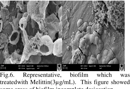
Treating of Biofilm with Melittin on stubs
Figure 6 is a biofilm with the same type
obtained from the first method. From the
figure shows that the biofilm was broken
and the drying process is not perfect.
Damage to the biofilm is still a big
question, whether caused by melittin
treatment or other factors that occur during
shooting process which uses SEM.
Jurnal Kedokteran Dan Kesehatan Volume 2 Nomor 4, Oktober 2015 538 Fig.6. Representative, biofilm which was
treatedwith Melittin(3µg/mL). This figure showed some areas of biofilm incomplete desiccation. Treating of Biofilm with Melittin in well Figure 7 shows the biofilms treated with melittin(5 µg/mL) in the well. C is a positive control samples, MA is a sample in treatment with melittin for 15 minutes, MB is in the sample treatment with melittin for 30 min and an MC sample in treatment with melittin for 60 minutes. Last color (shown in figure 7) was produced in accordance with the expectations of this experiment, namely;
•
Red
color
changed
to
yellow,
indicating the presence of cell
•
Red color or does not change color,
indicating nonexistence cells.
On other hand, if observed in tables 1 and 2 (the color change for each hour) it will show oddities (the sample MA, MB and MC). Peculiarities that arise, namely, the resulting red color of red as the primary color (after the addition of Bacteriological peptone medium were added phenol red) changed to orange and even some samples that have changed color to yellow back to red after standing for 24 hours. All samples (MA, MB and MC) back into the red.
Discussion
The images (Fig. 1, 2, & 3) above show
that the biofilm formation produces large
amounts of non-cellular material with very
few visible cells. The thickness of film
makes it impossible to know how many
cells may be embedded within. When
viewed from the biofilm formation
processing, this method using human
serum in order to make the cells can stick
to each other. Basically,MRSA cells able
to produce polysaccharides by themself to
make their inherent between cells with
other cells. Human serum is a protein
which is found in human blood plasma.
Essentially, human serum has the same
elements (hydrogen, oxygen, carbon, and
nitrogen) with polysaccharides which is
produced by cells MRSA. Based on these
explanations, possibility that the film
formed on the first experiments is human
serum,so it is notfilm which is produced
by MRSAcells. In figure 5, growth in
medium
supplemented
with
sucrose
resulted in a deeper biofilm than with
Bacteriological peptone as a regular
medium, or with ethanol added. Based on
previous research (Welch
et al., 2012)
describe rapid growth in a culture in the
presence of sucrose in base medium.
Therefore, sucrose was chosen as an
additive to the medium in this experiment.
On biofilm treatment process using melittin (3 µg/mL), the images show that the iofilm has been visibly damaged. However, this cannot be assumed to occur as a result of melittin. This form of damage could be a result of incomplete desiccation prior to the vacuum process associated with evaporative metal coating.According to Welch, Ken et al., (2012), the viability assay used in this experiment based on a metabolic activity assay (MAA), evaluates the amount of viable bacteria with the help of a pH indicator. The accumulation of metabolic acid products causes a drop in pH of the assay, which is indicated by a change in color of phenol red from red to yellow (Welch, Ken et al., 2012). The tables showing color-changes following melittin treatment showed irregularities. Controls showed a rapid change in color, with melittin treatments showing less rapid changes. At all-time variations melittin treatment process (15, 30 and 60 minutes) all showed weird occurrence. However, there is still one sample on melittin treatment with 60 minutes having stable color is red. This is consistent with the hypothesis of the experiment. But for another samples, the color is not stable. They changed, from red to orange to yellow and they became stable on red after overnight. From the latter observation suggests that the cells in the biofilm dead. Basically, this metabolic test is used to assess the number of viable bacteria in the test medium through production of metabolites. Metabolites produced by theJurnal Kedokteran Dan Kesehatan Volume 2 Nomor 4, Oktober 2015 538 biofilm or planktonic cultures will depend on
both, the individual bacterial metabolic activity and the number of bacteria that are marked with red color changes to yellow.When reviewed in images which were produced from the process of biofilm formation using sucrose medium, biofilm has formed two different cell shapes. It has the largest number of cells which were shaped similar to MRSA cells, but many kinds of other cells form unknown. Based on that information, the opinion raises that two different cell typesmust be have different metabolic activities. Therefore unstable discoloration caused by the metabolic activity of cells other than the metabolic activity of MRSA.
Conclusion
The resultsshowed that the procedures
used were capable of inducing this MRSA
strain to form a biofilm and melittin
(5µg/mL) able to treat MRSA with treating
of time 15, 30, or 60 minutes.
Suggestion
However, itis still not a perfect trial due to
process of checking the viability of cells
which were observed for each hour was
still a lot of samples that unstable
discoloration.Therefore,
for
the
foreseeable future will be carried out the
modified experiment and the experiment
should be done with the variation of
treatment and increasing the time of
treatment. It aims to get the best results in
the healing process of the disease which is
caused by MRSA.
Reference
Abe, Y., Shigemura, K., Yoshida, H.,
Fujisawa, M., & Arakawa, S.
(2012). Risk factors for anti-MRSA
drug resistance.
Int J Antimicrob
Agents,
40(5),
423-426.
doi:
10.1016/j.ijantimicag.2012.07.005
AkiyamaH, Yamasaki O, Kanzaki H, Tada
J, dan Arata J. Effects of Sucrose
and Silver on Staphylococcus
aureus
Biofilm.
Journal
of
Antimicrobial
Chemotherapy
Departement
of
Dermatology,
Okayama
University
Medical
School,
Shikata-Cho
2-5-1,
Okayama 700-8558, JPn.1998;42:
629-634.
Black JG. Microbiology. 8
th. International
Student Version. Singapore: John
Wiley and Sons. 2013:69-86.
Brooks GF, Butel JS, dan Morse SA. Mikr
obiologi Kedokteran:Jawetz,
Melnick & Adleberg’s Medical
Microbiology. Edisi 23.
2004:251-7,325.
Chamberlain NR. The Big Picture Medical
Microbiology. AmerikaSerikat:M
c Graw Hill Medical. 2009.
214-217.
Chang,Raymond. Kimia
Dasar:Konsep-Konsep Inti. Edisi 3. Jilid 1.
Jakarta:Erlangga. 2005:351.
Cowan MK. Microbiology
A
System
Approach. 3rd
Edition. New
York:McGraw Hill. 2012:88-95.
Dansese
PN, Pratt
LA,
dan
Klter
R. Expolysaccharide Productin is
Required for Development of
Escherichiaa coli K-12
Biofilm
Architecture. Departement of
Microbiology
and
Molecular
Genetics
Harvard
Medical.
2000;182(12):3593-3596.
Donlan RM.
Biofilm
formation
A
Clinically
Relevant
Microbiological
Process. Healthcare
Epidemiolgy. Clinical
Infectious
Disease 2001;33:1387-92.
Jurnal Kedokteran Dan Kesehatan Volume 2 Nomor 4, Oktober 2015 538
Gomez,MA., Galvez,JM., Hontoria,Ernest
o., dkk.
Influence
Ethanol
Concentration
on
Biofilm
Bacterial Composition from a
Denitrifying
Submerged
Filter
Used
for
Contaminated
Groundwater. Journal
of
Bioscience and Bioenginerering.
2003;95(3):245-251.
Harvey RA, Cornelisse CN, dan Fisher BD
. Lippicotts’s IllustratesdRewiews:
Microbiology. 3rd Edition. Philadel
phia:Wolter Kluwer
Health. 2013:111-115.
Lodish H,et al. Bacterial Metabolisme and
Genetic
Molecular
Cell
Biology. 6th ed. New York,WH
Freeman. 2007.
Mahon CR,Lehman DC, dan Manuselis,Ge
orge. Text Book of Diagnostic Mic
robiology. 4
thEdition. China:Saude
rs Elsevier.2011:13-14,758- 765.
Phillips PL, Wolcott RD, Fletcher J,dkk. B
iofilm
Made
Easy. Wounds
International. 2010;1(3):1-6.
Presscott H dan Klein. Microbiology. Edisi
V. California:McGrawHill.
2002:55-76.
Rose S, Keystone J, dan Hackett P.
International
Travel
Health
Guide. Travel medicine. 2015.
Syahrurachman
A,
dkk. Buku
Ajar
Mikrobiologi
Kedokteran. EdisiRevisi.
Jakarta:Binarupa
Aksara
Pubhliser. 1994:195-198.
Vu B, Chen M, Crawford RJ, dan Ivanova
E. Bacterial Extracellular Polysacc
harides Involved in Biofilm Format
ion. 2009;14(2535-
2554)1420-3049:1-20.