THE ACTIVE FRACTION FROM
Nigella sativa
AND ITS ACTIVITY AGAINST T47D CELL LINE
Heny Ekowati
1,*, Eka Prasasti
1, and Undri Rastuti
2 1Department of Pharmacy, Faculty of Medicine and Health Sciences, Jenderal Soedirman University, Karangwangkal, Purwokerto
2
Chemistry Department, Faculty of Science and Engineering, Jenderal Soedirman University, Karangwangkal Purwokerto
Received May 18, 2011; Accepted October 17, 2011
ABSTRACT
Breast cancer is one of the main causes of death in women. Cancer treatment with surgery, chemotherapy, and radiology often cause undesirable side effects. Therefore, alternative cancer treatment by using plants as traditional medicine was expected to reduce side effects. Nigella sativa is one of the plants used as anticancer empirically. This study conducted to examine the cytotoxic activity of Nigella sativa seeds and identify its components on T47D breast cancer cells. Petroleum ether, chloroform, ethyl acetate, and ethanol were used to extract N. sativa seeds. The extracts were tested their cytotoxic activity on T47D cell line using MTT method. The active compound was separated using column chromatography. Cytotoxic test on T47D cell line was perform for extracts of each separation stage. Data were analyzed by probit analysis to obtain IC50 values. Components identification was performed using GC-MS analysis. The results showed that chloroform extract has cytotoxic activity better than other extracts with IC50 of 124.206 µg/mL. The third fraction has cytotoxic activity better than other fractions with IC50 of 68.568 µg/mL. The GC-MS analysis showed that in the third fraction of the chloroform extract contain linoleat acid, the major compound and tryptamine.
Keywords:Nigella sativa, cytotoxic, T47D, GC-MS
INTRODUCTION
Breast cancer is the second leading cause of death from cancer in women in the United States after lung cancer. In Britain as many as 20% of cases of cancer were breast cancer, as well as causing the most deaths in women 35-55 years age group. Until now there is no accurate statistics data in Indonesia, but the hospital data indicate that breast cancer ranked first among other cancers in women [1]. Cancer is a condition in which cells have lost normal control mechanisms, so cells growth of abnormal, rapid, and uncontrolled. Breast cancer (mammary carcinoma) is a neoplastic disease derived from the parenchyma [2].
Cancer treatment can be done with surgery, chemotherapy, and radiology. Multiple drug therapy is cancer drugs cytostatica (methotrexate, doxorubicin, vinblastine), hormones (estrogen), and antihormone (tamoxifen). However, they pose cancer treatment side
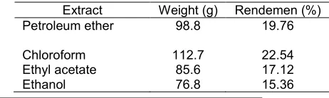
Table 1. Solvent-free condensed extract maceration results
Extract Weight (g) Rendemen (%)
Petroleum ether 98.8 19.76
Chloroform Ethyl acetate Ethanol
112.7 85.6 76.8
22.54 17.12 15.36
effects of which not only attack cancer cells but also attack normal cells [3]. As a result of which could arise in the form of bleeding, bone marrow depression which facilitates the occurrence of infection. In the gastro intestinal system can occurred nausea, vomiting, anorexia and gastrointestinal ulceration [4]. Alternative cancer treatment is to utilize compounds derived from plants, which have anti-tumor properties with minor toxicity and side effects [5].
which the third active ingredient is contained in the essential oil. The main active component of N. sativais also known has antioxidant activity which can inhibit the development of several types of cancer [13]. Research showed the biological effects of N. sativa seeds can reduce the formation of breast tumors [14].
In Indonesia, N. Sativa seeds was used as jamu. Therefore, this study conducted to examine the cytotoxic activity ofN. sativaseeds and identify its components on T47D breast cancer cells, so N. sativa able to use as herbal medicine or phytopharmaca.
EXPERIMENTAL SECTION
Materials
Plant materials, chemicals, cell line and culture Seeds of Nigella sativa, Petroleum ether, chloroform, Cell line T47D, RPMI 1640 medium (GIBCO), Fetal Bovine Serum (FBS) 10% (Sigma), 3% penicillin-streptomycin (Sigma), Fungison 1% (Gibco BRL), sodium bicarbonate, 4 (2-hydroxyethyl)-I-piperazine-ethane-sulphonic acid (HEPES) (Sigma), dimethyl sulfoxide (DMSO), aqua-bidest, 70% alcohol, ethyl acetate, methanol, acetic acid, vanillin-H2SO4, Yellow MTT, sodium dodecyl sulfate (SDS), and tamoxifen.
Instrumentation
CO2 incubator, liquid nitrogen tank, water bath (Laboratory Equipment Sydney), laminary air flow cabinet (NUAIRE), ELISA reader (SLT 240 ATC) microplate 96 wells (Nalge Nunc International, Denmark), tissue culture flasks (TCF) 75 cm2, inverted microscope, hemocytometer (Neubauer), centrifuge (Sigma), pH meter (TOA), light microscope, were used in cytotoxic test. A set of fractionation equipment were used to fractioned the extracts. Gas Chromatography-Mass Spectroscopy (GC-MS)-Agilent 7890A GC System with MSD Detector 5975 C series were used to determine the compounds in extract.
Procedure
Preparation of extract combination
Nigella sativa seeds are washed with water, dried and ground into powder. N. sativa seed powder were macerated using petroleum ether for 1 x 24 h. Extract was filtered and then evaporated by the evaporator. Then the filtrate was continued macerated with chloroform, ethyl acetate, and ethanol respectively, each for 3 x 24 h. The extract is filtered and then evaporated. Each extracts was tested for cytotoxic activities on T47D cell line.
Cell viability assay: 3-(4,5-dimethylthiazol-2-yl)-2,5-diphenyl tetrazolium bromide (MTT) assay
T47D cancer cells were cultured in a 96-well plates (Nalge Nunc International, Denmark) at a density of 2 × 10⁴ cells per well, incubated 24 h. The cells were then treated with varying concentrations of extract and tamoxifen. After 24 h incubation, the cells were washed and treated with 0.01 mL MTT per well. Plates were incubated at 37 °C in a 5% CO2 atmosphere for 4 h, and 0.1 mL of the extraction buffer (10% sodium dodecyl sulfate in 0.01% HCl) was added. After an overnight incubation at 37 °C, the absorbance was measured at 595 nm using a ELISA reader (Bio-Rad) and was compared with the control cultures without compound. To determine cell viability, percent viability was calculated as [(absorbance of drug-treated) sample/(control absorbance)] × 100.
Fractionation
The active extract was partitioned with n-hexane : chloroform (3:1, 2:1, 1:1) v/v}, chloroform, chloroform : ethyl acetate {(1:1, 1:2) v/v}, chloroform : ethyl acetate : methanol {(1:2:2, 1:1:4) v/v}, ethyl acetate : methanol {(1:2) v/v}, and methanol. The chemical composition of each fraction was monitored on thin layer chromatography, each fraction was tested for cytotoxicity test on T47D cell line.
RESULT AND DISCUSSION
Preparation of the extract was done by maceration method. Maceration was done using the appropriate solvent with several times shaking or stirring at room temperature [15]. Maceration is a method that suitable for compounds that do not withstand heating at high temperatures [16]. The aim is to attract the chemical components based on the principle of mass transfer of substance into the solvent component, where the movement began to occur at the interface layer and then diffuses into the solvent [17]. Petroleum ether is a non polar used to attract non-polar compounds such as wax, fat and lipids which can interfere during the separation process [18]. In addition, petroleum ether was used to eliminate the resins which can interrupt the extraction. After the filtrate macerated with petroleum ether, maserat aerated and then macerated with chloroform for 3 x 24 h. Chloroform is one of the semi-polar solvent commonly used to attract terpenoid compounds, polyphenols, fatty acids, fats, oils, essential oils, phenols and lipids [17].
Fig 1. Inhibition rate on T47D cell line with petroleum ether, chloroform, ethyl acetate, and ethanol extracts. T47D cells were treated with various doses of extracts, incubated for 24 h at 37 °C in humidified 5% CO2 atmosphere. Cell viability was determined by MTT assay, absorbance was read at 595 nm. Inhibition rate (%) was defined as : ((live cell in the control – live cell in the test group)/live cell in the control) x 100. Standard curve : y = 0.00001 x + 0.3411 (R2 = 0.9906). Results are average of three independent experiments (mean ± SD)
Fig 2.IC50of petroleum ether, chloroform, ethyl acetate, and ethanol extract on T47D cell line. Concentrations inhibiting 50% of the cell were determined by probit analysis using SPSS software
Extracts N. sativa had cytotoxic activity on T47D Cell Line
In our research showed that the increasing concentration of test material, the greater the percentage of inhibition (Fig. 1). This showed the increasing toxic effects of the test material.
Cytotoxic test of chloroform extract using the MTT method (3-(4.4-Dimethylthiazol-2,5dipheniltetrazolium bromide) by measuring the absorbance. T47D living cell will absorb MTT into formazan. This is because the MTT is only absorbed by dehydrogenase enzymes in mitochondria into formazan which is the result of tetrazolium salt metabolism of living cells that have terminated tetrazolium ring. Absorbance read with ELISA
reader at long wavelength (λ) 559 nm. Therefore,
absorbance describes the number of cells living. Absorbance is to be used to calculate the percentage of living cells as a response [22].
The material has a ability to inhibit T47D cell growth as shown by the IC50value Fig. 2 showed that. This indicates that extracts has cytotoxic activity against T47D cells. Meyer et al. [24] declares an extracts said to have anticancer activity if the IC50value of less than 1000 µg/mL after 24 h of contact time.
IC50is concentration that can inhibit cell growth by 50% cell line. The smaller the IC50of a compound the more toxic compound it was [23]. Cytotoxic activity of chloroform extract of Nigella sativa seeds is higher than petroleum ether extract. Research suggests that chloroform is one of the semi-polar solvent commonly used to attract terpenoid compounds, phenols, and lipids [17]. So it is possible thymoquinone largely attracted by the solvent chloroform. The results obtained prove that the IC50 petroleum ether and chloroform extracts of Nigella sativa seeds have anticancer activity in vitro. Extract was said to have anticancer activity when the IC50 value of less than 1000 µg/mL after 24 h contact time [24].
The extracts had cytotoxic activity against T47D cells. N. sativa contained thymoquinone which is a component of volatile oil that is responsible as anticancer [19,21]. Thymoquinone have a role in inhibiting the growth of cancer cells and induce apoptosis in cancer cells [19,21]. Thymoquinone also have activity as an antioxidant. Thymoquinone through reaction with reductase enzyme conjugate with glutathione (GSH) plays a role in protecting organs from oxidative damage agent [21]. Thymoquinone can improve the immune system by increasing the activity of interferon-γ, macrophages, and CD4 + T cell and NK cell function [9].
Research showed that the essential oils and ethyl acetate extract from black cumin seeds had anticancer activity in a variety of different tumour cells (P815, Vero, BSR, and ICO1 cell line). The results showed ethyl acetate extracts and essential oils have a cytotoxic effect on P815 cells (IC50(% v/v) = 0.6, 0.75), on Vero cells (IC50 (% v/v) = 0.22; 0.25), on BSR cells (IC50 (% v/v) = 0.2; 1.2%) [11]. This research with N. sativaseed showed differences in IC50values because of the differences of the target cell. In this study, extracts of which have the smallest IC50that is 124.829 µg/mL is the chloroform extract. Therefore, the chloroform extract is what will continue to fractionated.
Fractionation of Chloroform Extract
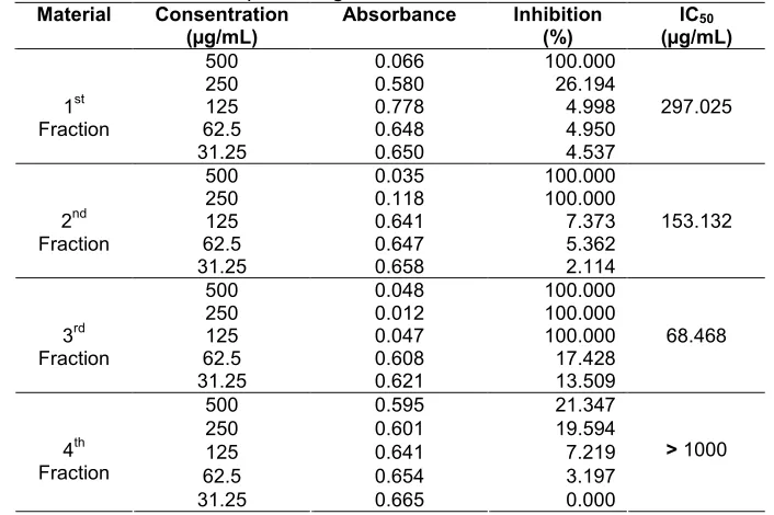
Table 2.The mean absorbance and the percentage inhibition of T47D cells after administration of test material
components of organic compounds in the sample. Hexane and chloroform was used with various comparisons and proceed according to different levels of polarity using the other solvent that was ethyl acetate and methanol. Elution process performed to form ribbons of yellow and brownish red in stationary phase. These tapes are eluted by the eluent until the exit of the column-eluate into the eluate separated and placed in tubes. The fractionation carried out until all samples were eluted by the eluent, eluent last used is a polar eluent is methanol. When the sample on the separation column has not happened and the eluate is colorless, then the fractionation discontinued.
Determination of the fraction containing the same components conducted by TLC method. Each faction identified with TLC method to see the pattern of separation. Fractions that have a pattern of separation and the same Rf value thought to contain the same components. Fractions with the stain color and the same Rf value pooled and evaporated the solvent. Based on the results of grouping with 3 fractions obtained by TLC, furthermore these three fractions were then tested for cytotoxic activity in T47D cells and identified the content of chemical compounds by GC MS.
Cytotoxic activity Fraction of Chloroform Extract
Cytotoxic activity test performed on fractions of fractions of chloroform extract of black cumin seeds. Chloroform fraction test results, shown in Table 2. Data Table 2 shows that with increasing concentration, the absorbance is smaller. This shows that the number of T47D cells with increasing concentrations of living
become less and less. Table 2 also shows that the fraction 1, 2, and 3 have cytotoxic activity against breast cancer cells T47D. This is shown by the IC50 value of 297.025, 153.132, 68.468 µg/mL. While the fraction 4 does not have toxic properties against breast cancer T47D cells, with IC50 of more than 1000 µg/mL. In this study, the most active fraction was fraction 3 with IC50 value of 68.468 µg/mL. Fraction 3 was determined the content of the compounds using GC-MS method.
Analysis of Active Compounds in fractions using GC-MS method
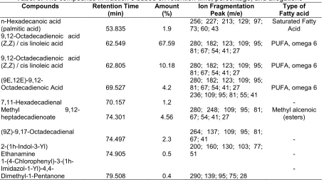
Identification of the structure using a mass spectrometer conducted to determine the compounds contained in the samples analyzed, this can be seen from the relative abundance of mass fragments of molecules (m/e) of the molecular ion (M +). The more stable a molecular fragment that is formed then the fragment will be at a relative abundance of large and have a longer lifespan [25]. Retention time, percentage and allegations of compounds in fraction 3 can be seen in Table 3.
Tabel 3.The compounds in fraction 3 based on retention time, percentage, and allegations
(palmitic acid) 53.835 1.9
256; 227; 213; 129; 97; 73; 60; 43
Saturated Fatty Acid 9,12-Octadecadienoic acid
(Z,Z) / cis linoleic acid 62.549 67.59 280; 182; 123; 109; 95;
81; 67; 54; 41; 27
PUFA, omega 6
9,12-Octadecadienoic acid
(Z,Z) / cis linoleic acid 62.805 10.18 280; 182; 123; 109; 95;
81; 67; 54; 41; 27
PUFA, omega 6
(9E,12E)-9,12-Octadecadienoic Acid 69.527 4.2
280; 182; 123; 109; 95;
Dimethyl-1-Pentanone 79.508 0.4 290; 139; 95; 75; 28
-Percentage of oil extraction, as well as physical and chemical properties of the crude oil, was influenced by the extraction methods. TLC and GLC analysis indicated that nigella seed oil contained significant amounts of sterols. Linoleic (C18:2), oleic (C18:1) and palmitic (C16:0), as in most of the common edible oils, are the main fatty acids [26].
The results of animal studies have performed that the consumption of omega-3 fatty acids can slow the growth of cancer xenografts, increase the efficacy of chemotherapy and reduce the side effects of the chemotherapy or of the cancer [27].
The effect of fish oils and their active omega-3 fatty acid constituents, docosahexaenoic acid (DHA) and eicosapentaenoic acid (EPA), were investigated on breast cancer growth. The results suggest that inhibition of breast cancer growth in nude mice by dietary fish oil and inhibition of breast cancer cell growth in culture by treatment with DHA and EPA was mediated by activation of N-SMYase [28].
CONCLUSION
Chloroform extract of black cumin have cytotoxic activity against T47D cells with IC50 of 124.206 µg/mL. The 3rdfraction was the most active one, with IC50values of 68.568 µg/mL. The GC-MS analysis showed that in the 3rd fraction of the chloroform extract contain linoleic acid, the major compound and tryptamine, an alkaloid.
ACKNOWLEDGEMENT
Funding support from Higher Directorate General Education, National Education Department 2010 with competitive grant scheme, is gratefully acknowledged.
REFERENCES
1. Hartanto, H., Darmaniah, N., and Wulandari, N., 2007, Buku Ajar Patologi Robbins, 7th Ed., EGC, Jakarta.
2. Anonymous, 2007, Indonesia Health Profile 2005, 70, Departemen Kesehatan Republik Indonesia, Pusat Data Kesehatan, Jakarta.
3. Stetler-Stevenson, W.G. and Kleiner, D.E., 2001, Molecular Biology of Cancer: Invasion and Metastases in Cancer Principle & Practice of Oncology, 6th Ed., Eds. DeVita, V.T., Hellman, S., dan Rosenberg, S.A., Lipicott Williams &Wilkins, Philadelphia, USA, 91–102.
4. Kurnianda, J., Hisyam, B., Wahyuningsih, E., and Hutajulu, S.H., 2005, Acta Med. Indones., 37, 4, 210–213.
5. Amin, A., Gali, M.H., Ocker, M., and Schneider, S.R., 2009,Int. J. Biomed. Sci.; 5, 1, 1–11.
6. Hanahan, G. and Weinberg, R.A., 2000, Cell, Vol. 100, 57–70.
7. Vajira, P.B., 2006,Ruhuna J. Sci.,1, 152–161. 8. Zaher, K.S., Ahmed, W.M., and Zerizer, S.N.,
9. Gilani, A.H., Jabeen, Q., and Khan, M.A.U., 2004,
Pak. J. Biol. Sci.,7, 4, 441–451.
10. Farah, I.O., 2005,Int. J. Environ. Res. Publ. Health, 2, 3, 411–419.
11. Mbarek, L.A., Mouse, H.A., Elabbaadi, N., Bensalah, M., Gamouh, A., Braz. J. Med. Biol. Res., 40, 6, 839–847.
12. Buyugahapitiya, V.P., 2006, Ruhuna J. Sci., 1, 152–161.
13. Randhawa, M.A., 2008, J. Ayub. Med. Coll. Abbottabad,20, 2,1–2
14. El-Aziz, M.A., Hassan, H.A., Mohamed, M.H., Meki, A.R., Abdel, G.S.K., and Hussein, M.R., 2005,Int. J. Exp. Pathol., 86, 6, 383–396.
15. Fathiyawati, 2008,Uji Toksisitas Ekstrak Daun Ficus racemosa L. terhadap Artemia salina Leach dan Profil Kromatografi Lapis Tipis, Skripsi, Fakultas Farmasi Universitas Muhammadiyah Surakarta. 16. Anonymous, 1985, Cara Pembuatan Simplisia,
Direktorat Jenderal Pengawas Obat dan Makanan, Departemen Kesehetan, Jakarta.
17. Harbone, J.B., 1989, Metode Fitokimia: Penuntun Cara Modern Menganalisis Tumbuhan, ITB, Bandung.
18. Dinasari, E., 2009, Identifikasi Senyawa Metabolit Sekunder dari Ekstrak Bunga Kecombrang (Nicolia speciosa Horan) dan Uji Toksisitasnya dengan metode Brine Shrimp Test, Skripsi, Fakultas Sains
dan Teknik jurusan MIPA Prodi Kimia Universitas Jenderal Soedirman Purwokerto, 17.
19. Ivankovic, S., Stojkovic, R., Jukic, M., Milos, M., and Jurin, M.M.M., 2006, Exp. Oncol., 28, 3, 220–224.
20. Nickavar, B., Mojab, F., Javidnia, K., and Amoli, M.A.R., 2003,Z. Naturforsch., 58c, 629–631. 21. Edris, A.E., 2009, Curr. Clin. Pharmacol., 4, 1,
43–46.
22. Sieuwerts, A.M., Klijn, J.G.M., Peters, H.A., and Foekens, J.A., 1995, Eur. J. Clin. Chem. Clin. Biochem., 33, 11, 813–823.
23. Doyle, A., and Griffiths, J.B., 2000,Cell and Tissue Culture for Medical Research, John Willey and Sons, Ltd., New York, 47.
24. Meyer, B.N., Ferigni, N.R., Putman, J.E., Cobsen, L.B., Nichols, D.E., and McLaughlin, J.L., 1982, Planta Med., 45, 5, 31-34.
25. Silverstain, R.M., Bassler, G.C. and Morrill, T.C., 1981,Penyidikan Spektrometrik Senyawa Organik. Erlangga, Jakarta.
26. Atta, M.B., 2003,Food Chem., 83, 1, 63–68. 27. Hardman, W.E., 2002, J. Nutr., 132, 11,
35085–35125.