Paediatrica Indonesiana
VOLUME 53 May• 2013 NUMBER 3
Original Article
Familial congenital heart disease in Bandung,
Indonesia
Sri Endah Rahayuningsih
Abstract
Background Congenital heart disease (CHD) may occur in
several members of a family. Studies have shown that familial genetic factor play a role in CHD.
ObjectiveTo identify familial recurrences of CHD in families with at least one member treated for CHD in Dr. Hasan SadikinHospital, Bandung Indonesia.
Methods In this descriptive study, subjects were CHD patients hospitalized or treated from January 2005 to December 2011. Weconstructed family pedigrees for five families.
Results During the study period, there were 1,779 patients with CHD. We found 5 families with 12 familial CHD cases, consisting of 8 boys and 4 girls. Defects observed in these 12 patients were
tetralogy of Fallot, transposition of the great arteries, persistent ductus arteriosus, ventricular septal defect, tricuspid atresia,pulmonary stenosis, and dilated cardiomyopathy. Persistent ductus arteriosus was the most frequently observed defect
(4 out of 12 subjects). None of the families had a history of consanguinity. The recurrence risk of CHD among siblings was calculated to be 0.67%, and the recurrence risk of CHD among cousins was 0.16%.ConclusionFamilial CHD may indicate the need for genetic counseling and further pedigree analysis.
[Paediatr Indones. 2013;53:173-6.]
Keywords: Familialgenetic, congenital heart disease, Indonesia
ne of the most frequently found congenital
anomalies is con
genital heart disease (CHD).
Congenital heart disease incidence has been
reported to be 8-10 of 1,000 births in nearly all
countries.
1In Indonesia, the birth rate is 4 million per
year,
2so the incidence of CHD has been estimated
to
be 32-40 thousand per year.
To date, it is unknown why CHD occurs. Past
studies reported that familial or genetic factors play a
role, since CHD may occur repeatedly in families.
3-5Furthermore, consanguinity may also increase the risk
of CHD.
6Familial factors may be due to chromosomal
anomalies or gene mutations.
5In addition to genetic
factors, maternal factors such as infection, metabolic
anomalies, immune disorders, obesity,
7consuming
drugs during pregnancy,
3ethnicity, and advance age
at pregnancy have been correlated with CHD.
8,9Other
factors such as engaging in sex, consuming alcohol or
smoking during pregnancy, climate, and environment
(urban, suburban, or rural)
9, 10have not been shown to
correlate to CHD.
11Therefore, CHD has been
associated with genetic and non-genetic factors, or
interactions between the two.
11The aim of this study was to identify the
familial recurrences of CHD in families that had a
member with CHD hospitalized in Dr. Hasan Sadikin
Hospital,
Bandung, Indonesia.
Methods
We retrieved data from patients’ medical records
From the Department of Child Health, Padjadjaran University Medical School/Dr. Hasan Sadikin General Hospital, Bandung, Indonesia.
Reprint requests to:Sri Endah Rahayuningsih, Department of Child Health, Dr. Hasan Sadikin General Hospital/Padjadjaran University, Jalan Pasteur 38 Bandung 40161, Indonesia. Tel. +62-816 487 0962, Fax. +62-222-034426.E-mail: seraning@yahoo.com
O
Sri Endah Rahayuningsih:Familial congenital heart disease in Bandung, Indonesia
hospitalized in Dr. Hasan Sadikin Hospital, Bandung,
Indonesia from January 2005 to December 2011.
Diagnosis
of
congenital
heart
disease
was
based
on
echocardiographic examination performed by pediatric
cardiology consultants using a General Electric Type
Logic 700 or General Electric Type Vivid 3 machines.
Based on this data, three-generation family pedigrees
were generated for each affected family.
Results
Family 2 was a mixed marriage between an
Indonesian mother and a Korean father with 2 affected
children (
Figure 2
). Child II.3 had TGA and was
diagnosed based on echocardiography at the age of 4
years, while child II.5 was diagnosed with VSD. One
child
with VSD underwent a closure at 5 months of age.
The other child’s with TGA defect was inoperable
due
to pulmonary vascular disease, and he is now 16
years
old. Hypospadias was a congenital malformation
found in individual II.6.
I
During a 7 year-study period, there were 1,779
patients with CHD in our hospital. Five families
had a
total of 12 familial CHD members, consisting of
8 boys and 4 girls. Defects observed in these subjects
were tetralogy of Fallot (TOF), transposition of the
great arteries (TGA), persistent ductus arteriosus
(PDA), ventricular septal defect (VSD), tricuspid
atresia,
pulmonary
stenosis,
and
dilated
cardiomyopathy.
Persistent ductus arteriosus was the
most common
defect in the subjects. No history of
consanguinity was found. A medical pedigree was
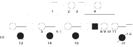
used to describe the families. The symbols used
were defined as
follows: squares represent boys
;circles represent girls
;solid black indicates CHD or
suspected CHD
;hatch lines indicate individuals
with other congenital malformations.
Figure 1
shows three individuals with CHD
and
one with suspected CHD in Family 1. Defects
found in these individuals were as follows: TOF+PDA
(II.9), VSD+PDA (III.14), TGA (III.15, died at age 2
months), and suspected CHD (III.16, died at age 2
months due to respiratory failure and cyanosis). Two
individuals, II.9 and III.14, underwent corrective
procedures. One individual had labiopalatoschisis
(III.13).
1 2
I I
3 4 5 6
Figure 2.
Family 2 pedigree
In family 3 there were 2 siblings with PDA,
II.3 and II.5, and 1 sibling with mental retardation,
II.6
(
Figure 3
).
I
1 2
I I
3 4 5 6
Figure 3.
Family 3 pedigree
Figure 4
shows an affected child (II.3) with
familial dilated cardiomyopathy, who is now 5
years
old. Her father (I.1) died at 30 years of age.
Figure 4.
Family 4 pedigree
Figure 5
shows two affected children in family
5,
one with tricuspid atresia + VSD (II.3) and the other
with pulmonary valve stenosis (II.5).
The recurrence risk of CHD among siblings
was calculated to be CHD/siblings = 0.67%. The
recurrence risk of CHD among cousins was
CHD/
cousins = 0.16%. None of the families had a
history
of consanguinity.
1 2
1 2 3 4
I I I
13 14 15 16
1 2
5 6 7 8 91011
Figure 1.
Family 1 pedigree
Sri Endah Rahayuningsih:Familial congenital heart disease in Bandung, Indonesia
3 4 5
Figure 5.
Family 5 pedigree
Discussion
Congenital heart disease is a common occurrence,
but its etiology is poorly understood. Genetic factors
have been correlated with CHD in studies carried
out on the role of gene mutations in CHD.
4The aim
of this study was to identify familial recurrences of
CHD to be used as a framework for further genetic
studies. We found that the recurrence risk for CHD
was 0.67%. Previous studies found familial recurrence
Table 1. Types of congenital heart disease in the five families
Type of CHD Total Tetralogy of Fallot 1
Transposition of great arteries 2
Persistent ductus arteriosus 4
Ventricular septal defect 3
Tricuspid atresia 1
Pulmonary stenosis 1
Dilated cardiomyopathy 2
Table 2. Extracardiac anomalies accompanying congenital heart disease
Extracardial anomaly Relationship n
Labiopalatoschisis Cousin 1
Mental retardation Elder brother 1
Hypospadia Younger brother 1
risks of CHD of 1-3%.
12The highest recurrence
risk is reportedly among siblings.
12Tetralogy of Fallot and TGA are conotruncal
defects of CHD. In TGA, embryological anomaly
may
occur on the conotruncal or ventricular outflow tract,
i.e., transposition of both great arteries, so that the
aorta moves out from the right ventricle and the
pulmonary artery from the left ventricle.
Such
anomalies were found in about 5% of all CHD
patients,
characterized by parallel unrelated systemic and
pulmonary circulations. The survival of a neonate
who was born with such an anomaly depends mostly
on good mixture of systemic venous return and
pulmonary venous return through the atrial
septal
defect (ASD), VSD, or PDA.
13,14Recurrence of TGA is rarely found because it
generally occurs sporadically.
14We found that different
defects occurred in a single family, as seen in Family 1
in
which the first child had TGA and the third child had a
VSD. Ventricular septal defect accounts for about
20%
of CHD defects, but in our study we found only
one patient
with VSD. A previous study suggested that familial
VSD is due to genetic factors.
15Tetralogy of Fallot is a cyanotic type of CHD.
Its
prevalence is about 5—10% of all CHD cases.
Tetralogy
of Fallot was first described by Arthur Louis Etienne
Fallot, who performed an autopsy on a patient with
cyanosis, known as “la maladie bleu.”
16Tetralogy of
Fallot consists of VSD, pulmonary arterial stenosis,
overriding aorta and right ventricular dilatation.
Currently this combination of the four anomalies is
referred to as TOF. The main cause of the four
anomalies is antero-cephalad deviation of the
muscular outlet septum insertion relative to the limbs
of the septomarginal trabeculation, coupled with an
arrangement of the septoparietal trabeculations
which produces a squeeze at the mouth of the
infundibulum.
16,17Previous studies revealed the recurrence
risk of TOF to be 2.5—3%.
18Tetralogy of Fallot was
also a recurrent defect in this study.
In our study, the most frequently recurring familial
defect was PDA. In contrast, previous studies showed
that PDA had a low risk for familial recurrence, at only
3%, because it is generally of a
polygenic etiology.
19Previous studies stated that ASD
was the CHD with
the highest rate of recurrence, at
40-100%.
However, no familial ASD was found in our subjects. In
Indonesia, a study on the GATA4 gene
mutation in
ASD found that spontaneous mutation had no
correlation with familial ASD.
20In conclusion, familial CHD may indicate the
need
for genetic counseling and further pedigree analysis.
The result of this study may be used as a framework
for identifying gene mutations affecting CHD.
References
1. Hoffman JIE, Kaplan S. The incidence of congenital heart disease. J Am Coll Cardiol. 2002;39:1890-900.
1 2
I
I I
Sri Endah Rahayuningsih:Familial congenital heart disease in Bandung, Indonesia
2. Indonesia Ministry of Health. Indonesia Health Profile. Jakarta: Indonesia Ministry of Health;2011.
3. Park MK. Pediatric cardiology for practitioners. 5th ed.
Philadelphia: Mosby;2008.
4. Okubo A, Miyoshi O, Baba K. A novel GATA4 mutation
completely segregated with atrial septal defect in a largeJapanese family. J Med Genet. 2004;41:e97.
5. Konig K, Will JC, Berger F, Muller D, Benson DW. Familial
congenital heart disease, progressive atrioventricular
block and the cardiac homeobox transcription factor gene NKX2.5: identification of a novel mutation. Clin Res Cardiol. 2006;95:499-503.
6. YunisK, Mumtaz G, Bitar F, Chamseddine F, Kassar M, Rashkidi J, et al. Consanguineous marriage and congenital heart defects: a case-control study in the neonatal period. Am J Med Genet A. 2006;140:1524-30.
7. Mills JL, Troendle J, Conley MR, Carter T, Druschel CM. Maternal obesity and congenital heart defects: a population-based study. Am J Clin Nutr. 2010;91:1543-9.
8. Botto LD, Cornea A, Erickson JD. Racial and temporal variations in the prevalence of heart disease. Pediatrics. 2001;107:E32.
9. Forrester MB, Merz RD. Descriptive epidemiology of selected congenital heart defects, Hawaii, 1986-1999. Paediatr Perinat Epidemiol. 2004;18:415-24.
10. Morales-Suarez-Varela MM, Bille C, Christensen K, Olsen J.
Smoking habits, nicotine use, and congenital malformations.Obstet Gynecol. 2006;107:51-7.
11. Clark EB. Etiology of congenital cardiovascular malforma
-tions: epidemiology and genetics. In: Allen HD, Gutgesell HP, Clark EB, Driscoll DJ, editors. Moss and Adam's heart disease in infants, children, and adolescents. 6th ed.
Philadelphia: William & Wilkins;2001. p. 64-79.
12. Calcagni G, Digilio MC, Sarkozy A, Dallapiccola B, Marino B. Familial recurrence of congenital heart disease: an overview and review of the literature. Eur J Pediatr. 2007;166:111-6.
13. Fyler DC. D-transposition of the great arteries. In: Fyler DC, editor. Nadas' pediatric cardiology.2nded. Philadelphia:
Hanley&Belfus;2006. p. 645-61.
14. Blue GM, Kirk EP, Sholler GF, Harvey RP, Winlaw DS. Congenital heart disease: current knowledge about causes and inheritance. Med J Aust. 2012;197:155-9.
15. Robinson SW, Morris CD, Goldmuntz E, Reller MD, Jones MA, Steiner RD, et al. Missense mutations in CRELD1 are associated with cardiac atrioventricularseptal defects. Am J Hum Genet. 2003;72:1047-52.
16. Frank LH, Kumar TK, Jonas RA, Donofrio MT. Tetralogy of Fallot with inverted great arteries {S, D, I1: case report, literature review, and discussion of embryology. Pediatr Cardiol. 2012;33:150-4.
17. Gittenberger-de Groot AC, Bartelings MM, Deruiter MC, Poelmann RE. Basics of cardiac development for the understanding of congenital heart malformations. Pediatr Res. 2005;57:169-76.
18. Digilio MC, Marino B, Giannotti A, Toscano A, Dallapiccola B. Recurrence risk figures for isolated tetralogy of Fallotafter screening for 22g11 microdeletion.
J Med Genet. 1997;34:188-190.
19. Hajj H, Dagle JM. Genetics of patent ductus arteriosus susceptibility and treatment. Semin Perinatol. 2012;36:98
104.
20. Hamanoue H, Rahayuningsih SE, Hirahara Y, Itoh J, Yokoyama U, Mizuguchi T,et al. Genetic screening of 104
patients with congenitally malformed hearts revealed a fresh mutation of GATA4 in those with atrial septal defects. Cardiol Young. 2009;19:482-5.