REVIEW
Epigenetics and its role in male infertility
Rima Dada&Manoj Kumar&Rachel Jesudasan& Jose Luis Fernández&Jaime Gosálvez&Ashok Agarwal
Received: 28 November 2011 / Accepted: 17 January 2012 #Springer Science+Business Media, LLC 2012
Abstract Male infertility is a common and complex prob-lem affecting 1 in 20 men. Despite voluminous research in this field, in many cases, the underlying causes are un-known. Epigenetic factors play an important role in male infertility and these have been studied extensively. Epige-netic modifications control a number of processes within the body, but this review will concentrate on male fertility and the consequences of aberrant epigenetic regulation/modifi-cation. Many recent studies have identified altered epige-netic profiles in sperm from men with oligozoospermia and
oligoasthenoteratozoospermia. During gametogenesis and germ cell maturation, germ cells undergo extensive epigenetic reprogramming that involves the establishment of sex-specific patterns in the sperm and oocytes. Increasing evidence sug-gests that genetic and environmental factors can have negative effects on epigenetic processes controlling implantation, pla-centation and fetal growth. This review provides an overview of the epigenetic processes (histone-to-protamine exchange and epigenetic reprogramming post-fertilization), aberrant epigenetic reprogramming and its association with fertility, possible risks for ART techniques, testicular cancer and the effect of environmental factors on the epigenetic processes.
Keywords Epigenetics . Male infertility . ART . Chromatin remodeling . Imprinting . Protamines
Introduction
Male infertility is a complex problem where not only the genes, but also the epigenetic factors play a crucial role. There is an enormous interest in one potential cause of male infertility—the aberrant epigenetic reprogramming in male germ cells that can lead to sperm abnormalities. A number of studies have explored the causes of male infertility and now there is sufficient information supporting the idea that epigenetic changes contribute to male infertility.
What is epigenetics?
The term epigenetics refers to changes in the phenotype caused by mechanisms other than changes in DNA sequences, hence the name epi- (above or over)- genetics. Waddington [1] reintroduced the term to explain that gene action and expres-sion that give rise to the phenotype [2]. Epigenetic changes encompass an array of molecular modifications of DNA or Capsule Epigenetic mechanisms regulate germ cell development and
differentiation. Sperm epigenome is critical for optimal embryogenesis. Sperm from infertile men are prone to epigenetic instability and this may lead to increased incidence of imprinting defects in children conceived by advanced assisted reproductive procedures.
R. Dada
:
M. KumarLaboratory for Molecular Reproduction and Genetics,
Department of Anatomy, All India Institute of Medical Sciences, New Delhi 110029, India
R. Jesudasan
Centre for Cellular and Molecular Biology, Hyderabad 500007, India
J. L. Fernández Unidad de Genética,
Complejo Hospitalario Universitario A Coruña (INIBIC), As Xubias 84,
15006 A Coruña, Spain
J. Gosálvez Unidad de Genética, Departamento de Biología, Universidad Autónoma de Madrid, 28049 Madrid, Spain
A. Agarwal (*)
Center for Reproductive Medicine,
Cleveland Clinic 9500 Euclid Avenue, Desk A19.1, Cleveland, OH 44195, USA
histones that are intimately associated with DNA. DNA wraps around histones to form nucleosomes. Nucleosomes are pack-aged into a higher order of structures called chromatin; mod-ifications in chromatin control gene-expression in a spatio-temporal manner [3–6]. The genome-wide approach to study-ing epigenetics is defined as epigenomics.
Epigenetic mechanism of gene regulation
Two major modifications that occur in chromatin are DNA methylation and post-translational histone modifications [3, 4]. DNA methylation is a biochemical process which involves addition of a methyl group to the 5′ position of the cytosine pyrimidine ring typically occurring in a CpG dinucleotide [4]. DNA methylation occurs as a result of DNA methyltransferase (DNMT) activity. There are 3 main DNMTs: i) DNMT1 [7]—which plays a key role in main-tenance of methylation; ii) DNMT 3a and iii) 3b, which are de novo methyltransferases that methylate the genomic DNA during early embryonic development [8]. The changes are acquired in a gradual rather than by an abrupt process [6, 9]. CpG islands are genomic regions that are approximately 500 base pairs long, which have a high frequency of CpG sites (CG to GC ratio >55%) [10]. These stretches of DNA are located within the promoter region of about 40% of mammalian genes which, when methylated, cause stable heritable transcriptional silencing. Hypomethylation and hypermethylation can occur simultaneously at different regions in the genome [11].
Histones are basic proteins in eukaryotic nuclei, and they package DNA into nucleosomes. H2A, H2B, H3 and H4 histones are integral part of nucleosomes. Histone modifi-cations, such as acetylation, methylation, ubiquitylation and phosphorylation, have emerged as the main players in epi-genetic regulatory mechanisms. An intricate interplay exists between modifications of the histone tails of H3 and H4, some of which act antagonistically to regulate the conver-sion from an active chromatin state to an inactive one termed the histone code [12]. Generally, the acetylation of histones marks active, transcriptionally competent regions, whereas hypoacetylated histones are found in transcription-ally inactive euchromatic or heterochromatic regions. In contrast, histone methylation can be a marker for both active and inactive regions of chromatin. Methylation of lysine 9 on the N terminus of histone H3 (H3-K9) is a feature of silent DNA and is globally distributed throughout hetero-chromatic regions. On the other hand, methylation of lysine 4 of histone H3 (H3-K4) denotes activity and is found predominantly at the promoters of active genes. H3-K9 methylation is a prerequisite for DNA methylation in fungi and plants [13,14]. DNA methylation can also trigger H3-K9 methylation [15], as has been documented in mammals.
Epigenetic gene regulation during germ-cell development
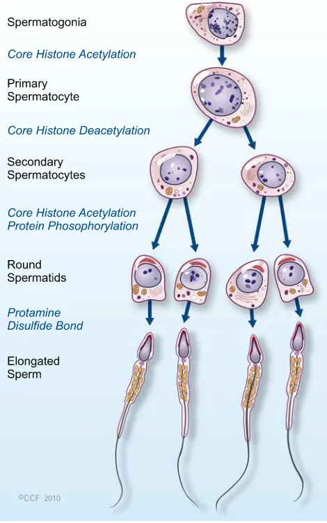
Epigenetic mechanisms regulate DNA accessibility through-out an organism’s lifetime as specific sets of genes are active at any stage of development. Each cell type has its own epige-netic signature that reflects the developmental history and environmental influences, and is ultimately reflected in the phenotype of the cell and organism. At the time of fertilization paternal genome delivered by the mature sperm has a haploid genome and is packaged densely with protamines, whereas maternal genome arrested at metaphase II is packaged with histones. Upon fertilization, protamines are rapidly replaced by histones and oocyte completes the second metaphase, releasing the polar body. The H3 and H4 histones that associ-ate with the passoci-aternal chromatin are more acetylassoci-ated than those present in the maternal chromatin [16,17].
spermatocyte stage. Histone methylation in spermatogenesis is carried out by the H3-K4 and H3-K9 methyltransferase.
It has been reported that hyperacetylation of histone H4 is associated with a histone-to-protamine exchange in haploid spermatids [31–33]. Recently, Govin et al. [34] reported that the double bromodomain-containing protein, BRDT (bro-modomain testis specific) binds hyperacetylated histone H4 before accumulating in condensed chromatin and helps in
organizing the spermatozoon’s genome by mediating a gen-eral histone acetylation-induced chromatin compaction and maintaining a differential histone acetylation of specific regions.
A factor named BORIS (brother of regulator of imprinted sites), specifically expressed in male gonads, could be di-rectly involved in the resetting of methylation marks during male germ cell differentiation [35]. The domains of BORIS Elongated
Spermatid
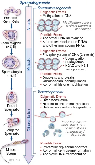
Spermatogenesis
Spermatocytogenesis
Spermiogenesis
Mitosis
Possible Errors
• Abnormal DNA methylation
• Altered expression of mRNAs and other non-coding RNAs
Epigenetic Events • Methylation of DNA
Modification occurs while structure is condensed
Transition occurs while structure is open; histones
removed and degraded Epigenetic Events
• Phosphorylation of DNA (2 events)
• Ubiquitylation
• Sumoylation
• H2AZ and H3.3 incorporation
Epigenetic Events • Hyperacytelation
• Histone to protamine transition
• Histone removal and degradation
Possible Errors
• Protamine replacement errors
• Abnormal centrosome formation
• Apoptotic DNA fragmentation
Possible Errors
• Double strand breaks
• Chromosomal nondisjunction
• Abnormal histone modification
Meiosis
Diff
erentiation
Mature Sperm Round Spermatid Spermatocyte
(I & II) Spermatogonia
(A & B) Primordial Germ Cells
2010
Fig. 1 Epigenetic events during spermatogenesis. In primodial germ cells (mitosis), DNA methylation occurs to set up the paternal specific imprints. Phosphorylation (in meiotic cell) occurs to assist in both recombination and XY body formation. Ubiquitylation, sumoylation and incorporation of H2AZ and H3.3 variants are all involved in XY body formation. Hyperacetylation occurs during spermiogenesis to assist in the
have the same 11 Zinc Finger as CTCF (CCCTC binding factor: a somatic regulator for expression of imprinted genes), which binds to specific target DNA sequences and plays an important role in the maintenance of differential methylation patterns in somatic cells [36]. CTCF is present in both somatic and germ cells whereas BORIS is expressed specifically in the male germ line. Studies [35, 37] have shown that BORIS is linked with both methylases mediating de novo methylation and demethylases mediating erasure of imprinting marks.
Paternal impact on early embryogenesis
A growing body of evidence suggests that both genetic and epigenetic abnormalities may contribute to idiopathic male infertility, which may affect the outcome of in vitro fertiliza-tion (IVF) [38]. Advanced maternal age is one of the obvious contributors to poor fecundity [39], but little is known about the effect of paternal age. We know that advanced paternal age is associated with decreased semen volume, sperm morphol-ogy, and sperm motility, but no significant reduction in sperm concentration [40]. A number of studies have documented age-dependent changes in the testis [41,42].
High DNA fragmentation is associated with diminished sperm count, motility, and morphology [42,43]. Increased DNA fragmentation also decreases fertilization and implan-tation rates. The influence of sperm DNA methylation on
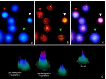
pregnancy was done on mice after using methylation deple-tion by use of 5-aza-deoxycytidine. This molecule is base analog that when incorporated into DNA decreases the level of DNA methylation varying the gene expression [43]. One of the main problems of solving or throwing some light about the actual role of DNA modifications on fertilization is the absence of reliable techniques which allow easy and reproducible analysis of the level of DNA modification in each gamete. A simple Sperm Chromatin Dispersion (SCD) based technology which offered stable results can be used to analyze the amount of methylated DNA residues and the level of DNA damage in each sperm (Fig.2). This technique consisted of using MABs against 5-Met-Citosine on a par-tially denatured DNA molecule once the SCD test was performed. The methodology is mainly based on previous results where the SCD and FISH were used to characterize the level of DNA damage and the presence of aneuploidies (CITA). Figure 2 shows a panel of some results where different levels of DNA methylation could be simultaneous-ly correlated with the level of sperm DNA in each sperm. In some cases DNA fragmentation fully correlates with an abnormal DNA molecule while in other cases this does not occurs. This points to the fact that probably methylation and its consequence are characteristically displayed by each sperm although its variations shall be associated to certain pathological cases. Interestingly, adaptation of this tech-nique may allow semiquantitative assesment of the level of methylation cell by cell.
Fig. 2 Sperm DNA fragmentation using the SCD test (Halosperm) (panela), combined with the
simultaneous visualization of sperm methylation (panelb) using anti-5metyl-C. Panel 2c
was produced after merginga
Histones are considered the best candidates for the trans-mission of epigenetic information because of their influence on the modification of chromatin structure, and access of transcriptional machinery to genes [44, 45]. The methyl-transferases facilitate gene silencing by mono-, di- or trime-thylation of lysine or arginine [3, 46]. It has yet to be determined whether modified histones play a crucial role in gene expression during early embryogenesis or if abnor-mal histone modifications in the sperm are associated with diminished embryo development. Alterations in methylation patterns can effect biallelic expression or repression of imprinted genes resulting in various pathologies [47]. Im-paired spermatogenesis has been associated with aberrant H4 acetylation [48]. H4 hyperacetylation was also observed in infertile men exhibiting Sertoli cell only (SCO) syn-drome. Sperm of patients with asthenozoospermia and ter-atozoospermia have decreased levels of DNA methylation [49]. Recently, researchers have begun to look at the contri-bution to early embryogenesis by spermatozoa beyond those of genetic factors. There is evidence of epigenetic contribu-tion to complex diseases.
Epigenetic changes important for male gametes
Gonadal sex determination and testis development occur between embryonic days 12 and 15 (E12 to E15) in the rat (after midgestation in the human) and are initiated by the differentiation of precursor Sertoli cells in response to the testis-determining factor SRY. Aggregation of the precursor Sertoli cells, PGCs, and migrating mesonephros cells (pre-cursor peritubular myoid cells) promotes testis morphogen-esis and cord formation. The fetal testis contains steroid receptors and is a target for endocrine hormones. The an-drogen receptor (AR) and estrogen receptor-b (ERb) are present in Sertoli cells as well as in precursor peritubular myoid cells and germ cells at the time of cord formation (E14). Although the testis does not produce steroids at this stage of development, estrogens and androgens have the ability to influence early testis cellular functions. Treatment with endocrine disruptors vinclozolin and methoxychlor, at a critical time during gonadal sex determination (E8 to E15 in the rat), promotes an adult testis phenotype with de-creased spermatogenic capacity and male infertility. This study shows that external factors can induce an epigenetic transgenerational phenotype through an apparent reprog-ramming of the male germ line [50]. It is not clear whether steroidal factors acting inappropriately at the time of gonad-al sex determination reprogram the germ line epigeneticgonad-ally (altered DNA methylation) to cause the transgenerational transmission of an altered phenotype or genetic trait.
Many epigenetic modifiers, including DNA methyltrans-ferases, histone-modification enzymes and their regulatory
proteins play essential roles in germ-cell development. Some of these are specifically expressed in germ cells whereas others are more widely expressed. The crucial roles of germ-cell-specific genes such as Dnmt3L and Prdm9 were revealed by conventional knockout studies [51–53]. A recent report showed that there are numerous intra- and inter-individual differences in DNA methylation in human sperm samples [54] which could contribute to phenotypic differences in the next generation. Furthermore, it has been reported that sperm samples from oligospermic patients often contain DNA-methylation defects at imprinted loci [55,56].
Epigenetics and protamine abnormalities
Marques et al. [54] suggested an association between aber-rant sperm epigenetic modifications and altered spermato-genesis. During differentiation of the male gamete, the genome undergoes major changes that not only affect the DNA sequence and genetic information by homologous recombination but also alter its nuclear structure and epige-netic information. One of the challenging issues is to under-stand how the specific nucleoprotamine/nucleohistone structure of the sperm nucleus conveys epigenetic informa-tion and how it controls early embryonic events.
Cho et al. [57] have shown that both protamines 1 and 2 are essential for sperm function, and the haploinsufficiency of either protamine 1 (P1) or protamine 2 (P2) results in a reduced amount of the respective protein. The phosphoryla-tion of protamines has also been shown to be very impor-tant. Indeed, mutation of the calmodulin-dependent protein kinase Camk4, which phosphorylates protamine 2, results in defective spermiogenesis and male sterility [58,59]. The P1/ P2 ratio in fertile men lies close to 1.0 [60–62] and ranges from 0.8 to 1.2 [60,61]. Perturbation of this ratio, in either direction, is characterized by poor semen quality, increased DNA damage, and decreased fertility [63–68].
of temporal uncoupling of transcription and translation dur-ing spermatogenesis [71].
The study of two relatively novel areas—sperm epige-netics and sperm transcriptome could be of particular inter-est in men with protamine expression abnormalities. Abnormal protamine incorporation into chromatin may af-fect transcription of other genes. For example, in mice, deregulation of protamines causes precocious chromatin condensation, transcription arrest, and spermatogenic failure [72]. The human sperm nucleus, which retains 10–15% of its original histone content, distributes it in a heterogeneous manner within the genome [73]. Various studies concluded that histones that are retained bind specific regions, to con-vey epigenetic information to the early embryo. If so, there are obvious and profound implications for sperm with ab-normal protamine replacement, and for the use of such sperm, or any immature sperm, for intracytoplasmic sperm injection (ICSI). Future research will classify and character-ize the role of retained histones throughout the sperm
genome in mature sperm from fertile men as well as in patients with known chromatin abnormalities.
Epigenetics and ART
In humans, the use of assisted reproductive technology (ART) has been shown to induce epigenetic alterations and to affect fetal growth and development. There is an open debate about the influence ART on certain pathol-ogies, especially those that are dependent on genomic imprinting such as the Beckwith-Wiedemann or Angel-man syndrome [74]. Possible epigenetic risks linked to ART techniques may result from either the use of sperm with incomplete reprogramming or from in vitro embryo procedures performed at a time of epigenetic reprogram-ming [75–78]. Loss of gene imprinting may occur during preimplantation under certain conditions of gamete han-dling [79]. Although epigenetic states are relatively sta-ble, it has been estimated that the loss of epigenetic control (epimutation) may be one or two orders of mag-nitude greater than that of somatic DNA mutation [80]. Recent reports from various studies have shown that epimutations not only lead to inappropriate expression of the affected gene but may also expose hidden genetic variation [81]. It is possible that some sub-fertile couples have a genetic predisposition to epigenetic instability, which makes their offspring more susceptible to epige-netic changes, independently of whether or not they are conceived by ART. Epimutations affecting imprints can arise during imprint erasure, imprint establishment or imprint maintenance. It has been suggested by animal model studies that loss-of-function mutations of DNA methyltransferases affect all imprinted domains as well as other chromosomal regions. For example, mutations of Dnmt3a (de-novo methylase) and maintenance methylase (Dnmt) could lead to loss of imprinting and embryonic lethality [82]. Another study has shown that targeted disruption of Dnmt3L in mice caused azoospermia in homozygous mutant males, and heterozygous progeny of homozygous females died before midgestation. Anoth-er study suggested that imprinting defects and subfAnoth-ertility can have a common and possibly genetic cause and that super-ovulation instead of ICSI may further increase the risk factor of conceiving a child with an imprinting defect. Based on this study and the study by Chang et al. [83] it is tempting to speculate that super-ovulation leads to the maturation of epigenetically imperfect oocytes that would not have developed without treatment and may disturb the process of DNA methylation in the oocyte. It is therefore suggested that imprinting errors can lead to spontaneous abortions. The influence of sperm DNA methylation on pregnancy was documented
in one study where 5-aza-deoxycytidine (5-azaC) was used to induce methylation depletion in mice. DNA methylation level decreases when 5-azaC (a base analog) is incorporated into DNA [84]. It has been proposed that the level of DNA methylation in human sperm could be linked to their ability to initiate pregnancy by assisted reproduction [85].
Epigenetics and testicular cancer
Testicular germ cells tumor (TGCT) represents approxi-mately 98% of all testicular neoplasms and is the most common malignancy among young males [86]. Epigenetic changes that deregulate gene expression are frequently ob-served during the development of cancer. The epigenetic equilibrium of the normal cell is disrupted during tumori-genesis. In human neoplasms, at least two types of DNA methylation defects are found: hypomethylation, character-ized by a global loss of 5-methylcytosine, and hypermethy-lation of regulatory regions of promoters, associated with the silencing of tumor suppressor genes. Hypomethylation was the first epigenetic abnormality to be identified in cancer cells [87–89]. Studies in mouse models have indicat-ed a causal relation between rindicat-educindicat-ed levels of 5-methylcytosine and tumor formation [90]. In contrast to the mere handful of oncogenes activated by DNA hypome-thylation, a long list of tumor suppressor genes is transcrip-tionally silenced by DNA hypermethylation in cancer cells. TGCTs are believed to arise from primordial germ cells (PGCs)—where DNA methylation and parental imprints are erased and totipotency is restored [19, 91, 92]. A genome-wide DNA methylation study using restriction landmark genome scanning (RLGS) showed that the ge-nome of seminomas was extensively hypomethylated and virtually completely devoid of CpG island hypermethylation [93]. In contrast, the nonseminoma group was less exten-sively hypomethylated and revealed variable CpG island hypermethylation levels, which were comparable with tumors of other tissues [94]. From embryonic studies in mice, a wave of demethylation immediately after fertiliza-tion has been shown to erase the majority of methylafertiliza-tion marks in the genome, with the exception of some imprinted genes and repeat sequences [92], leading to totipotency. High expression of the de novo methyltransferases DNMT3A and DNMT3B, as well as their homologue DNMT3L, is significantly associated with the embryonal carcinoma subtype [95]. The presumptive testis-specific chromatin regulator CTCFL (BORIS) and the pluripotency marker POU5F1 (OCT3/4) have recently been proposed to share properties with the cancer/testis associated genes in being hypermethylated in somatic tissue and hypomethy-lated in normal testis tissue [96,97].
The effect of epigenetic sperm abnormalities on early embryogenesis
Imprinting errors in the developing fetus have been identi-fied and shown to cause severe pathologies. Some studies have also suggested that the use of ART increases the risk of imprinting diseases. A study by Marques et al. [56] sug-gested that an increase in abnormal methylation of the H19 gene in oligospermic men is associated with Beckwith-Wiedemann syndrome. A decreased genome-wide methyla-tion pattern in sperm has also been identified with poor embryo quality in rats and decreased IVF pregnancy rates in humans [49]. Benchaib et al. [43] used 5-methyl-cytosine immunostaining as an indicator of genome-wide methyla-tion pattern in sperm. He showed that decreased global methylation in semen samples from normospermic men is related to a poor pregnancy outcome during IVF [85] sug-gesting that global methylation status independently affects embryogenesis. Mitchell et al. [98] found a correlation be-tween the frequency of P1 transcripts and pregnancy rates in men undergoing testicular sperm extraction (TESE) for ICSI. This could suggest that epigenetic regulation of DNA via nuclear packaging in the sperm is related to the function of the mature sperm. Current knowledge of genetic and epigenetic factors in sperm contributing to poor em-bryogenesis is limited. Both the complex path of sperm production and the delicate balance of epigenetic and genet-ic factors during sperm maturation contribute to the forma-tion of a mature sperm with the ability to fertilize an oocyte and contribute to the developing embryo. A defect at any step may manifest as male infertility.
Epigenetic regulation and nutrition
Epigenetic programming is tightly regulated, both tempo-rally and spatially, during fetal development and lactation [19,20,99]. Dietary supplementation with a methyl donor during pregnancy increases the proportion of pups carrying a methylated IAP (Intracisternal-A particles) sequence [100, 101]. There are many environmental and metabolic factors that can influence patterns of histone acetylation and DNA methylation, two major epigenetic marks. Metabolic factors influencing these epigenetic modifications include intranu-clear levels of acetyl-CoA for HAT activity, NAD+for Sir2 deacetylases, ATP for the deacetylation of chromatin sub-strates by at least some HDACs and methyl donors of SAM provided by the folate-methionine pathway. Once the spe-cific epigenetic patterns corresponding to ‘labile’ and
silencing, which may be associated with treatment failure [103,104].
Most of these studies have looked at modifications of the pattern of DNA methylation, as it is the easiest epigenetic mark to study. Nutrition during early development can in-fluence DNA methylation because one-carbon metabolism is dependent on dietary methyl donors and on co-factors such as methionine, choline, folic acid and vitamin B-12 [104]. The availability of dietary methyl donors and cofac-tors is therefore very critical during development. The epi-genetic change, caused by a decrease in DNMT1 activity, [105] can be prevented by folate supplementation [106]. Although not directly regulated by nutrition, maternal be-havior also programs the epigenetic regulation (DNA meth-ylation and histone acetmeth-ylation) of the gluco-corticoid receptor gene in the hippocampus and determines the stress responses of the offspring [107,108].
It was recently reported that a methyl-donor-deficient diet in postnatal life can permanently affect the expression of IGF2, resulting in growth retardation [109]. This suggests that the effects of nutrition are not only limited to the fetal stage but nutrition during postnatal development can also permanently alter the epigenetic regulation of imprinted genes. In humans, diet has been shown to affect the DNA methylation status of patients with hyperhomocysteinaemia. This disease is characterized by the accumulation of S-adenosylhomocysteine (an inhibitor of DNA methyltrans-ferases). The impact of diet, nutrients or drugs on early epigenetic programming must be seriously considered to achieve a directed epigenetic regulation in spermatogenesis.
The role of environmental factors in epigenetic modifications
Epigenetic modifications provide a putative link between the environment and alterations in gene expression that might lead to disease phenotype. An increasing body of evidence from animal studies support the role of environ-mental epigenetics in disease susceptibility. Environenviron-mental exposures to nutritional, chemical and physical factors have the potential to alter gene expression and therefore, modify adult disease susceptibility in various ways through changes in the epigenome. These genomic targets contain CpG islands and other DNA sequences, although in some cases the status of histone modifications in the same region, determine levels of gene expression.
Monozygotic twins provide an interesting model for studying the role of environmental factors in epigenetic modifications [110]. A large epigenetic study on monozy-gotic twins [111] recently showed that twins are epigeneti-cally concordant at birth in most cases, and that epigenetic differences (DNA methylation and histone modifications)
accumulate with age in monozygotic twins. Remarkably, the twins displaying the greatest epigenetic differences were found to be those who had lived together for the smallest amount of time. This finding underlines the relative impor-tance of environmental factors in addition to intrinsic factors.
Future perspectives
Whether common diseases have an epigenetic basis is still open to speculation, but if they do, this holds great promise for medicine. Knowledge of genetic and epigenetic modifi-cations of germ cells is necessary for the production of functional gametes and for overcoming infertility. Categori-zation of infertile men using a more detailed analysis of DNA methylation patterns might reveal a new level of reduced fertilization, implantation or pregnancy rates. Epi-genetic studies offer an important window to understanding the role of environmental interactions with the genome in causing disease, and in modulating those interactions to improve human health. Our increasing knowledge over last 10 years is beginning to be translated into new approaches to molecular diagnosis and targeted treatments across the clinical spectrum.
References
1. Waddington CH. Canalization of development and the inheri-tance of acquired characters. Nature. 1942;150:563–5.
2. Gilbert SF, Sarkar S. Embracing complexity: organicism for the 21st century. Dev Dyn. 2000;219(1):1–9. doi:10.1002/1097-0177 (2000) 9999:9999<::AID-DVDY1036>3.0.CO;2-A.
3. Li E. Chromatin modification and epigenetic reprogramming in mammalian development. Nat Rev Genet. 2002;3(9):662–73. doi:10.1038/nrg887nrg887.
4. Talbert PB, Henikoff S. Spreading of silent chromatin: inaction at a distance. Nat Rev Genet. 2006;7(10):793–803. doi:10.1038/ nrg1920.
5. Klose RJ, Bird AP. Genomic DNA methylation: the mark and its mediators. Trends Biochem Sci. 2006;31(2):89–97. doi:10.1016/ j.tibs.2005.12.008.
6. Richardson BC. Role of DNA methylation in the regulation of cell function: autoimmunity, aging and cancer. J Nutr. 2002;132(8 Suppl):2401S–5S.
7. Bestor T, Laudano A, Mattaliano R, Ingram V. Cloning and sequencing of a cDNA encoding DNA methyltransferase of mouse cells. The carboxyl-terminal domain of the mammalian enzymes is related to bacterial restriction methyltransferases. J Mol Biol. 1988;203(4):971–83.
8. Okano M, Xie S, Li E. Cloning and characterization of a family of novel mammalian DNA (cytosine-5) methyltransferases. Nat Genet. 1998;19(3):219–20. doi:10.1038/890.
10. Takai D, Jones PA. Comprehensive analysis of CpG islands in human chromosomes 21 and 22. Proc Natl Acad Sci U S A. 2002;99(6):3740–5. doi:10.1073/pnas.052410099052410099. 11. Issa JP. CpG-island methylation in aging and cancer. Curr Top
Microbiol Immunol. 2000;249:101–18.
12. Lachner M, O’Sullivan RJ, Jenuwein T. An epigenetic road map for histone lysine methylation. J Cell Sci. 2003;116(Pt 11):2117– 24. doi:10.1242/jcs.00493116/11/2117.
13. Tamaru H, Selker EU. A histone H3 methyltransferase controls DNA methylation in Neurospora crassa. Nature. 2001;414 (6861):277–83. doi:10.1038/3510450835104508.
14. Malagnac F, Bartee L, Bender J. An Arabidopsis SET domain protein required for maintenance but not establishment of DNA methylation. EMBO J. 2002;21(24):6842–52.
15. Tariq M, Saze H, Probst AV, Lichota J, Habu Y, Paszkowski J. Erasure of CpG methylation in Arabidopsis alters patterns of his-tone H3 methylation in heterochromatin. Proc Natl Acad Sci U S A. 2003;100(15):8823–7. doi:10.1073/pnas.14329391001432939100. 16. Adenot PG, Mercier Y, Renard JP, Thompson EM. Differential H4 acetylation of paternal and maternal chromatin precedes DNA replication and differential transcriptional activity in pronuclei of 1-cell mouse embryos. Development. 1997;124(22):4615–25. 17. Santos F, Hendrich B, Reik W, Dean W. Dynamic reprogramming of
DNA methylation in the early mouse embryo. Dev Biol. 2002;241 (1):172–82. doi:10.1006/dbio.2001.0501S0012160601905019. 18. Surani MA, Hayashi K, Hajkova P. Genetic and epigenetic
reg-ulators of pluripotency. Cell. 2007;128(4):747–62. doi:10.1016/j. cell.2007.02.010.
19. Morgan HD, Santos F, Green K, Dean W, Reik W. Epigenetic reprogramming in mammals. Hum Mol Genet. 2005;14(Spec No 1):R47–58. doi:10.1093/hmg/ddi114.
20. Allegrucci C, Thurston A, Lucas E, Young L. Epigenetics and the germline. Reproduction. 2005;129(2):137–49. doi:10.1530/ rep.1.00360.
21. Kimmins S, Sassone-Corsi P. Chromatin remodelling and epige-netic features of germ cells. Nature. 2005;434(7033):583–9. doi:10.1038/nature03368.
22. McLaren A. Primordial germ cells in the mouse. Dev Biol. 2003;262(1):1–15.
23. Seki Y, Hayashi K, Itoh K, Mizugaki M, Saitou M, Matsui Y. Extensive and orderly reprogramming of genome-wide chromatin modifications associated with specification and early develop-ment of germ cells in mice. Dev Biol. 2005;278(2):440–58. doi:10.1016/j.ydbio.2004.11.025.
24. Payne C, Braun RE. Histone lysine trimethylation exhibits a distinct perinuclear distribution in Plzf-expressing spermatogonia. Dev Biol. 2006;293(2):461–72. doi:10.1016/j.ydbio.2006.02.013. 25. Peters AH, O’Carroll D, Scherthan H, Mechtler K, Sauer S,
Schofer C, Weipoltshammer K, Pagani M, Lachner M, Kohlmaier A, Opravil S, Doyle M, Sibilia M, Jenuwein T. Loss of the Suv39h histone methyltransferases impairs mammalian het-erochromatin and genome stability. Cell. 2001;107(3):323– 37.
26. Namekawa SH, Park PJ, Zhang LF, Shima JE, McCarrey JR, Griswold MD, Lee JT. Postmeiotic sex chromatin in the male germline of mice. Curr Biol. 2006;16(7):660–7. doi:10.1016/j. cub.2006.01.066.
27. Turner JM, Mahadevaiah SK, Ellis PJ, Mitchell MJ, Burgoyne PS. Pachytene asynapsis drives meiotic sex chromosome inactivation and leads to substantial postmeiotic repression in spermatids. Dev Cell. 2006;10(4):521–9. doi:10.1016/j.devcel.2006.02.009. 28. Martianov I, Brancorsini S, Catena R, Gansmuller A, Kotaja N,
Parvinen M, Sassone-Corsi P, Davidson I. Polar nuclear localization of H1T2, a histone H1 variant, required for spermatid elongation and DNA condensation during spermiogenesis. Proc Natl Acad Sci U S A. 2005;102(8):2808–13. doi:10.1073/pnas.0406060102.
29. Rousseaux S, Caron C, Govin J, Lestrat C, Faure AK, Khochbin S. Establishment of male-specific epigenetic information. Gene. 2005;345(2):139–53. doi:10.1016/j.gene.2004.12.004.
30. Oakes CC, La Salle S, Smiraglia DJ, Robaire B, Trasler JM. A unique configuration of genome-wide DNA methylation patterns in the testis. Proc Natl Acad Sci U S A. 2007;104(1):228–33. doi:10.1073/pnas.0607521104.
31. Meistrich ML, Trostle-Weige PK, Lin R, Bhatnagar YM, Allis CD. Highly acetylated H4 is associated with histone displacement in rat spermatids. Mol Reprod Dev. 1992;31(3):170–81. doi:10.1002/mrd.1080310303.
32. Hazzouri M, Pivot-Pajot C, Faure AK, Usson Y, Pelletier R, Sele B, Khochbin S, Rousseaux S. Regulated hyperacetylation of core histones during mouse spermatogenesis: involvement of histone deacetylases. Eur J Cell Biol. 2000;79(12):950–60.
33. Sonnack V, Failing K, Bergmann M, Steger K. Expression of hyperacetylated histone H4 during normal and impaired human spermatogenesis. Andrologia. 2002;34(6):384–90.
34. Govin J, Lestrat C, Caron C, Pivot-Pajot C, Rousseaux S, Khochbin S. Histone acetylation-mediated chromatin compaction during mouse spermatogenesis. Ernst Schering Res Found Workshop. 2006;57:155–72.
35. Klenova EM, Morse 3rd HC, Ohlsson R, Lobanenkov VV. The novel BORIS + CTCF gene family is uniquely involved in the epigenetics of normal biology and cancer. Semin Cancer Biol. 2002;12(5):399–414.
36. Schoenherr CJ, Levorse JM, Tilghman SM. CTCF maintains differential methylation at the Igf2/H19 locus. Nat Genet. 2003;33(1):66–9. doi:10.1038/ng1057ng1057.
37. Loukinov DI, Pugacheva E, Vatolin S, Pack SD, Moon H, Chernukhin I, Mannan P, Larsson E, Kanduri C, Vostrov AA, Cui H, Niemitz EL, Rasko JE, Docquier FM, Kistler M, Breen JJ, Zhuang Z, Quitschke WW, Renkawitz R, Klenova EM, Feinberg AP, Ohlsson R, Morse 3rd HC, Lobanenkov VV. BORIS, a novel male germ-line-specific protein associ-ated with epigenetic reprogramming events, shares the same 11-zinc-finger domain with CTCF, the insulator protein in-volved in reading imprinting marks in the soma. Proc Natl Acad Sci U S A. 2002;99(10):6806–11. doi:10.1073/ pnas.09212369999/10/6806.
38. Emery BR, Carrell DT. The effect of epigenetic sperm abnormal-ities on early embryogenesis. Asian J Androl. 2006;8(2):131–42. doi:10.1111/j.1745-7262.2006.00127.x.
39. Hassold T, Hunt P. To err (meiotically) is human: the genesis of human aneuploidy. Nat Rev Genet. 2001;2(4):280–91. doi:10.1038/3506606535066065.
40. Kidd SA, Eskenazi B, Wyrobek AJ. Effects of male age on semen quality and fertility: a review of the literature. Fertil Steril. 2001;75(2):237–48.
41. Kuhnert B, Nieschlag E. Reproductive functions of the ageing male. Hum Reprod Update. 2004;10(4):327–39. doi:10.1093/ humupd/dmh030dmh030.
42. Wyrobek AJ, Eskenazi B, Young S, Arnheim N, Tiemann-Boege I, Jabs EW, Glaser RL, Pearson FS, Evenson D. Advancing age has differential effects on DNA damage, chromatin integrity, gene mutations, and aneuploidies in sperm. Proc Natl Acad Sci U S A. 2006;103(25):9601–6. doi:10.1073/pnas.0506468103.
43. Benchaib M, Ajina M, Lornage J, Niveleau A, Durand P, Guerin JF. Quantitation by image analysis of global DNA methylation in human spermatozoa and its prognostic value in in vitro fertiliza-tion: a preliminary study. Fertil Steril. 2003;80(4):947–53. 44. Ooi SL, Henikoff S. Germline histone dynamics and epigenetics.
Curr Opin Cell Biol. 2007;19(3):257–65. doi:10.1016/j. ceb.2007.04.015.
46. Biermann K, Steger K. Epigenetics in male germ cells. J Androl. 2007;28(4):466–80. doi:10.2164/jandrol.106.002048.
47. Schaefer CB, Ooi SK, Bestor TH, Bourc’his D. Epigenetic deci-sions in mammalian germ cells. Science. 2007;316(5823):398–9. doi:10.1126/science.1137544.
48. Faure AK, Pivot-Pajot C, Kerjean A, Hazzouri M, Pelletier R, Peoc’h M, Sele B, Khochbin S, Rousseaux S. Misregulation of histone acetylation in Sertoli cell-only syndrome and testicular cancer. Mol Hum Reprod. 2003;9(12):757–63.
49. Benchaib M, Braun V, Lornage J, Hadj S, Salle B, Lejeune H, Guerin JF. Sperm DNA fragmentation decreases the pregnancy rate in an assisted reproductive technique. Hum Reprod. 2003;18 (5):1023–8.
50. Anway MD, Cupp AS, Uzumcu M, Skinner MK. Epigenetic trans-generational actions of endocrine disruptors and male fertility. Sci-ence. 2005;308(5727):1466–9. doi:10.1126/science.1108190. 51. Bourc’his D, Xu GL, Lin CS, Bollman B, Bestor TH. Dnmt3L
and the establishment of maternal genomic imprints. Science. 2001;294(5551):2536–9. doi:10.1126/science.10658481065848. 52. Hata K, Okano M, Lei H, Li E. Dnmt3L cooperates with the
Dnmt3 family of de novo DNA methyltransferases to establish maternal imprints in mice. Development. 2002;129(8):1983–93. 53. Hayashi K, Yoshida K, Matsui Y. A histone H3 methyltransferase
controls epigenetic events required for meiotic prophase. Nature. 2005;438(7066):374–8. doi:10.1038/nature04112.
54. Flanagan JM, Popendikyte V, Pozdniakovaite N, Sobolev M, Assadzadeh A, Schumacher A, Zangeneh M, Lau L, Virtanen C, Wang SC, Petronis A. Intra- and interindividual epigenetic variation in human germ cells. Am J Hum Genet. 2006;79(1):67– 84. doi:10.1086/504729.
55. Kobayashi H, Sato A, Otsu E, Hiura H, Tomatsu C, Utsunomiya T, Sasaki H, Yaegashi N, Arima T. Aberrant DNA methylation of imprinted loci in sperm from oligospermic patients. Hum Mol Genet. 2007;16(21):2542–51. doi:10.1093/hmg/ddm187. 56. Marques CJ, Carvalho F, Sousa M, Barros A. Genomic
imprint-ing in disruptive spermatogenesis. Lancet. 2004;363 (9422):1700–2. doi: 10.1016/S0140-6736(04)16256-9S0140-6736(04)16256-9.
57. Cho C, Willis WD, Goulding EH, Jung-Ha H, Choi YC, Hecht NB, Eddy EM. Haploinsufficiency of protamine-1 or -2 causes infertility in mice. Nat Genet. 2001;28(1):82–6. doi:10.1038/ 8831388313.
58. Wu JY, Means AR. Ca(2+
)/calmodulin-dependent protein kinase IV is expressed in spermatids and targeted to chromatin and the nuclear matrix. J Biol Chem. 2000;275(11):7994–9.
59. Wu JY, Ribar TJ, Cummings DE, Burton KA, McKnight GS, Means AR. Spermiogenesis and exchange of basic nuclear pro-teins are impaired in male germ cells lacking Camk4. Nat Genet. 2000;25(4):448–52. doi:10.1038/78153.
60. Balhorn R, Cosman M, Thornton K, Krishnan VV, Corzett M, Bench G, Kramer C, Lee IV J, Hud NV, Allen M, Priety M, Meyer-IIse W, Brown J, Kirz J, Zhang X, Bradbury E, Maki G, Braun R, Breen W. Protamine mediated condensation of DNA in mammalian sperm. In: Gagnon C, editor. The male gamete: from basic knowledge to clinical applications. Vienna: Cache River Press; 1999. p. 55–70.
61. Carrell DT, Liu L. Altered protamine 2 expression is uncommon in donors of known fertility, but common among men with poor fertilizing capacity, and may reflect other abnormalities of sper-miogenesis. J Androl. 2001;22(4):604–10.
62. Corzett M, Mazrimas J, Balhorn R. Protamine 1: protamine 2 stoichiometry in the sperm of eutherian mammals. Mol Reprod Dev. 2002;61(4):519–27. doi:10.1002/mrd.10105.
63. Chevaillier P, Mauro N, Feneux D, Jouannet P, David G. Anom-alous protein complement of sperm nuclei in some infertile men. Lancet. 1987;2(8562):806–7.
64. Balhorn R, Reed S, Tanphaichitr N. Aberrant protamine 1/prot-amine 2 ratios in sperm of infertile human males. Experientia. 1988;44(1):52–5.
65. Belokopytova IA, Kostyleva EI, Tomilin AN, Vorob’ev VI. Hu-man male infertility may be due to a decrease of the protamine P2 content in sperm chromatin. Mol Reprod Dev. 1993;34(1):53–7. doi:10.1002/mrd.1080340109.
66. Carrell DT, Emery BR, Liu L. Characterization of aneuploidy rates, protamine levels, ultrastructure, and functional ability of round-headed sperm from two siblings and implications for intra-cytoplasmic sperm injection. Fertil Steril. 1999;71(3):511–6. 67. Razavi S, Nasr-Esfahani MH, Mardani M, Mafi A, Moghdam A.
Effect of human sperm chromatin anomalies on fertilization out-come post-ICSI. Andrologia. 2003;35(4):238–43.
68. Aoki VW, Liu L, Carrell DT. Identification and evaluation of a novel sperm protamine abnormality in a population of infertile males. Hum Reprod. 2005;20(5):1298–306. doi:10.1093/humrep/deh798. 69. Weber M, Hellmann I, Stadler MB, Ramos L, Paabo S, Rebhan
M, Schubeler D. Distribution, silencing potential and evolution-ary impact of promoter DNA methylation in the human genome. Nat Genet. 2007;39(4):457–66. doi:10.1038/ng1990.
70. Baarends WM, Hoogerbrugge JW, Roest HP, Ooms M, Vreeburg J, Hoeijmakers JH, Grootegoed JA. Histone ubiquitination and chromatin remodeling in mouse spermatogenesis. Dev Biol. 1999;207(2):322–33. doi:10.1006/dbio.1998.9155.
71. Carrell DT, Emery BR, Hammoud S. Altered protamine expression and diminished spermatogenesis: what is the link? Hum Reprod Update. 2007;13(3):313–27. doi:10.1093/humupd/dml057. 72. Kleene KC. Patterns, mechanisms, and functions of translation
regulation in mammalian spermatogenic cells. Cytogenet Genome Res. 2003;103(3–4):217–24. doi:10.1159/00007680776807. 73. Gatewood JM, Cook GR, Balhorn R, Bradbury EM, Schmid CW.
Sequence-specific packaging of DNA in human sperm chromatin. Science. 1987;236(4804):962–4.
74. Gosden R, Trasler J, Lucifero D, Faddy M. Rare congenital disor-ders, imprinted genes, and assisted reproductive technology. Lancet. 2003;361(9373):1975–7. doi:10.1016/S0140-6736(03)13592-1. 75. Lucifero D, Mann MR, Bartolomei MS, Trasler JM.
Gene-specific timing and epigenetic memory in oocyte imprinting. Hum Mol Genet. 2004;13(8):839–49. doi:10.1093/hmg/ ddh104ddh104.
76. Niemitz EL, Feinberg AP. Epigenetics and assisted reproductive technology: a call for investigation. Am J Hum Genet. 2004;74 (4):599–609. doi:10.1086/382897S0002-9297(07)61887-4. 77. Thompson JR, Williams CJ. Genomic imprinting and assisted
reproductive technology: connections and potential risks. Semin Reprod Med. 2005;23(3):285–95. doi:10.1055/s-2005-872457. 78. Horsthemke B, Buiting K. Imprinting defects on human
chromo-some 15. Cytogenet Genome Res. 2006;113(1–4):292–9. doi:10.1159/000090844.
79. Mann MR, Lee SS, Doherty AS, Verona RI, Nolen LD, Schultz RM, Bartolomei MS. Selective loss of imprinting in the placenta following preimplantation development in culture. Development. 2004;131(15):3727–35. doi:10.1242/dev.01241dev.01241. 80. Bennett-Baker PE, Wilkowski J, Burke DT. Age-associated
acti-vation of epigenetically repressed genes in the mouse. Genetics. 2003;165(4):2055–62.
81. Sollars V, Lu X, Xiao L, Wang X, Garfinkel MD, Ruden DM. Evidence for an epigenetic mechanism by which Hsp90 acts as a capacitor for morphological evolution. Nat Genet. 2003;33 (1):70–4. doi:10.1038/ng1067ng1067.
assisted reproductive technology: a case series of 19 patients. F e r t i l S t e r i l . 2 0 0 5 ; 8 3 ( 2 ) : 3 4 9–5 4 . d o i :1 0 . 1 0 1 6 / j . fertnstert.2004.07.964.
84. Kelly TL, Li E, Trasler JM. 5-aza-2′-deoxycytidine induces alter-ations in murine spermatogenesis and pregnancy outcome. J Androl. 2003;24(6):822–30.
85. Benchaib M, Braun V, Ressnikof D, Lornage J, Durand P, Niveleau A, Guerin JF. Influence of global sperm DNA methylation on IVF results. Hum Reprod. 2005;20(3):768–73. doi:10.1093/humrep/ deh684.
86. Bosl GJ, Motzer RJ. Testicular germ-cell cancer. N Engl J Med. 1997;337(4):242–53. doi:10.1056/NEJM199707243370406. 87. Feinberg AP, Tycko B. The history of cancer epigenetics. Nat Rev
Cancer. 2004;4(2):143–53. doi:10.1038/nrc1279nrc1279. 88. Feinberg AP, Vogelstein B. Hypomethylation distinguishes genes
of some human cancers from their normal counterparts. Nature. 1983;301(5895):89–92.
89. Ehrlich M. The controversial denouement of vertebrate DNA methylation research. Biochemistry (Mosc). 2005;70(5):568–75. 90. Gaudet F, Hodgson JG, Eden A, Jackson-Grusby L, Dausman J, Gray JW, Leonhardt H, Jaenisch R. Induction of tumors in mice by genomic hypomethylation. Science. 2003;300(5618):489–92. doi:10.1126/science.1083558300/5618/489.
91. Bestor TH. Cytosine methylation and the unequal developmental potentials of the oocyte and sperm genomes. Am J Hum Genet. 1998;62(6):1269–73. doi:10.1086/301891.
92. Reik W, Dean W, Walter J. Epigenetic reprogramming in mam-malian development. Science. 2001;293(5532):1089–93. doi:10.1126/science.1063443293/5532/1089.
93. Lind GE, Skotheim RI, Lothe RA. The epigenome of testicular germ cell tumors. APMIS. 2007;115(10):1147–60. doi:10.1111/ j.1600-0463.2007.apm_660.xml.x.
94. Smiraglia DJ, Szymanska J, Kraggerud SM, Lothe RA, Peltomaki P, Plass C. Distinct epigenetic phenotypes in seminomatous and nonseminomatous testicular germ cell tumors. Oncogene. 2002;21 (24):3909–16. doi:10.1038/sj.onc.1205488.
95. Almstrup K, Hoei-Hansen CE, Nielsen JE, Wirkner U, Ansorge W, Skakkebaek NE, Rajpert-De Meyts E, Leffers H. Genome-wide gene expression profiling of testicular carcinoma in situ progression into overt tumours. Br J Cancer. 2005;92(10):1934– 41. doi:10.1038/sj.bjc.6602560.
96. Hoffmann MJ, Muller M, Engers R, Schulz WA. Epigenetic control of CTCFL/BORIS and OCT4 expression in urogenital malignancies. Biochem Pharmacol. 2006;72(11):1577–88. doi:10.1016/j.bcp.2006.06.020.
97. de Jong J, Looijenga LH. Stem cell marker OCT3/4 in tumor biology and germ cell tumor diagnostics: history and future. Crit Rev Oncog. 2006;12(3–4):171–203.
98. Mitchell V, Steger K, Marchetti C, Herbaut JC, Devos P, Rigot JM. Cellular expression of protamine 1 and 2 transcripts in testicular spermatids from azoospermic men submitted to TESE-ICSI. Mol Hum Reprod. 2005;11(5):373–9. doi:10.1093/molehr/gah169. 99. Lee J, Inoue K, Ono R, Ogonuki N, Kohda T, Kaneko-Ishino T,
Ogura A, Ishino F. Erasing genomic imprinting memory in mouse
clone embryos produced from day 11.5 primordial germ cells. Development. 2002;129(8):1807–17.
100. Rakyan VK, Chong S, Champ ME, Cuthbert PC, Morgan HD, Luu KV, Whitelaw E. Transgenerational inheritance of epigenetic states at the murine Axin(Fu) allele occurs after maternal and paternal transmission. Proc Natl Acad Sci U S A. 2003;100 (5):2538–43. doi:10.1073/pnas.04367761000436776100. 101. Waterland RA, Jirtle RL. Early nutrition, epigenetic changes at
transposons and imprinted genes, and enhanced susceptibility to adult chronic diseases. Nutrition. 2004;20(1):63–8.
102. Egger G, Liang G, Aparicio A, Jones PA. Epigenetics in human disease and prospects for epigenetic therapy. Nature. 2004;429:457– 63.
103. Egger G, Liang G, Aparicio A, Jones PA. Epigenetics in human disease and prospects for epigenetic therapy. Nature. 2004;429 (6990):457–63. doi:10.1038/nature02625nature02625.
104. MacLennan NK, James SJ, Melnyk S, Piroozi A, Jernigan S, Hsu JL, Janke SM, Pham TD, Lane RH. Uteroplacental insufficiency alters DNA methylation, one-carbon metabolism, and histone acetylation in IUGR rats. Physiol Genomics. 2004;18(1):43–50. doi:10.1152/physiolgenomics.00042.200400042.2004.
105. Lillycrop KA, Slater-Jefferies JL, Hanson MA, Godfrey KM, Jack-son AA, Burdge GC. Induction of altered epigenetic regulation of the hepatic glucocorticoid receptor in the offspring of rats fed a protein-restricted diet during pregnancy suggests that reduced DNA methyltransferase-1 expression is involved in impaired DNA meth-ylation and changes in histone modifications. Br J Nutr. 2007;97 (6):1064–73. doi:10.1017/S000711450769196X.
106. Lillycrop KA, Phillips ES, Jackson AA, Hanson MA, Burdge GC. Dietary protein restriction of pregnant rats induces and folic acid supplementation prevents epigenetic modification of hepatic gene expression in the offspring. J Nutr. 2005;135(6):1382–6. 107. Weaver IC, Cervoni N, Champagne FA, D’Alessio AC, Sharma
S, Seckl JR, Dymov S, Szyf M, Meaney MJ. Epigenetic program-ming by maternal behavior. Nat Neurosci. 2004;7(8):847–54. doi:10.1038/nn1276nn1276.
108. Meaney MJ, Szyf M, Seckl JR. Epigenetic mechanisms of peri-natal programming of hypothalamic-pituitary-adrenal function and health. Trends Mol Med. 2007;13(7):269–77. doi:10.1016/j. molmed.2007.05.003.
109. Waterland RA, Lin JR, Smith CA, Jirtle RL. Post-weaning diet affects genomic imprinting at the insulin-like growth factor 2 (Igf2) locus. Hum Mol Genet. 2006;15(5):705–16. doi:10.1093/ hmg/ddi484.
110. Poulsen P, Esteller M, Vaag A, Fraga MF. The epigenetic basis of twin discordance in age-related diseases. Pediatr Res. 2007;61(5 Pt 2):38R–42R. doi:10.1203/pdr.0b013e31803c7b98.
111. Fraga MF, Ballestar E, Paz MF, Ropero S, Setien F, Ballestar ML, Heine-Suner D, Cigudosa JC, Urioste M, Benitez J, Boix-Chornet M, Sanchez-Aguilera A, Ling C, Carlsson E, Poulsen P, Vaag A, Stephan Z, Spector TD, Wu YZ, Plass C, Esteller M. Epigenetic differences arise during the lifetime of monozygotic twins. Proc Natl Acad Sci U S A. 2005;102(30):10604–9. doi:10.1073/ pnas.0500398102.