* Corresponding author. Tel. 081360114230
Fax : +(62)(651) 53498; Email: [email protected]
PREPARATION OF CHITOSAN-GOLD NANOPARTICLES:
PART 2. THE ROLE OF CHITOSAN
Adlim
1,*and Mohamad Abu Bakar
2 1Jurusan Kimia FKIP Universitas Syiah Kuala Darussalam, Banda Aceh, 23111 Indonesia 2
School of Chemical Sciences, Universiti Sains Malaysia, 11800 Minden, Penang, Malaysia
Received 30 April 2008; Accepted 22 May 2008
ABSTRACT
Colloidal gold nanoparticles prepared by employing chitosan as the stabilizer in solvent of methanol-acetic acid solution were stable for months without precipitation. The mole ratio of chitosan-gold ions of 5:1 – 30:1 gave dispersed and fine gold particles in range of 9.4-10.4 nm. Gold reduction in chitosan matrix was faster at higher chitosan concentration, and molar ratio of chi : Au, from 5:1 to 40:1. Higher acidity of acetic acid (pH 2-6) led to faster reduction of gold ions. The intensity of gold metal colloid plasmon band increased at higher concentration of acetic acid. Chitosan functioned both as a stabilizer and a reducing agent for gold ions. Gold colloidal particles immobilized on chitosan coated TiO2 as the solid support gave more dispersed and smaller particles (4.6 nm) compared with gold particles supported on TiO2 without chitosan coating.
Keywords: gold nanoparticles, chitosan, chitosan coated TiO2
INTRODUCTION
In previous article, the effect of reducing technique on the dispersion and size of gold particles stabilized by chitosan have been discussed [1]. In this paper the role of chitosan on preparation for gold nanoparticles will be elaborated. This study includes the investigation of the dual role of chitosan in the catalyst preparation, as a stabilizer and a reducing agent. Interaction of gold with chitosan has interesting feature since gold ions are easily reduced while chitosan contains some hydroxyl groups which are potential as reducing agent for gold ions as known for reducing sugar in Fehling test. Gold metal is stable and chitosan is a biomolecules, both have potential uses in biological systems. Most of colloidal metal nanoparticles were fabricated using synthetic polymers or chemicals such as sodium alkyl sulphate, PVA (polyvinyl alcohol), ethylene glycol, DMF (N,N’-Dimethyl formamide), alkanethiols as the stabilizer [2-11]. Although gold nanoparticles have been immobilized in solid supports employing synthetic polymer as a template, chitosan –coated TiO2 has not
been much explored as a solid support of gold nanoparticles [5]. This paper will also discuss the use of chitosan-coated TiO2 for gold nanoparticles
immobilization.
EXPERIMENTAL SECTION
Material
The following commercially available materials were used without further purification: TiO2 (anatase,
99.9%, Aldrich, USA), N,N’-Dimethyl formamide (99.8%, Merck, USA), polyvinyl alcohol (MW 22000, 98%, BDH), sodium borohydride (95 %, Reidel de Haen), chitosan of
medium molecular weight (400000, Fluka, Switzerland), acetic acid 99.8% (BDH, England), HAuCl4.3H2O (99.5%, Sigma, USA) and methanol
(Systerm, A.C.S certified grade, Malaysia).
Equipment
TEM (transmission electron microscope) model Philip CM 12 was used to record micrographs which is used for determination of particle size and size distribution. The sample preparation technique has been verified in the previous report [12]. The average particle size and size distribution were obtained from > 300 particles.
Infra-red spectrophotometer (FTIR) Model : Perkin Elmer FT-IR 2000 was employed to verify the change of the functional groups of chitosan. The dried chitosan-gold sol was placed in KBr window and recorded as the FTIR spectrum.
Procedure
The effect of stabilizer
Gold ions were reduced in aqueous formic acid (for) and citrict acid (cit) or acetic acid with and without chitosan as stabilizer. Refluxing in methanol, addition of NaBH4 or photo-irradiation/microwave irradiation
were employed as reducing agent and reducing technique for the gold ions. Molar ratio of chitosan:metal was designated as 5:1, 10:1 or 15:1 etc.
Preparation of chitosan-stabilized gold colloids (chi-Au)
The chitosan-stabilized metal colloids were prepared with modification of the previously reported methods [12] and described in our previous article [1].
The effect of chitosan concentration on gold reduction was studied to compare the rate of gold reduction at various chitosan-Au molar ratio (5:1 – 40:1). The solution were photo-irradiated and the reduction was followed by examining the absorbance of the chitosan-gold plasmon band (525-550 nm) by periodically sampling the solution, and cold down in an ice bath before UV-Vis spectrophotometry analyses. Similar characterization method was verified to study the effect acetic acid concentration on gold reduction. One mL of chitosan solution (3.7 x 10⁻⁵ mol) and 1 mL of gold ion solution (7.4 x 10⁻⁶ mol) were dissolved into 6 various concentrations of acetic acid ( from 0.026 M to 0.0955 M) at 36 mL of each to obtain the total volume of 38 mL. Microwave irradiation method and pH effect was studied by adjusting the pH of the solution to 2.8, 3.5, 4, 4.5, and 5.5 with addition of NaOH solution. All solutions were irradiated in a microwave oven for 240 seconds and the absorbance of each of the solution were recorded.
Absorption of gold on chitosan beads
Chitosan solution was prepared by dissolving chitosan flake in aqueous acetic acid. Chitosan beads were obtained by neutralizing chitosan solution with sodium hydroxide [13]. Chitosan beads were suspended in distilled water at various pH (2-5.5). Then, certain amount of gold (III) ion was mixed with the suspension for 30 min. The amount of gold absorbed on the beads was calculated from the remaining concentration of gold ion in the solution as determined with atomic absorption spectrometry (AAS).
Chitosan coated TiO2 as a support for Au
nanoparticles
Preparation of Au particles supported on chitosan-coated TiO2 (TiO2-chi-Au) was carried out by
modification of a previously reported method [11]. A 0.2080 g of chitosan was dissolved in 346 mL of aqueous acetic acid and diluted with 300 mL of methanol. To avoid high viscosity of the chitosan solution, the chitosan concentration was set to 0.6 mg mL-1 of aqueous acetic acid. The solution was stirred until a clear solution was obtained. 10 g of TiO2
(anatase) was suspended in 200 mL of aqueous acetic acid-methanol. The slurry was added dropwise to the chitosan solution with vigorous stirring. The mixture was neutralized by addition of sodium hydroxide solution and the precipitate was filtered and washed with distilled water until neutral. The solid was dried in an oven at 50oC overnight and the dried solid of TiO2–
chi was ground and dried again in an oven at 110 oC for 1 h.
Typically, 1.000 g of TiO2–chi was suspended in
100 mL of methanol-water (1:1 v/v) with stirring. Exactly 0.1000 g (2.54 x 10⁻⁴ mol) of HAuCl4.3H2O was
dissolved in distilled water and added into the chi-TiO2
suspension. The solution was stirred vigorously at room temperature for 24 h. The TiO2-chi-Au was
reduced either by reflux, photo-irradiation, or addition of NaBH4. The residue was filtered and washed several
times with water and subsequently with methanol, dried in an oven at 50 oC for 24 h and at 110 oC for 1 h. The solid residue was found to have about 2% of gold based on AAS analysis.
RESULT AND DISCUSSION
The effect of Stabilizers
Ethylene glycol, DMF (N,N’-Dimethyl formamide) or aqueous PVA where used as stabilizers and reducing agents for gold ions since these compounds
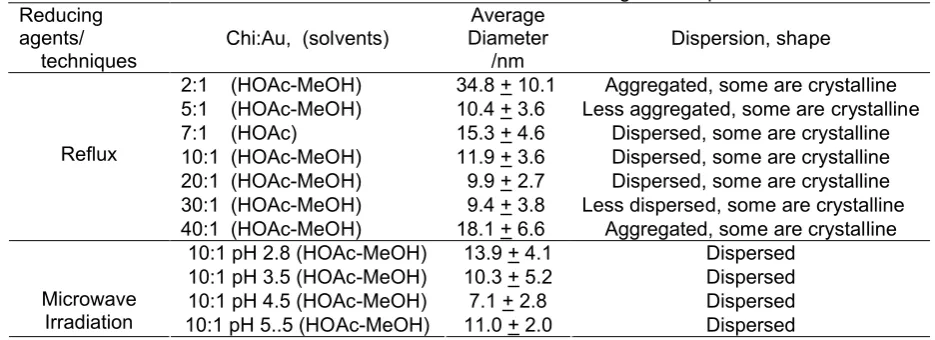
Table 1. Size and distribution of chitosan-stabilized gold nanoparticles Reducing
agents/ techniques
Chi:Au, (solvents)
Average Diameter
/nm
Dispersion, shape
2:1 (HOAc-MeOH) 34.8 + 10.1 Aggregated, some are crystalline 5:1 (HOAc-MeOH) 10.4 + 3.6 Less aggregated, some are crystalline
7:1 (HOAc) 15.3 + 4.6 Dispersed, some are crystalline
10:1 (HOAc-MeOH) 11.9 + 3.6 Dispersed, some are crystalline 20:1 (HOAc-MeOH) 9.9 + 2.7 Dispersed, some are crystalline 30:1 (HOAc-MeOH) 9.4 + 3.8 Less dispersed, some are crystalline Reflux
40:1 (HOAc-MeOH) 18.1 + 6.6 Aggregated, some are crystalline
10:1 pH 2.8 (HOAc-MeOH) 13.9 + 4.1 Dispersed
10:1 pH 3.5 (HOAc-MeOH) 10.3 + 5.2 Dispersed
10:1 pH 4.5 (HOAc-MeOH) 7.1 + 2.8 Dispersed
Microwave
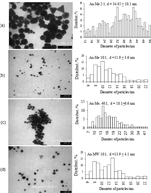
Fig. 1. TEM images of chi-Au prepared in methanol (Me); Chi:metal mol ratio, (a) 2 :1 (reflux), (b) 10 :1 (reflux), (c) 40 :1 (reflux) (d) 10 :1 prepared with microwave irradiation (MW)
Au-Me 10:1, d =11.9 + 3.6 nm
0 5 10 15 20
6
8
1
0
1
2
1
4
1
6
1
8
2
0
2
2
Diameter of particles/nm
D
is
tr
ib
u
t.
/%
Au-Me 2:1, d = 34.85 + 10.1 nm
0 1 2 3 4 5 6
1
2 16 20 24 28 23 36 40 44 48 54
Diameter of particles/nm
D
is
tr
ib
u
t.
/%
Au-Me, 40:1, d = 18.1+6.6 nm
0
5
10
15
7
1
0
1
3
1
6
1
9
2
2
2
7
3
0
3
4
4
1
Diamter of particles/nm
D
is
tr
ib
u
t.
/%
Au-MW 10:1, d =13.9 + 4.1 nm
0 5 10 15 20
6
8
1
0
1
2
1
4
1
6
1
8
2
0
2
2
Diameter of particles/nm
D
is
tr
ib
u
t.
/%
(a)
(b)
(c)
0.0
contain hydroxyl or amide groups which were potential to reduce and stabilize gold ions. These solutions were refluxed without addition of acetic acid, methanol and chitosan. After reflux, the solution color changed to purple, indicating gold ions have been reduced to gold metal or gold sol. However the sol was only standing for 3-5 hours before precipitate out. Thus, the further study for these experiments were terminated.
When the stabilizer was substituted with chitosan in methanol and acetic acid solution, the gold sol was stable for months without precipitation. In most cases, it is generally observed that the particle size of colloidal gold decreased and the dispersion increased with increasing chitosan concentration as summarized in Table 1. The representative TEM monographs were nanoparticles and thereby reduced the size.
In the case of chitosan, the optimum molar ratio of the chitosan:Au, should be in the range of 5:1 to 30:1. The ratio of 20:1 gave the well dispersed and finest gold colloid. It is understood that the presence of larger amount of the stabilizer (chitosan) shall lead to higher dispersion of the metal ions (gold) on the polymer matrix hence the metal clusters formed.
The Effect of pH
The effect of pH on gold particle size and distribution prepared with microwave irradiation is shown in Table 1. The particle size tended to decrease with increasing pH from 2.8 to 4.5 and then increased again, but the size distribution was continuously narrowed up to pH 5.0. In addition, the particles look more dispersed, and more spherical at higher pH.
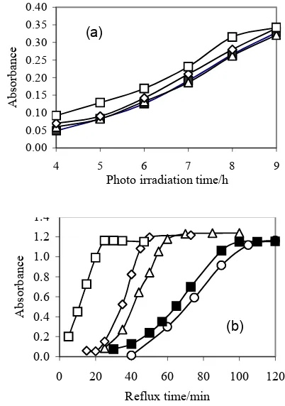
Chi-Au at different pHs showed different absorbance intensity when they were prepared by microwave irradiation as shown in Fig. 2. The intensity of plasmon band was observed in the order of pH 3.5 4 > pH 5 >> pH 6. The original pH of the chi-Au in aqueous acetic acid solution was 2.8. Neutralization of the acetic acid at higher pH decreased the solubility of chitosan hence decreased the stability. Fig. 2 displayed that gold ion reduction was faster at low pH which is pH of 4.
Fig 2. Effect of pH on UV-Vis absorption intensity of Chi-Au colloid as a function of time of microwave irradiation.
Fig. 3 (a-b). The effect of chitosan concentration on UV-Vis absorption Intensity of chitosan-colloidal gold as a function of time prepared with (a) Photo-irradiation at 30oC (100 W tungsten lamp),(b) Reflux (oil bath) at temperature of 90oC, molar ratio Chi:Au; O = 5:1, ■ =10:1, ∆ = 20:1, ◊ = 30:1, = 40:1.
The Role of Chitosan
The effect of chitosan concentration on colloidal gold was first observed in the study of the effect of acetic acid. The UV-Vis absorption with respect to time of refluxing or photo-irradiation of colloidal gold with various chitosan concentrations is shown in Fig. 3 (a-b). Higher chitosan:Au ratio exhibits faster reductions of
(a)
0.0
photo-irradiation for 6 h reflux for 3 h
Fig 4. FT-IR spectra of dried chitosan bead (prepared by neutralizing aqueous-acetic acid-chitosan solution); (A) before and (B) after redox reaction of gold-chitosan.
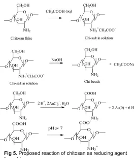
Fig 5. Proposed reaction of chitosan as reducing agent for gold
gold (III) ions when refluxed although less prominent for photo-irradiation. Also in the absence of acetic acid and methanol, chitosan could still reduce gold (III) ion but it was slower than in aqueous acetic acid solution. Apparently, chitosan functioned not only as the stabilizer but also as the reducing agent which is consistent with previous report [14]. The supporting evidence was from FT-IR of chitosan showing a strong new band at 1522 cm-1 after the redox reaction as shown in Fig. 4. Such band (1522 cm-1) was in absorption region of either ionized carboxyl or unconjugated nitro group [15-16]. However, the existence of unconjugated nitro group was
Fig 6. Changes in UV-Vis absorption intensity of Chi-Au as a function of acetic acid concentration prepared with reflux (3 h) or photo-irradiation (6 h, 100 W tungsten lamp) at 30oC.
not likely since the amino group of chitosan was not as easily oxidized as compared to the hydroxyl group [17-19].
Instead, in acetic acid solution, this leave the primary alcohol at C6 position of chitosan to be oxidized to carboxylic group without disturbing the amino group as reported previously [20]. When the oxidized-chitosan was washed up to neutral pH, the carboxyl group can be ionized and gives the observe IR signal at 1522 cm-1 as common case in amino acid [19]. The reaction was proposed by making analogy with earlier reports [4,20,22]. The proposed reactions was shown in Fig. 5.
The intensity of the gold plasmon band increased with increasing acetic acid concentration after 6 h of photo-irradiation or 3 h of refluxing as shown in Fig. 6. In this case the change in pH with increasing acetic acid concentration was small thereby it might not give a significant effect on the gold reduction. Slight decrease of gold absorbance at higher acetic acid concentration could be due to gold particle precipitation. When the ratio of Chi:Au is 3:1, the gold colloid precipitated out in the acetic acid of 0.026 M, but it formed stable colloid at 0.052 M, and precipitated again when acetic acid 0.079 M. Nevertheless, when Chi:Au ratio was doubled (6:1), all of the samples gave stable gold colloids.
This can be explained from both the solubility of chitosan and process of stabilization of gold. Since the solubility of chitosan is affected by the acetic acid concentration, the increase of acetic acid concentration led to better dispersion of chitosan and subsequently gave stable gold colloid [23]. Whereas, the stabilization process of gold started with interaction between gold ions with the amino group of chitosan as known for chitosan-metal ion adsorption [24]. At higher concentration of acetic acid, such interaction was presumably sterically hindered since more amino groups were surrounded by H+ and CH3COO
and leading to a destabilized gold colloid [20].
(a) (b)
(c) (d)
(e) (f)
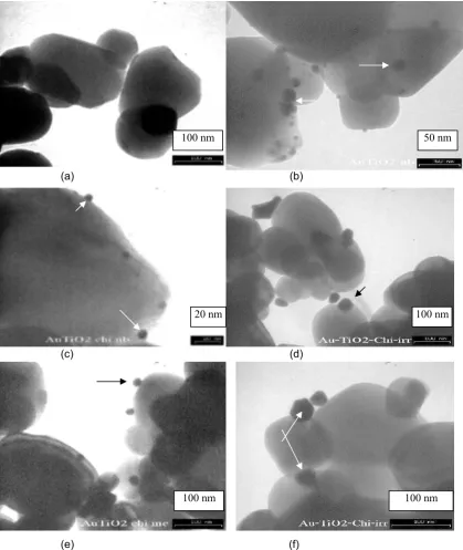
Fig 7. TEM images of chi-TiO2 and chitosan-coated TiO2-Au prepared with various methods, (a) TiO2 (b) TiO2
-Au-nb (c) TiO2-chi-Au-nb (d) TiO2-chi-Au-ir (e) TiO2-chi-Au-me (f) Crystalline gold on the surface of chitosan coated
TiO2. Some of gold metal particles were shown by arrows.
Gold Nanoparticles Immobilized on Chitosan Coated TiO2
Having known that chitosan could stabilize nanoparticulate gold, it is interesting to prepare some solid heterogenised chitosan-gold nanoparticles. Since chitosan could not be isolated as fine crystalline particles from the solution, chitosan coated TiO2 was used as a
support for gold nanoparticles. To study the gold adsorption on chitosan coating, chitosan beads without TiO2 was prepared . It was found that the amount of
adsorbed gold ion on chitosan beads was 0.8875 mg per g of chitosan after an equilibrium time of 25 min. In a separate experiment, 0.1 g of HAuCl4.3H2O was
impregnated onto chitosan-coated TiO2. After
reduction, the gold metal content was 2%.
100 nm 50 nm
100 nm 20 nm
As shown in Fig 7 (a) TEM image of chi-TiO2 show
a regular shape of chi-TiO2 particles with very clean
surfaces, no fragment of TiO2 was observed even at
high magnification. 7(c-d) shows that the particles of TiO2-chi-Au-nb were more dispersed with smaller
particle size and distribution (4.6 + 1.1 nm) compared with on TiO2-Au-nb without chitosan (5.8 + 2.1) nm as
displayed in Fig. 7(b), but gold particles on these solid support were larger and have a broader size distribution compared with the colloidal solution. TiO2-chi-Au-me and
TiO2-chi-Au-ir gave very large particles around 15-30 nm
with very broad size distribution with some of crystalline gold particles observed on the surface of the TiO2-chi as
presented in Fig. 7 (e-f). Apparently, chitosan functioned as a support to segregate the gold particles and leading dispersion of gold metal clusters on the surface of titania.
CONCLUSION
Chitosan stabilized gold nanoparticles were stable for months without precipitation. The optimum mol proportion of chitosan-gold metal ions in methanol acetic acid solution (5:1 to 30:1) gave fine gold particles at range of 9.4-10.4 nm. Gold ion reduction was faster in higher molar ratio of chi-Au. pH solution affected the gold reduction rate and the gold particle size with some exception. Chitosan functioned as the stabilizer and the reducing agent for gold colloidal particles. Gold nanoparticles supported on chitosan coated TiO2 gave
smaller size and narrower size distribution as compared to gold particles supported on TiO2 without chitosan
coating.
REFERENCES
1. Adlim and Abu Bakar, M., 2007, Indon. J. Chem., 8, 2
2. Sun, Y and Xia, Y., 2002, Science, 298, 2176-2179.
3. Santos, I. P and Marzan, M. L., 2002, Langmuir, 18, 2888-2894.
4. Lee C., Wan, C and Wang, Y., 2001, Adv. Funct. Mater., 11, 344-347.
5. Yan, X., Liu H and Liew, K. Y., 2001, J. Mater. Chem., 11, 3387-3391.
6. Biella, S., Castiglioni, G. L., Fumagalli, C., Prati, L and Rossi, M., 2002, Catal. Today, 72, 43-49. 7. Luo, J., Maye, M. M., Lou, Y., Han, L., Hepel, M
and Zhong, C. J., 2002, Catal. Today, 77, 127-138.
8. Haruta, M and Daté, M., 2001, Appl. Catal. A: General, 222, 427-437.
9. Porta, F., Prati, L., Rossi, M., Coluccia, S and Martra, G., 2000, Catal. Today, 61, 165-172. 10. Carotenuto, G and Nicolais, L., 2003, J. Mater.
Chem., 13, 1038-1041.
11. Adlim, 2006, Indonesian Journal of Chemistry, 6, 1-10.
12. Adlim, M., Bakar, M. A., Liew. K. Y., Ismail, J., 2004, J. Mol. Catal. A: Chem. 212, 1–149.
13. Wan Ngah, W. S., Ghani, S. Ab., and Hoon, L. L., 2002, J. Chin. Chem. Soc., 49, 625-628.
14. Huang, H and Yang, X., 2004, Carbohydr. Res., 339, 2627-2631
15. Lide, D. R (Chief Ed), 1994, CRC Handbook of Chemistry and Physic, 74th edition, CRC Press, Boca Renton.
16. Pouchert, C. J., 1970, The Aldrich Library of Infrared Spectra, Aldrich Chemical. Co. Inc., USA. 17. Mayya, K. M., Jain, N., Gole, A., Langevin, D and
Sastry, M., 2004, J. Colloid Interface Sci., 270, 133-139.
18. Gole, A., Kumar, A., Phadtare, S., Mandale, A. B and Sastry, M., 2001, Phys. Chem. Commun., 19, 1-8.
19. Larock, R. C., 1999, Comprehensive Organic Transformation, A guide to Functional Group Preparation, 2nd edition, Wiley-VCH, New York. 20. Hoston, D and Just, E. K, 1973, Carbohydr. Res.,
29, 173-179.
21. Bloomfield, M. M, 1992, Chemistry and Living Organism, 5th edition, John Wiley & Sons, New York.
22. Muzzarelli, R. A.A., 1977, Chitin, Pergamon Press, Oxford.
23. Roberts, G.A.F. (Ed.), 1992, Chitin Chemistry, Hongkong: MacMillan