PA0305 of
Pseudomonas aeruginosa
is a quorum
quenching acylhomoserine lactone acylase
belonging to the Ntn hydrolase superfamily
Mariana Wahjudi,
1,23
Evelina Papaioannou,
13
Oktavia Hendrawati,
1Aart H. G. van Assen,
1Ronald van Merkerk,
1Robbert H. Cool,
1Gerrit J. Poelarends
1and Wim J. Quax
1Correspondence Wim J. Quax [email protected]
Received 20 July 2010 Revised 17 February 2011 Accepted 28 February 2011
1Department of Pharmaceutical Biology, University of Groningen, 9713AV Groningen, The Netherlands
2Faculty of Technobiology, University of Surabaya, Indonesia
ThePseudomonas aeruginosaPAO1 genome has at least two genes,pvdQandquiP,encoding acylhomoserine lactone (AHL) acylases. Two additional genes,pa1893andpa0305, have been predicted to encode penicillin acylase proteins, but have not been characterized. Initial studies on apa0305transposon insertion mutant suggested that the gene is not related to the AHL growth phenotype ofP. aeruginosa. The close similarity (67 %) ofpa0305to HacB, an AHL acylase of Pseudomonas syringae, prompted us to investigate whether the PA0305 protein might also function as an AHL acylase. Thepa0305gene has been cloned and the protein (PA0305) has been overproduced, purified and subjected to functional characterization. Analysis of the purified protein showed that, likeb-lactam acylases, PA0305 undergoes post-translational processing resulting ina- andb-subunits, with the catalytic serine as the first amino acid of theb-subunit, strongly suggesting that PA0305 is a member of the N-terminal nucleophile hydrolase superfamily. Using a biosensor assay, PA0305his was shown to degrade AHLs with acyl side chains ranging in length from 6 to 14 carbons. Kinetics studies usingN-octanoyl-L-homoserine lactone (C8-HSL) andN-(3-oxo-dodecanoyl)-L-homoserine lactone (3-oxo-C12-HSL) as
substrates showed that the enzyme has a robust activity towards these two AHLs, with apparent Kcat/Kmvalues of 0.14¾104M”1s”1towards C8-HSL and 7.8¾104M”1s”1towards 3-oxo-C12
-HSL. Overexpression of thepa0305gene inP. aeruginosashowed significant reductions in both accumulation of 3-oxo-C12-HSL and expression of virulence factors. A mutantP. aeruginosa
strain with a deletedpa0305gene showed a slightly increased capacity to killCaenorhabditis eleganscompared with theP. aeruginosaPAO1 wild-type strain and the PAO1 strain carrying a plasmid overexpressingpa0305. The harmful effects of theDpa0305strain on the animals were most visible at 5 days post-exposure and the mortality rate of the animals fed on theDpa0305 strain was faster than for the animals fed on either the wild-type strain or the strain overexpressing pa0305.In conclusion, thepa0305gene encodes an efficient acylase with activity towards long-chain homoserine lactones, including 3-oxo-C12-HSL, the natural quorum sensing signal
molecule inP. aeruginosa,and we propose to name this acylase HacB.
INTRODUCTION
Pseudomonas aeruginosa is an opportunistic pathogen, often detected in immune-compromised patients and
hospital-acquired infections. A high percentage of cystic fibrosis patients acquire chronic P. aeruginosa infections leading to high mortality rates within this group (Lyczak
et al., 2000; Tattersonet al., 2001).P. aeruginosaemploys a complex network of quorum sensing (QS) systems neces-sary to control expression of density-dependent genes, including genes encoding virulence factors. Induction of these genes depends on production, secretion and detec-tion of the 3-oxo-C12-HSL and the C4-HSL signal
molec-ules[full abbreviations for all the acylhomoserine lactones (AHLs) used in this study are given in Table 1]. The high
Abbreviations:AHL, acylhomoserine lactone; BrMMC, 4-bromomethoxy-7-methyl coumarin; HSL, homoserine lactone; QS, quorum sensing; RLU, relative light units.
3These authors contributed equally to this work.
resistance of this bacterium to common antibiotics has been linked to biofilm formation and to the regulation of the QS system (de Kievit, 2009). Inhibition of QS, a process referred to as quorum quenching, is thought to provide
means of fighting P. aeruginosa infections (Papaioannou
et al., 2009).
One of the ways quorum quenching inP. aeruginosacan be
achieved is by degrading the 3-oxo-C12-HSL and C4-HSL
signal molecules, which can occur via two different enzymes: acylases or lactonases. A number of studies reported the identification of acylases capable of degrading AHLs produced by Gram-negative bacteria. These acylases all belong to the Ntn-hydrolase superfamily.Ralstoniasp. strain XJ12B produces the AiiD acylase, degrading 3-oxo-C10-HSL
(Linet al., 2003).P. aeruginosaPAO1 produces at least two AHL acylases, PA2385 (PvdQ) (Huanget al., 2003; Sioet al.,
2006) and PA1032 (QuiP) (Huang et al., 2006), both of
which have been experimentally proven to degrade
long-chain AHLs (Huang et al., 2006; Sio et al., 2006). The
recently determined 3D crystal structure of PvdQ in
complex with 3-oxo-C12has confirmed the Ntn-hydrolysis
mechanism typical of this class of acylases (Bokhoveet al., 2010). Interestingly, this enzyme has a large hydrophobic
binding pocket that can accommodate the 3-oxo-C12acyl
chain, which is consistent with the observed quenching of the 3-oxo-C12-HSL endogenous toP. aeruginosa.
In theP. aeruginosaPAO1 genome, besidespvdQandquiP,
two additional genes, pa1893 and pa0305, have been
identified as genes encoding hypothetical penicillin acylase proteins belonging to the Ntn-hydrolase superfamily. The
pa0305 gene is predicted to encode a 795 amino acid polypeptide that shows 26 % sequence similarity to PvdQ and 29 % similarity to QuiP. PA0305 also has 67 % sequence similarity to HacB, an AHL acylase ofPseudomonas syringae
that was initially annotated as penicillin acylase (Shepherd &
Lindow, 2009). The function of the pa0305 gene and its
product has not been experimentally tested to date, to our knowledge. Initial studies on apa0305transposon insertion
mutant strain did not demonstrate any defect in growth on C10-HSL compared with the wild-type strain, thus
suggest-ing that the gene is not involved in the metabolism of AHLs (Huanget al., 2006).
Based on the high sequence similarity between PA0305 and HacB, we reasoned that the PA0305 protein might also function as an AHL acylase. The aim of this study was to investigate whether PA0305 is able to degrade acylhomo-serine lactones and whether it functions as a quorum quenching acylase. Herein, we report that the PA0305
protein ofP. aeruginosaPAO1 degrades medium- to
long-chain AHLs with high efficiency resulting in a reduction of production of quorum-sensing-dependent virulence fac-tors in P. aeruginosa. We also, for the first time to our knowledge, report kinetic data for an AHL acylase.
METHODS
Bacterial strains and growth media. Escherichia coli and P. aeruginosa strains were routinely grown and maintained in Luria– Bertani (LB) broth (0.5 % sodium chloride, 1 % tryptone and 0.5 % yeast extract, buffered with 50 mM MOPS, pH 7.0), unless otherwise indicated. Where necessary, gentamicin (10mg ml21for E. coli and 100mg ml21forP. aeruginosa) and ampicillin (100mg ml21forE. coli
and 300mg ml21forP. aeruginosa)were added to maintain the plasmids.
AHLs (Table 1) used in this study were purchased from Sigma and Fluka.
Cloning of thepa0305gene and expression inE. coliandP. aeruginosa. Chromosomal DNA was isolated from an overnight culture of P. aeruginosaPAO1 using a GenElute bacterial Genome DNA kit (Sigma). The ORF encoding the acylase gene (bases 346 690– 344 303) (Stoveret al., 2000) was amplified from chromosomal DNA by PCR using the primer pair ForA and RevA (Table 2). The PCR product was digested withNdeI andHindIII restriction enzymes and ligated to the similarly digested pET-20b plasmid, containing a 6-His tag (Novagen).The resulting pET-pa0305his plasmid was used for the construction of pUCP-pa0305his by reamplifying (using primers ForB and RevB) and subcloning the pa0305 fragment containing a C-terminal 6-His tag (Table 2). For purification and enzyme activity assays the pUCP-pa0305his construct was transformed into E. coli
Origami. pUCP-pa0305was constructed in a similar fashion, but now omitting the 6-His tag. For pa0305 overexpression in PAO1 and
pa0305 complementation in PAO1 Dpa0305 strains, the ORF was amplified by PCR fromP. aeruginosaPAO1 chromosomal DNA using the ForB and RevC primers (Table 2). After digestion of the PCR product with SacI and XbaI restriction enzymes, the product was ligated to the pMMB67EH vector in which gene expression is under the control of a tac promoter. The resulting construct was then transformed intoP. aeruginosacells using the protocol described by Smith & Iglewski (1989).
Purification of PA0305his protein.E. coliOrigami cells containing the pUCP-pa0305his plasmid were grown in 26tryptone yeast extract
(TY) medium (0.5 % sodium chloride, 0.6 % tryptone and 1 % yeast extract) supplemented with 100mg ampicillin ml21and 0.1 % glycerol, for 40 h at 37uC. The culture was induced with 0.4 mM IPTG at OD6000.8–1.0. Cells were harvested by centrifugation, sonicated in lysis
buffer (50 mM Tris/HCl pH 8.0, 2 mM EDTA, 0.1 % Triton X-100) and centrifuged (30 min, 20 000g, 4uC). The cell-free extract was then used for purification of PA0305his on an Ni-NTA agarose gravity column (Qiagen). The protein was eluted with 250 mM imidazole in 50 mM Tris/HCl pH 8.0 and afterwards desalted using 50 mM Tris/ HCl (pH 8.0) on a PD10 desalting column (GE Healthcare) (Otten Table 1.Different acylhomoserine lactones (AHLs) used in this
study
Full name of AHL Abbreviation
N-Butanoyl-DL-homoserine lactone C4-HSL
N-Hexanoyl-DL-homoserine lactone C6-HSL
N-b-Ketocaproyl-DL-homoserine lactone 3-oxo-C6-HSL
N-Heptanoyl-DL-homoserine lactone C7-HSL
N-Octanoyl-DL-homoserine lactone C8-HSL
N-3-Oxo-octanoyl-L-homoserine lactone 3-oxo-C8-HSL
N-Decanoyl-DL-homoserine lactone C10-HSL
N-3-Oxo-decanoyl-L-homoserine lactone 3-oxo-C10-HSL
N-Dodecanoyl-DL-homoserine lactone C12-HSL
N-3-Oxo-dodecanoyl-L-homoserine lactone 3-oxo-C12-HSL
N-Tetradecanoyl-DL-homoserine lactone C14-HSL
et al., 2007). The purified protein was analysed on an SDS-PAGE gel. A Western blot was then carried out for immunodetection of the protein with Ni-NTA HRP conjugate (Qiagen).
Protein identification. To characterize the purified PA0305his protein, thea-subunit and theb-subunit-his were extracted from the gel, trypsin digested and analysed by using MALDI-TOF MS. Protein identification based on mass spectra was done by peptide mass fingerprinting using Mascot software (Matrix Science).
For N-terminal sequencing the purified PA0305his was loaded onto an SDS-PAGE gel. After separation, proteins were transferred onto a PVDF membrane (Sequi-Blot PVDF membrane, Bio-Rad) by semi-dry electroblotting. The membrane was then stained with Coomassie Briliant Blue (CBB) R-250 and the bands corresponding to thea-subunit and theb-subunit-his of PA0305his were excised. The first six residues of the subunits were determined by ABI Procise 494 sequencing (Alphalyse).
Preparation of mRNA and real-time PCR (QRT-PCR).Cells were harvested at the late-exponential growth phase. Total RNA was isolated
and purified using the RNeasy mini kit (Qiagen), followed by DNase (Qiagen) on-column digestion according to the manufacturer’s instructions. RNA was quantified by the A260:A280 ratio using a
NanoDrop (ND-1000) spectrophotometer. cDNA was synthesized with 1mg total RNA as a template using the iScript cDNA Synthesis kit (Bio-Rad). Control reactions for detection of DNA contamination contained the RNA template but lacked iScript reverse transcriptase. The expression of the pa0305 gene in P. aeruginosa was quantified by QRT-PCR using the primer pair ForD and RevD (Table 2) with iQ SYBR Green Supermix (Bio-Rad) in an iCycler (Bio-Rad). Amplification was performed at 95uC (10 min), followed by 40 cycles of 95uC (15 s), 62uC (30 s), followed by melting curve analysis. The
rpoDgene was chosen as an internal housekeeping gene for the baseline control in the evaluation ofpa0305expression. The relative expression of pa0305 transcript was normalized to the rpoD gene and was quantified using the Pfaffl equation (Pfaffl, 2001). For each condition studied, two separate experiments were performed in triplicate.
Bio-activity assay.An aliquot (2ml) of 0.5 mg C12-HSL ml21in
acetonitrile was placed in a 96-well microtitre plate (Lumitrac600, Table 2.Strains, plasmids and oligonucleotides used in this study
Strain, plasmid or oligonucleotide Relevant characteristic or sequence (5§A3§) Source or reference
Strains
P. aeruginosaPAO1 Wild-type Holloway collection
P. aeruginosaPAO1Dpa0305 Deletedpa0305, PAO1 derivative This study
E. coliS17-1lpir Dara-leu7697DlacX74DphoAPvuII phoR araD139 ahpC galE galK rpsL(Smr)
F9[lac+(lacIq)pro]gor522: : Tn10(Tcr)trxB: : kan
Simonet al.(1989)
E. coliOrigami Dara–leu7697DlacX74DphoAPvuII phoR araD139 ahpC galE galK rpsL
F9[lac+(lacIq)pro]gor522: : Tn10(TcR)trxB: : kan
Novagen
E. coliJM109 : : pSB401 Biosensor for detecting C6-, C7- and C8-HSL and its 3-O-form Swiftet al.(1997)
E. coliJM109 : : pSB536 Biosensor for detecting C4-HSL Swiftet al.(1997)
E. coliJM109 : : pSB1075 Biosensor for detecting C10- to C14-HSL and their 3-O-forms Winsonet al.(1998)
Plasmids
pET-20b Expression vector Novagen
pET-20b-pa0305his pET20b containingpa0305with a six-His tag at the C-terminus This study pUCP18 E. coli–Pseudomonasshuttle vectors derived from pUC18, pMB1, pRO1600
replicon,lacZa,bla, ApRCbR
Westet al.(1994)
pUCP-pa0305his pUCP18 containingpa0305with a six-His tag at the C-terminus This study
pUCP-pa0305 pUCP18 containingpa0305 This study
pMMB67EH AmpR IncQ,tacpromoter Fu¨rsteet al.(1986)
pMMB-pa0305 pMMB67EH containingpa0305 This study
pEX18Gm Gmr,oriT+sacB+, gene replacement vector Hoanget al.(1998)
pEX-Dpa0305 pEX18Gm containing flanking regions ofpa0305 This study
GreinerBio). Acetonitrile was allowed to evaporate and the AHL was reconstituted in 100ml PBS buffer pH 7.4 (Gibco) containing 5ml of either cell-free extract or purified enzyme. The plate was incubated at 30uC, at 70 r.p.m. After 4 h, 100ml of a 1 : 100 dilution of the lux-basedE. colipSB1075 biosensor was added to each well and the plate was further incubated for 6 h at 30uC. Bioluminescence was detected on a FLUOstar Omega platereader (BMG labtech). As a control, reactions with only PBS mixed with the enzyme were conducted in the same way. All of the assays were performed in triplicate.
To determine the specificity of AHL-degradation activity, acetonitrile stocks of AHLs (Table 1) were transferred in a 96-well microtitre plate at a final concentration of 100, 10, 1 and 0.1mg ml21in the
reaction mixture. The solvent was allowed to evaporate and the AHLs were reconstituted in 100ml PBS pH 7.4 containing 2.5mg purified enzyme. The plate was incubated at 30uC, at 70 r.p.m. Next to this, control reactions using inactive enzyme were performed in the same way. The enzyme was inactivated by boiling a mixture of PBS with 2.5mg enzyme at 100uC for 15 min. After 4 h of incubation, 100ml of a 1 : 100 dilution of the appropriate biosensor (E. coli pSB401, pSB536 or pSB1075) was added to each well and the plate was further incubated for 6 h at 30uC. Bioluminescence was detected on a FLUOstar Omega platereader (BMG labtech). The experiments were performed in triplicate. The differences in relative light units (RLU) between experimental samples and controls were used to determine the degree of degradation of each AHL.
Analysis of C8-HSL deacylase activity by HPLC.The assays were
carried out using C8-HSL as described previously (Urozet al., 2008).
The reaction mixture consisted of 1 mM C8-HSL and 25mg
(~0.286mM) of the enzyme in 1 ml PBS buffer pH 7.4 (Invitro-gen). The mixture was incubated at 30uC and 70 r.p.m. for 4 h. Three samples of 750ml were taken at 0 and 4 h and processed for detection of the residual substrate, the released octanoic acid and the HSL. For detection of the substrate, residual C8-HSL in the reaction
mixture was extracted twice with 750ml ethyl acetate. Octanoic acid in the sample was extracted three times with hexane and dried under nitrogen streaming. Derivatization of the octanoic acid was then performed by addition of 4-bromomethoxy-7-methyl coumarin (BrMMC) reagent as described previously (Wolf & Korf, 1990). The presence of free HSL was determined by addition of 750ml dansyl chloride (2.5 mg ml21) in acetone to the reaction mixture followed by overnight incubation at 37uC (Linet al., 2003). The mixture was dried under nitrogen streaming and neutralized using 50ml 0.2 M HCl. After dilution with 250ml acetonitrile, the sample was analysed by reverse-phase HPLC separation. As a control, reference HSL (final concentration 2 mM) was dansylated in parallel with the same reagents. The presence of C8-HSL residue and dansylated HSL was
analysed by reverse-phase HPLC separation (Uroz et al., 2008; Lin
et al., 2003).
HPLC analysis was performed using a Shimadzu-VP system. The column used was a LichroCART 125-4 Superspher 100 RP-18 endcapped. The mobile phase was water (solvent A) and acetonitrile (solvent B). The C8-HSL was detected at 219 nm, whereas the
dansylated HSL was detected at 267 nm and the BrMMC-derivative of octanoic acid at 328 nm.
Kinetic studies of PA0305his on C8-HSL and 3-oxo-C12-HSL
substrate.Kinetic studies were performed in 0.1 M phosphate buffer pH 7.5 at 30uC (Linet al., 2003). For C8-HSL kinetics, six different
concentrations of the substrate (0.02–0.20 mM) were used, at which the substrate was completely soluble. For each of these concentra-tions, the substrate was incubated with 25mg PA0305his (ml reaction volume)21for 2 min. A 100ml sample of the reaction mixture was taken and mixed directly with 100ml o-phthalaldehyde (OPA) solution in a 96-well microtitre plate and the A340was measured
spectrophotometrically. To obtain a calibration curve of the OPA-derivative of the HSL reference, the same procedure was performed in an enzymic mixture containing HSL. The calibration curve is a straight line that could be fitted with the following equation:
y51.959x+0.6966 (R250.9882). The initial rates (mM s21) were plotted versus the concentration of substrate (mM), which gives a straight line. Assuming that[S],,Km, the slope of the line equals
(Kcat6[E])/Km. Thus, dividing the slope by the enzyme concentration
results in a value for the apparentKcat/Km.
For 3-oxo-C12-HSL kinetics, the substrate (at seven different
concentrations, 0.01–0.12 mM, where the substrate was soluble) was incubated with 10mg PA0305his (ml reaction volume)21 for
1 min. A 1 ml sample was mixed directly with 1 ml ethyl acetate to stop the reaction and extracted three times with the same volume of ethyl acetate. The amount of HSL product released was quantified as described above.
Construction of precise gene deletions in PAO1. A precise chromosomal deletion of pa0305 was constructed as follows. The flanking regions of the target gene were amplified with PAO1 chromosomal DNA as a template, using the primer pair ForE and RevE and the primer pair ForF and RevF. The two resulting fragments were joined together in a second PCR round using ForA and RevB primers. The fusion product was then digested with HindIII and
EcoRI and cloned into the similarly digested pEX18-Gm (Hoanget al., 1998). The subsequent deletion procedure was performed as described previously (Papaioannou et al., 2009). The sequences of the primers and probes (ForG and RevG) are listed in Table 2. A double deletion (DpvdQ,DquiP) and a triple deletion strain (DpvdQ,
DquiP, Dpa0305) were constructed with the same approach using similar primer sets.
Elastase assay.Elastolytic activity of extracellular supernatant was assayed in the following manner. Supernatant samples of 0.1 ml were added in Eppendorf tubes containing 20 mg Elastin Congo red (ECR; Sigma) suspended in 0.90 ml ECR buffer (100 mM Tris, 1 mM CaCl2, pH 7.5). After 4 h incubation with agitation, insoluble ECR
was removed by centrifugation (20 000g, 5 min, 4uC). The absorbance of the supernatants measured at 495 nm was divided by the OD600of the culture. LB medium was used as a negative control
(Ohmanet al., 1980).
Pyocyanin assay. Pyocyanin was extracted from the supernatants and measured by the method previously described by Essar et al.
(1990). A 5 ml supernatant of culture grown in LB was extracted with 3 ml chloroform. The chloroform layer was transferred to a fresh tube and re-extracted with 1 ml 0.2 M HCl. After centrifugation, theA520
of the HCl fraction was measured. Concentrations, expressed as micrograms pyocyanin produced per millilitre culture supernatant, were determined as (OD520617.072)/OD600(Essaret al., 1990).
Quantification of 3-oxo-C12-HSL, C4-HSL and
2-heptyl-3-hydroxy-4(1H)-quinolone (PQS) production. The amount of 3-oxo-C12-HSL produced by the PAO1 parental strain and the
PAO1DpvdQDquiP and PAO1DpvdQDquiPDpa0305 mutant strains was determined using the biosensor E. coli (pSB1075), which produces light in response to long AHLs (Winson et al., 1998). 3-Oxo-C12-HSL concentrations were measured 4, 15, 27 and 50 h after
by the biosensor was read every hour during a 24 h time-course in a multifunctional microplate reader (FLUOstar Omega, BMG Labtech). Data points obtained immediately prior to the maximum light production were used for comparisons (approximately 9 h after initiation of the bioassay). For quantification of the accumulation levels of 3-oxo-C12-HSL and C4-HSL by TLC, 2ml of the acidified
supernatants from the same amount of cells was spotted on a silica gel 60 F254 TLC plate (Merck) for 3-oxo-C12-HSL and a reverse-phase
TLC plate for C4-HSL. The separation and detection were performed
as described by Yateset al.(2002). For quantification of accumulation levels of PQS, the compound was detected as previously described (Diggleet al., 2003).
Quorum quenching activity of expressed PA0305 inP. aerugi-nosa using a Caenorhabditis elegans model. Slow-killing kinetics ofC. elegansby PAO1 and its derivatives were determined by using the procedure described previously (Papaioannou et al., 2009). Briefly,Pseudomonasstrains were grown overnight at 37uC in LB broth supplemented with appropriate antibiotics and then diluted 100-fold into fresh broth. Nematode growth medium plates (59 mm diameter) were then spread with 80ml of the respective culture. After the plates were incubated at 37uC for 24 h and allowed to equilibrate to room temperature for 30 min, 50 larval stage 4 (L4) nematodes from stock plates were transferred onto theP. aeruginosalawn (PAO1, PAO1Dpa0305, PAO1DlasRlasI). The plates were then incubated at 24uC and scored for living and dead worms every 3–4 h for 7 days. For statistical purposes, a minimum of four replicates per trial was performed.E. coliOP50 was used as a negative control to evaluate background levels of worm death. A worm was considered to be dead when it failed to respond to plate tapping or gentle touch with a platinum wire. Worms that died as a result of getting stuck to the wall of the plate were excluded from the analysis. Results are presented as the percentage of living nematodes on the killing plates compared with their survival on the E. coli OP50 control strain. Statistical analysis was performed using the two-sample Student’st-test in the statistical programming language R. The results were plotted using PyLab, a python package for data analysis and visualization.
RESULTS
Expression and purification of PA0305
The P. aeruginosa pa0305 gene was amplified and cloned into the pUCP18 vector. Using this construct, expression of the gene in E. coliOrigami resulted in the synthesis of a protein of about 60 kDa as shown by SDS-PAGE with
Coomassie staining (Fig. 1, lane 2). The size of this protein matches the predicted size of theb-subunit of the putative acylase encoded by pa0305. Because of the relatively
low-level expression of the gene, overproduction of the
a-subunit was not observed. To facilitate protein detection and purification, the recombinant enzyme was also
produced in E. coli Origami as a C-terminal His-tagged
protein. In comparison with native PA0305 (without a fusion tag), however, the His-tagged PA0305 protein was produced at even lower levels and could not be detected by Coomassie staining (Fig. 1, lane 5). Nonetheless, the His-tagged protein could be efficiently purified by a Ni-based immobilized-metal-affinity chromatography protocol. SDS-PAGE analysis of the purified protein revealed three protein bands (Fig. 1, lane 7). The band of about 65 kDa represents the b-subunit, whereas the two bands of about 26 and
28 kDa represent different non-mature forms of the
a-subunit (as discussed below). The identity of the 65 kDa band as the His-taggedb-subunit of the PA0305 protein was confirmed by Western blotting with anti-His antibody (Fig. 1, lane 11). Expression of thepa0305gene from the vector
pMMB67EH inP. aeruginosaresulted in very low amounts
of PA0305 (data not shown) and purification from P.
aeruginosawas not further pursued.
Characterization of purified PA0305his
The purified PA0305his protein was subjected to MS and protein sequencing to establish the identity of the three protein bands observed on the SDS-PAGE gel (Fig. 1, lane 7). MALDI-TOF MS analysis of the protein band of about
65 kDa revealed that this protein is the b-subunit of
PA0305. N-terminal sequencing revealed that the first six amino acid residues of theb-subunit are, as predicted, Ser-Asn-Ala-Trp-Val-Val. From these observations, it can be
concluded that the b-subunit of PA0305 is correctly
formed inE. coli, resulting in a protein with an N-terminal serine.
MALDI-TOF MS analysis of the two smaller proteins revealed similar MS spectra, indicating that both proteins
correspond to thea-subunit. The spectrum of the 26 kDa
protein band is consistent with that expected for the C-terminally processeda-subunit (i.e. with signal peptide but without spacer peptide), whereas the spectrum of the
28 kDa protein band is consistent with thea-subunit that
includes both the signal peptide and the spacer peptide. N-terminal sequencing of the smallest (26 kDa) protein band revealed the sequence Met-Lys-Arg-Thr-Leu-Thr. This observation further confirms that the signal peptide indeed
remains attached to the a-subunit. Apparently, the signal
peptide is not efficiently removed inE. coli, which suggests that the protein is not properly secreted.
Analytical gel filtration was performed as described in Methods with thyroglobulin (670 kDa), ferritin (440 kDa) and aldolase (158 kDa) as reference proteins. PA0305his eluted with a retention time corresponding to a molecular mass of approximately 400 kDa (data not shown), a size that corresponds to a tetra-heterodimer.
PA0305 is an AHL acylase
The lux-basedE. colipSB1075 biosensor was used to test cell-free extracts for the presence of AHL-degrading activity. C12
-HSL was used as the substrate since it resembles the P.
aeruginosa signal molecule, 3-oxo-C12-HSL. Incubation of
C12-HSL with cell-free extract prepared from E. coli cells
overproducing PA0305his resulted in the conversion of C12
-HSL, as detected by a clear reduction in bioluminescence (Fig. 2). Cell-free extracts prepared from the negative control strain (E. coli cells containing an empty plasmid) did not show this reduction in bioluminescence (Fig. 2). These data clearly indicate that PA0305 is a C12-HSL-degrading enzyme.
Having established that PA0305 exhibits activity towards C12-HSL, various AHLs with alkyl chains ranging from C4
to C14 (for abbreviations see Table 1) were tested as
potential substrates for purified PA0305his. In these activity assays, purified PA0305his was mixed with four different concentrations of AHLs, ranging from 0.01 to
10 mg ml21and the conversion of the AHLs was followed
by measuring the quenching of light using a panel of three biosensor strains (Table 3). At each AHL concentration, the activity of the purified enzyme was compared with heat-inactivated protein. The PA0305his protein showed activity towards AHL with alkyl chains ranging from C6to
C14. The highest degradation rate (93-fold decrease in
substrate concentration) was observed on C8-HSL at 10mg
ml21. At the lowest concentration, 0.01 mg ml21, the
degradation rate was the highest on 3-oxo-C12-HSL
(64-fold). Activity against the 3-oxo- forms was detected from 3-oxo-C10-HSL to 3-oxo-C14-HSL (Table 3).
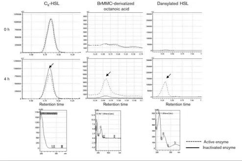
To investigate whether PA0305 functions as a lactonase or an acylase, the products of the conversion of C8-HSL were
subjected to HPLC analysis. The reaction was performed by exposing C8-HSL substrate to PA0305his for 4 h. C8-HSL
was detected as a specific peak at the retention time of 5.85 min (Fig. 3). Compared with a negative control with inactivated enzyme, PA0305his caused a significant reduc-tion of the C8-HSL concentration at 4 h. Detection of HSL
liberated by acylase activity was performed by dansylating the free amine of HSL. Fig. 3 shows the presence of dansylated HSL in the 4 h sample, resulting from the cleavage of the amide bond between the HSL and the acyl chain. The specific peak was detected at a retention time of 6.33 min. This dansylated product, clearly visible in the reaction mixture with active PA0305his, was absent in the control, indicating that degradation of the substrate was caused by acylase activity. To confirm the presence of octanoic acid released by acylase activity, derivatization of the fatty acid using BrMMC was performed. HPLC analysis showed that the specific peak of the BrMMC-octanoic acid
derivative was present at 12.59 min in C8-HSL samples
exposed to active PA0305 for 4 h. This peak was absent in the control samples with inactivated enzyme. These results clearly indicate that PA0305 is an AHL acylase that
converts C8-HSL to HSL and octanoic acid.
Kinetic studies of PA0305his on AHLs
Having established that PA0305 functions as an AHL acylase, kinetic parameters were determined for the
PA0305-catalysed conversion of C8-HSL and 3-oxo-C12
-HSL. The C8-HSL was chosen because it shows the highest
activity with PA0305his at high concentrations and 3-oxo-C12-HSL was chosen since it is one of the signal molecules
produced by P. aeruginosa. The initial rates of these
reactions were dependent on the concentration of the respective substrate. However, saturation with C8-HSL and
3-oxo-C12-HSL was not achieved for PA0305 because the
low solubility of these AHLs in aqueous buffer does not permit rate measurements at high substrate concentrations.
Hence, Kcat/Km values were determined using seven low
Fig. 2. Activity of cell-free extract ofE. coli Origami producing PA0305his on C12-HSL substrate. After incubation of C12-HSL
with extracts, the non-degraded substrate was analysed using the biosensor strain pSB1075 as described in Methods. The amount of light produced in response to C12-HSL was quantified as
relative light units (RLU). Lanes: 1, C12-HSL incubated without cell
extract; 2, C12-HSL incubated with cell-free extract ofE. coliwith
empty vector; 3, C12-HSL incubated with cell-free extract ofE. coli
substrate concentrations where the substrate is still soluble and the detection of HSL formed was performed using the derivatization method as described above. A comparison of the values showed that PA0305 is 56-fold more efficient in catalysing the conversion of 3-oxo-C12-HSL (Kcat/Km57.86104 M21s21) than C8-HSL (Kcat/ Km50.146104 M21 s21).
Deletion ofpa0305inP. aeruginosa results in
accumulation of 3-oxo-C12-HSL
The kinetic studies with purified PA0305 have shown that the enzyme is quite efficient at degrading 3-oxo-C12-HSL,
which is a natural QS signal molecule inP. aeruginosa. This observation prompted us to investigate whether deletion of thepa0305gene inP. aeruginosawould affect the ability of this strain to accumulate 3-oxo-C12-HSL. In order to detect
the accumulation of 3-oxo-C12-HSL in cell cultures, P.
aeruginosacells grown in LB were harvested at specific time points. The supernatants were subjected to detection of
3-oxo-C12-HSL using biosensor E. coli JM109 : : pSB1075
(Fig. 4a). The kinetics of 3-oxo-C12-HSL accumulation
revealed that P. aeruginosa wild-type and P. aeruginosa
Dpa0305 produced similar amounts of 3-oxo-C12-HSL
within 7 h post-inoculation. However, from 24 to 50 h
post-inoculation, 3-oxo-C12-HSL was much more abundant
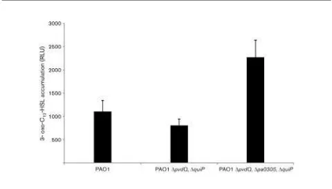
in theDpa0305strain compared with the wild-type strain as seen after separation on TLC plates (see Supplementary Fig. S1, available with the online version of this paper). In order to exclude any influence from other AHL acylases encoded by P. aeruginosa, a triple deletion strain (DpvdQ, DquiP,
Dpa0305) was constructed and compared with the double deletion strain (DpvdQ, DquiP). After 50 h, an
approxi-mately 250 % increase in accumulation of 3-oxo-C12-HSL
was observed (Fig. 5). This suggests that PA0305 has a
significant role in 3-oxo-C12-HSL degradation in P.
aeruginosa cells grown in LB medium. Notably, while
deletion of the pa0305 gene affects 3-oxo-C12-HSL
accu-mulation, no influence on the accumulation of the QS signal
molecules C4-HSL and PQS was observed (Fig. 4b).
Table 3.Specificity of purified PA0305his for AHL substrates
Degradation is expressed as RLU, generated by three lux-based biosensors, which produce light in response to the AHLs. The initial concentration of AHL (0.01, 0.1, 1 or 10mg ml21) is given. The degradation assay was carried out in PBS buffer (pH 7.4) with 25mg of either active or inactivated enzyme ml21and incubated at 30uC with shaking. The remaining amount of AHLs was detected by a suitable lux-based biosensor at 30uC for 6 h.
AHL substrate Purified PA0305his protein Degradation (10”3¾RLU)
0.01 0.1 1 10
*BiosensorE. coliJM109 : : pSB536.
DBiosensorE. coliJM109 : : pSB401.
The effects of PA0305 onP. aeruginosa QS
The AHL hydrolysing capability of PA0305, bothin vitroand
in vivo, suggests that this enzyme may function as a quorum
quencher in P. aeruginosa. To determine the ability of
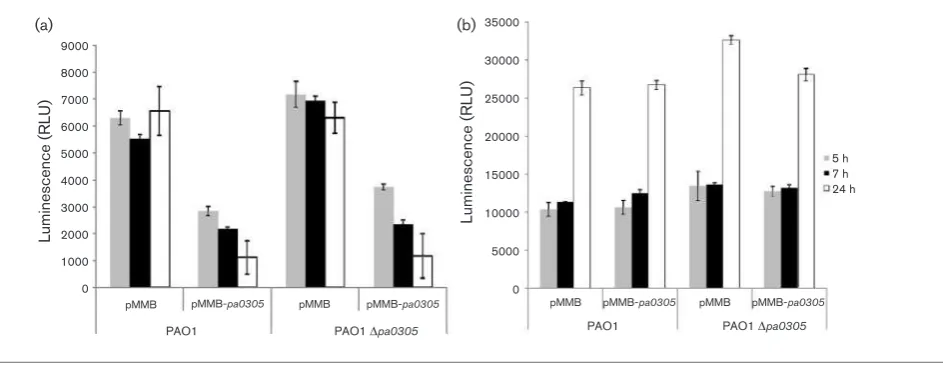
overexpressed PA0305 to interfere with the expression of QS-regulated functions, the P. aeruginosawild-type andP. aeruginosa Dpa0305 strains were transformed with the pMMB-pa0305construct. As controls, the same strains were transformed with the empty plasmid (pMMB67EH). Gene expression was either constitutive or induced with IPTG.
Overexpression of the pa0305 gene (LB growth, IPTG
induction) in P. aeruginosa resulted in a significant
reduction in elastolytic (LasB) activity throughout the growth phase, from 5 to 24 h post-inoculation (Fig. 6a). Pyocyanin production was also reduced after 5 h post-inoculation, but the reduction did not last until 24 h of
growth (Fig. 6b). The accumulation of 3-oxo-C12-HSL was
significantly reduced in all PA0305-overproducing strains and the amounts remained at low levels even after 24 h of
growth (Fig. 7a). However, C4-HSL accumulation levels
were not altered and this signal molecule was abundant throughout the 24 h growth period (Fig. 7b). This observation is consistent with the finding that PA0305 is not active against C4-HSL.
To prove that the reduced 3-oxo-C12-HSL accumulation
was correlated with the transcription of the pa0305 gene,
mRNA levels of the pa0305 gene in all strains were
quantified using QRT-PCR. All isolated total RNA samples were of good quality as shown by analysis of the samples on a formaldehyde agarose gel (data not shown). Further-more, the RNA samples were free of chromosomal DNA, as indicated by the finding that no cDNA could be generated from the RNA samples in the absence of iScript
tran-scriptase. The rpoD housekeeping gene was used as a
control for comparison of the quantity and quality of all Fig. 3. Identification of the products of the PA0305his-catalysed conversion of C8-HSL. C8-HSL was incubated with
PA0305his for 4 h. Samples were taken at time 0 (upper panels) and after 4 h (middle panels) and were then treated for quantification by HPLC analysis of the residual C8-HSL and the octanoic acid and HSL released. Reduction of C8-HSL levels
upon exposure to active PA0305his was corroborated with the occurrence of free octanoic acid and HSL, confirming that PA0305his is an acylase that cleaves the amide bond linking the HSL ring and the acyl chain. C8-HSL samples were extracted
with ethyl acetate and injected into an RP18 column as described in Methods. For efficient detection, octanoic acid was
BrMMC-derivatized and HSL was dansylated, as described in Methods. Arrows indicate the specific peaks that were subjected to UV analyses (lower panels). The spectra of C8-HSL, BrMMC-derivatized octanoic acid and dansylated HSL are identical to
mRNAs. The expression ofpa0305 was normalized to the
rpoD gene which had a similar expression profile in all
strains. The amount of pa0305 transcript present in the
wild-type culture at 5 h was low (Table 4). The mRNA levels ofpa0305were significantly higher in both the
wild-type strain and theDpa0305 strain harbouring thepa0305
expression plasmid when compared with the control strains harbouring an empty plasmid.
Quorum quenching activity of overproduced
PA0305 inP. aeruginosa using theC. elegans
infection model
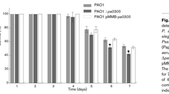
To assess whether either the deletion or overexpression of the pa0305 gene in P. aeruginosa have any effect on the
pathogenicity of the bacterium, the killing kinetics of the
wild-type PAO1 strain, theDpa0305strain and thepa0305
overexpressing PAO1 strain were determined using a C.
elegans infection model and a slow killing assay as previously described by Tan & Ausubel (2000). In the slow killing assay, the LT50value (time taken for half of the C. elegansto die) for PAO1 was shown to be around 68 h. We chose to screen our mutants with this method as it is highly sensitive and allows discrimination between strains that only slightly differ in their ability to kill C. elegans. During the first 4 days there were no differences observed between worms feeding on the three different strains. Up to the completion of the assay no significant differences could be seen between the PAO1 parental strain and the derived PA0305-overexpressing strain (P.0.05). However, between days 5 and 7 after exposure, the harmful effects of the
deletion of pa0305 were more profound. At day 7
post-exposure only about 41 % of the worms feeding on the deletion strain were alive compared with about 57 % of the
worms fed on the PAO1 wild-type or pa0305
over-expressing strain (Fig. 8). Statistical analysis showed a significant difference between the PA0305 deletion strain
and the PAO1 mutant for day 7 of the assay (P,0.05).
These observations give an insight into the effect of the
pa0305 gene on tempering the virulence of P. aeruginosa
PAO1.
DISCUSSION
In recent years, numerous genes encoding new members of the Ntn-hydrolase superfamily have been discovered in the genomes of prokaryotes. The first characterized members of this superfamily were described as penicillin acylases and recently they have been more broadly classified as
members of the family of b-lactam acylases. At present
it is clear that b-lactam deacylation is unlikely to be
the natural function of this class of enzymes, as this Fig. 4.Quantification of AHLs produced in a time lapse experiment. Supernatant of LB cultures of the respective strains grown for 0–5 h was extracted as described previously (Yateset al., 2002) and quantified in the bioluminescence assay. (a) Results of analysis with biosensorE. coliJM109 : : pSB1075 detecting 3-oxo-C12-HSL. (b) Results of analysis with biosensorE. coli
JM109 : : pSB536 detecting C4-HSL. It can be seen that the pMMB-pa0305-harbouring strains accumulate much less
3-oxo-C12-HSL over time as compared with the wild-type PAO1 strain and theDpa0305strain, whereas the accumulation of C4-HSL
is similar for all strains. X, Wild-type empty;&, PAO1 pMMB-pa0305;m, PAO1 pEx-Dpa0305empty;¾, PAO1
pMMB-pa0305–pEx-Dpa0305.
Fig. 5.Accumulation of 3-oxo-C12-HSL in HSL-acylase deletion
strains. The PAO1 parental strain, theP. aeruginosaDpvdQ,DquiP double mutant and theP. aeruginosaDpvdQ,DquiP,Dpa0305triple mutant were screened for 3-oxo-C12-HSL accumulation levels after
50 h of growth in LB medium. The graph illustrates the higher 3-oxo-C12-HSL levels present in the DpvdQ, DquiP, Dpa0305 triple
conversion does not inactivate penicillins or cephalospor-ins (Krzeslak et al., 2007; Sio & Quax, 2004) and, hence, does not give a competitive advantage. For example, the paradigm of penicillin acylases, PGA fromE. coli, generates a molecule with even higher antibiotic activity upon conversion of penicillin-G into 6-APA (Meevootisomet al., 1983; Schumacheret al., 1986). It has been argued that this conversion is therefore not likely to be the natural function
of this industrially important enzyme. The P. aeruginosa
PAO1 complete genome sequence revealed the presence of four genes with significant sequence similarity to penicillin and cephalosporin acylases. By now it has been established
that two of these genes encode products, PvdQ and QuiP, that can deacylate long-chain AHLs resulting in quorum quenching, which points to a major physiological role for
these enzymes (Huang et al., 2006; Sio et al., 2006).
Phenotypic analysis of a quiP (quorum signal utilization
and inactivation protein) transposon mutant showed that a strain carrying the transposon insertion was impaired in growth on decanoyl-HSL when compared with the parental
strain. QuiP complementation revealed that, when quiP
was constitutively expressed from a plasmid, AHL degrading activity potential was restored. A remarkable
decrease in 3-oxo-C12-HSL accumulation levels was
1.2
pMMB-pa0305 pMMB-pa0305 pMMB-pa0305 pMMB-pa0305
PAO1 Dpa0305 PAO1 Dpa0305
Fig. 6.Effects of PA0305 overexpression on virulence factor production in P. aeruginosaPAO1 and PAO1Dpa0305. The production of virulence factors was tested in wild-type andDpa0305strains carrying either an empty vector (pMMB) or the pa0305-expressing vector (pMMB-pa0305). The elastolytic activity (a) and pyocyanin production (b) in the supernatants of the respective strains are shown. Error bars,SD.
9000
pMMB-pa0305 pMMB-pa0305 pMMB-pa0305 pMMB-pa0305
PAO1 Dpa0305 PAO1 Dpa0305
Fig. 7. Effects of PA0305 overproduction on accumulation of QS signal molecules in P. aeruginosa PAO1 and PAO1
Dpa0305. The same strains as described in Fig. 6 were also tested for the accumulation of 3-oxo-C12-HSL as detected by
luminescence measured withE. colipSB1075 biosensor. (a) Overproduction of PA0305 reduces the accumulation of 3-oxo-C12-HSL in wild-type andDpa0305supernatant. (b)E. coli pSB536, which is sensitive to C4-HSL, was incubated with the
same cell extracts. It can be concluded that overproduction of PA0305 does not reduce the accumulation of C4-HSL. Error
detected when quiP was constitutively expressed in P. aeruginosa. Interestingly, even though QuiP has been proven to be involved in AHL utilization, this gene was not classified in microarray analysis as a QS-regulated gene. Similarly, PvdQ also effectively degrades AHL side chains ranging from 11 to 14 carbons and overexpression of this gene, as well as exogenous addition of purified protein in growingP. aeruginosacultures, delays the accumulation of the 3-oxo-C12-HSL molecule and as a result decreases the
expression of virulence factors such as elastase and pyocyanin (Sioet al., 2006).
Different organisms were also found to produce enzymes acting on autoinducer molecules and disrupting the QS
systems. Apart from the AHL acylases produced by P.
aeruginosa,Ralstoniastrain XJ12B produces the AiiD AHL
acylase that when expressed in P. aeruginosa results in
reduction of virulence (Linet al., 2003). In addition to the AHL acylases, a second group of enzymes that act on autoinducer molecules have been identified. These enzymes are characterized as AHL lactonases and they inactivate AHL signals by hydrolysing the lactone ring, thus yielding the corresponding acylhomoserine. Examples are the AiiA (autoinducer inactivator) AHL lactonase from the
Gram-positive bacterium Bacillus sp. 240B1 (Dong et al.,
2001) and the AiiB AHL lactonase produced by
Agro-bacterium tumefaciens(Liuet al., 2007). It has already been shown that transgenic plants expressing AHL lactonase
displayed an enhanced resistance to infection byErwinia
carotovora(Donget al., 2001).
InP. aeruginosatwo additional genes,pa1893andpa0305, have been annotated as genes possibly encoding penicillin acylases (Krzeslaket al., 2007) although they have not been characterized. The results of this study clearly demonstrate that PA0305 has acyl-HSL degrading activity similar to that of PvdQ and QuiP.
P. aeruginosaPAO1pa0305is predicted to encode a protein
of 795 amino acids in length (Stover et al., 2000) and
the polypeptide shows the striking features of post-translational processing typical of the members of the N-terminal nucleophile aminohydrolases (Ntn hydrolases) superfamily. The gene is transcribed as a single polypeptide and as a result of autocatalytic cleavage a mature active enzyme consisting of two dissimilar subunits is formed. The strongest evidence for PA0305 being an Ntn hydrolase is the presence of the conserved Ser 1 residue, the first
residue of theb-subunit. The N-terminal amino acids 1–25
of PA0305 are predicted to compose a signal sequence peptide (LipoP v.1.0, Phobius, SignalP v.3.0 Hidden
Markov Models) (Winsor et al., 2009), which is in line
with the observation that most bacterial Ntn hydrolases are secreted. However, determination of the N-terminal
sequence of the a-subunit purified from E. coli revealed
that the signal peptide is not cleaved off from thea-subunit under the experimental conditions used, indicating that the protein may not be translocated to the periplasm inE. coli
or that theE. colisignal peptidase is less capable of cleaving this sequence. The finding that the protein migrates as a tetra-heterodimer on a gel filtration column is unique in comparison with other characterized AHL acylases, but is not surprising considering that some cephalosporin acylases, also members of the Ntn-hydrolase superfamily,
Table 4. Fold changes of pa0305 gene expression in P. aeruginosastrains
Cells were harvested at the late-exponential growth phase (5 h post-inoculation). The pa0305 expression levels of all strains were compared with the expression of PAO1Dpa0305strain harbouring an empty plasmid, which is given the value 1. All of the values were normalized to the housekeeping generpoD.
P. aeruginosa Plasmid Fold change
PAO1 wild-type pMMB67EH 205±35 pMMB-pa0305 144488±19344
PAO1Dpa0305 pMMB67EH 1±0
pMMB-pa0305 93335±9506
Fig. 8. The effect of overexpressing and deleting thepa0305gene on the virulence of P. aeruginosa in the C. elegans model. C. elegans nematodes were fed on a layer of Pseudomonas cells as described previously (Papaioannou et al., 2009). Wild-type P. aeruginosa PAO1, P. aeruginosa PAO1
have a tetra-heterodimer structure (Kwon et al., 2000). PA0305 has been purified to homogeneity fromE. coliand it was used for further enzymic characterization revealing that it has activity towards medium- and long-chain HSLs. When compared with PvdQ and QuiP, which show 26 and 29 % sequence similarity, respectively, PA0305 shows broader substrate specificity. However, in spite of the 67 % sequence similarity with HacB fromP. syringae, PA0305 is
unable to degrade C4-HSL. Interestingly the ability to
degrade C4-HSL, the second major autoinducer, seems to be
absent from all AHL acylases of P. aeruginosa PAO1
described to date (Huang et al., 2006). Judging from
conversion rates at high concentrations of different acyl-HSLs it may be concluded that C8-HSL is the best substrate
for PA0305 (93-fold decrease at 10mg ml21; Table 3). This must be considered with care, however, as the solubility of long-chain HSLs may become limiting at high concentra-tions. In fact this is demonstrated by comparing the conversion rates of C8-HSL at 0.01 and 10mg ml21 with
the conversion rates of 3-oxo-C12-HSL at these respective
concentrations (Table 3). The deacylation rate of the enzyme at low concentrations appears to be best for 3-oxo-C12-HSL
and not for C8-HSL (64-fold versus 1.3-fold). To explain
these observations, one likely possibility is that at high substrate concentrations (10mg ml21) of 3-oxo-C12-HSL,
higher order assemblages (e.g. micelles) may form, limiting the enzyme activity. Nonetheless, our data suggest that at physiological concentrations the natural autoinducer of P. aeruginosais the best substrate for the PA0305 enzyme. To further substantiate this conclusion kinetic studies were performed using HPLC detection of derivatized products. For the first time, to our knowledge, we report an apparent
Kcat/Kmvalue for an acyl-HSL acylase. Indeed the catalytic
efficiency of PA0305 is higher for 3-oxo-C12-HSL in
comparison with C8-HSL, which is consistent with
3-oxo-C12-HSL being one of the natural substrates of this enzyme.
Although individual Kcat and Km values have not been
assessed, the apparent Kcat/Km value of 7.86104 M21 s21
implies that the enzyme has a high catalytic efficiency
towards 3-oxo-C12-HSL, which would require an efficient
acyl binding pocket in the enzyme similar to the substrate binding pocket described for PvdQ (Bokhoveet al., 2010).
The deletion of thepa0305gene from chromosomal DNA
resulted in an increase of the 3-oxo-C12-HSL levels in the
growth medium, which became most apparent after 50 h incubation. This phenotype was evident in both the
wild-type and the PAO1DpvdQDquiPbackground. The increase
in 3-oxo-C12-HSL did not result in alterations of the C4
-HSL accumulation levels, the growth rate (data not shown) nor the morphology of the mutant. This is all in agreement
with previous observations with HacB mutants of P.
syringae (Shepherd & Lindow, 2009). It seems that the apparent 3-oxo-C12-HSL concentration is close to
satura-tion level for regulating the AHL-responsive system in both
the wild-type and Dpa0305mutant.
Overproducing PA0305 in both PAO1 wild-type and
Dpa0305 did reduce, but not completely abolish, the
accumulation of 3-oxo-C12-HSL (Fig. 4a). This may be due
to the localization of PA0305, which is predicted to be restricted to the periplasm, whereas the substrate,
3-oxo-C12-HSL, is dispersed from the cytoplasm to the
extra-cellular medium.
Interestingly the reduction of the 3-oxo-C12-HSL levels
showed the highest effect on pyocyanin production after 24 h, whereas the reduction in elastase was most pronounced after 6 h and the effect on reducing C4-HSL was absent. This
difference in response probably reflects the difference in response regulators involved and their interaction with the regulatory elements of the respective genes.
According to mRNA quantification by RT-PCR, in rich media such as LB without exogenous addition of signal molecules,pa0305is expressed in low amounts at the late-exponential phase, at about 5 h post-inoculation. Previous studies using transposon insertions onpa0305revealed that this gene is not crucial for cell growth on C10-HSL (Huang et al., 2006). In addition, microarray data published pre-viously (Wagneret al., 2003, 2004) did not categorizepa0305
as a QS-regulated gene. In this study, we show that the
pa0305gene is expressed in cells grown in LB medium and is involved in the degradation of theP. aeruginosa las-regulon
signal molecule, 3-oxo-C12-HSL. From the overexpression
studies we conclude that the level ofpa0305transcript can be 50–100-fold increased and it may be interesting to study the
regulation of the pa0305 expression under different
experimental conditions, including infection models.
Unlike pvdQ, which is located in the pyoverdin operon
(Lamont & Martin, 2003),pa0305does not seem to be part of an operon and there is no suggestion of a function from the genetic surroundings ofpa0305.To verify the predicted activity of PA0305 as a penicillin acylase, we tested a
number of potential substrates. Using p
-dimethylamino-benzaldehyde for the detection of 6-aminopenicillanic acid (Sio et al., 2003) and 7-aminocephalosporanic acid (Sio
et al., 2002), we were able to determine that PA0305 has low activity towards penicillin V, a slight activity on penicillin-G and no activity on glutaryl 7-ACA (data not shown). However, based on the fact that the conversion of b-lactams will not provide an advantage to P. aeruginosa,
we believe that the deacylation of b-lactams is not the
natural function of PA0305 (Krzeslaket al., 2007).
The broad activity of PA0305 on different chain-length AHLs suggests a possible function of this protein under natural conditions. In addition to adding to the under-standing of bacterial signal communication, the availability of purified PA0305 offers a potential agent that can be used to reduce virulence of pathogenic P. aeruginosastrains or other pathogens by quenching the QS system. Initial
experiments in this study using the C. elegans infection
engineering or byin vitroformulation of purified enzyme in order to obtain a more stable acylase that might be used for more significant reductions in pathogenicity (Sio & Quax, 2004; Verhaertet al., 1997). The finding that at least three of the four Ntnhydrolases ofP. aeruginosaare capable of degrading AHLs and are thus capable of interfering with crucial gene regulatory circuits provides an explanation for the widespread occurrence of this class of enzymes in the genomes of prokaryotes. In view of the observed function
of PA0305 and its homology to P. syringae HacB, we
propose to renamepa0305 ashacB(AHLacylase B).
ACKNOWLEDGEMENTS
We gratefully acknowledge Miguel Ca´mara and Paul Williams (University of Nottingham) for providing the biosensor strains pSB1075, pSB536 and pSB401. We thank Stephan Heeb (University of Nottingham) for the gift of pEX18-Gm and Bert-Jan Baas (University of Groningen) for help with calculating enzyme kinetic parameters. In addition we thank Eric Daniel Fraenkel (University of Groningen) for the help in the statistical analysis of theC. elegansassay data. M. W. was a recipient of a Bernoulli grant from the University of Groningen. This research was partly funded by EU grant ANTIBIOTARGET MEST-CT-2005-020278.
REFERENCES
Bokhove, M., Nadal Jimenez, P., Quax, W. J. & Dijkstra, B. W. (2010).
The quorum-quenchingN-acyl homoserine lactone acylase PvdQ is an Ntn-hydrolase with an unusual substrate-binding pocket. Proc Natl Acad Sci U S A107, 686–691.
de Kievit, T. R. (2009).Quorum sensing inPseudomonas aeruginosa
biofilms.Environ Microbiol11, 279–288.
Diggle, S. P., Winzer, K., Chhabra, S. R., Worrall, K. E., Ca´mara, M. & Williams, P. (2003).The Pseudomonas aeruginosa quinolone signal molecule overcomes the cell density-dependency of the quorum sensing hierarchy, regulates rhl-dependent genes at the onset of stationary phase and can be produced in the absence of LasR.Mol Microbiol50, 29–43.
Dong, Y. H., Wang, L. H., Xu, J. L., Zhang, H. B., Zhang, X. F. & Zhang, L. H. (2001).Quenching quorum-sensing-dependent bacterial infec-tion by anN-acyl homoserine lactonase.Nature411, 813–817.
Essar, D. W., Eberly, L., Hadero, A. & Crawford, I. P. (1990).
Identification and characterization of genes for a second anthranilate synthase inPseudomonas aeruginosa: interchangeability of the two anthranilate synthases and evolutionary implications.J Bacteriol172, 884–900.
Fu¨rste, J. P., Pansegrau, W., Frank, R., Blo¨cker, H., Scholz, P., Bagdasarian, M. & Lanka, E. (1986). Molecular cloning of the plasmid RP4 primase region in a multi-host-range tacP expression vector.Gene48, 119–131.
Hoang, T. T., Karkhoff-Schweizer, R. R., Kutchma, A. J. & Schweizer, H. P. (1998).A broad-host-range Flp–FRT recombination system for site-specific excision of chromosomally-located DNA sequences: application for isolation of unmarked Pseudomonas aeruginosa
mutants.Gene212, 77–86.
Huang, J. J., Han, J. I., Zhang, L. H. & Leadbetter, J. R. (2003).
Utilization of acyl-homoserine lactone quorum signals for growth by a soil pseudomonad andPseudomonas aeruginosaPAO1.Appl Environ Microbiol69, 5941–5949.
Huang, J. J., Petersen, A., Whiteley, M. & Leadbetter, J. R. (2006).
Identification of QuiP, the product of gene PA1032, as the second acyl-homoserine lactone acylase ofPseudomonas aeruginosa PAO1.
Appl Environ Microbiol72, 1190–1197.
Krzeslak, J., Wahjudi, M. & Quax, W. J. (2007).Quorum quenching acylases inPseudomonas aeruginosa. InPseudomonas: a Model System in Biology, vol. 5, pp. 429–449. Edited by J.-L. Ramos & A. Filloux. Netherlands: Springer.
Kwon, T. H., Rhee, S., Lee, Y. S., Park, S. S. & Kim, K. H. (2000).
Crystallization and preliminary X-ray diffraction analysis of glutaryl-7-aminocephalosporanic acid acylase fromPseudomonas sp. GK16.
J Struct Biol131, 79–81.
Lamont, I. L. & Martin, L. W. (2003).Identification and characteriza-tion of novel pyoverdine synthesis genes inPseudomonas aeruginosa.
Microbiology149, 833–842.
Lin, Y. H., Xu, J. L., Hu, J., Wang, L. H., Ong, S. L., Leadbetter, J. R. & Zhang, L. H. (2003).Acyl-homoserine lactone acylase fromRalstonia
strain XJ12B represents a novel and potent class of quorum-quenching enzymes.Mol Microbiol47, 849–860.
Liu, D., Thomas, P. W., Momb, J., Hoang, Q. Q., Petsko, G. A., Ringe, D. & Fast, W. (2007).Structure and specificity of a quorum-quenching lactonase (AiiB) from Agrobacterium tumefaciens. Biochemistry 46, 11789–11799.
Lyczak, J. B., Cannon, C. L. & Pier, G. B. (2000).Establishment of
Pseudomonas aeruginosainfection: lessons from a versatile opportunist.
Microbes Infect2, 1051–1060.
Meevootisom, V., Somsuk, P., Prachaktam, R. & Flegel, T. W. (1983).
Simple screening method for isolation of penicillin acylase-producing bacteria.Appl Environ Microbiol46, 1227–1229.
Ohman, D. E., Cryz, S. J. & Iglewski, B. H. (1980).Isolation and characterization of Pseudomonas aeruginosa PAO mutant that produces altered elastase.J Bacteriol142, 836–842.
Otten, L. G., Sio, C. F., Reis, C. R., Koch, G., Cool, R. H. & Quax, W. J. (2007). A highly active adipyl-cephalosporin acylase obtained via rational randomization.FEBS J274, 5600–5610.
Papaioannou, E., Wahjudi, M., Nadal-Jimenez, P., Koch, G., Setroikromo, R. & Quax, W. J. (2009).Quorum-quenching acylase reduces the virulence ofPseudomonas aeruginosain aCaenorhabditis elegansinfection model.Antimicrob Agents Chemother53, 4891–4897.
Pfaffl, M. W. (2001). A new mathematical model for relative quantification in real-time RT-PCR.Nucleic Acids Res29, e45.
Schumacher, G., Sizmann, D., Haug, H., Buckel, P. & Bo¨ck, A. (1986).Penicillin acylase fromE. coli: unique gene–protein relation.
Nucleic Acids Res14, 5713–5727.
Shepherd, R. W. & Lindow, S. E. (2009). Two dissimilar N -acyl-homoserine lactone acylases ofPseudomonas syringaeinfluence colony and biofilm morphology.Appl Environ Microbiol75, 45–53.
Simon, R., Quandt, J. & Klipp, W. (1989). New derivatives of transposon Tn5suitable for mobilization of replicons, generation of operon fusions and induction of genes in Gram-negative bacteria.
Gene80, 161–169.
Sio, C. F. & Quax, W. J. (2004).Improvedb-lactam acylases and their use as industrial biocatalysts.Curr Opin Biotechnol15, 349–355.
Sio, C. F., Riemens, A. M., van der Laan, J. M., Verhaert, R. M. & Quax, W. J. (2002).Directed evolution of a glutaryl acylase into an adipyl acylase.Eur J Biochem269, 4495–4504.
Sio, C. F., Otten, L. G., Cool, R. H. & Quax, W. J. (2003).Analysis of a substrate specificity switch residue of cephalosporin acylase.Biochem Biophys Res Commun312, 755–760.
quenching by anN-acyl-homoserine lactone acylase fromPseudomonas aeruginosaPAO1.Infect Immun74, 1673–1682.
Smith, A. W. & Iglewski, B. H. (1989). Transformation of
Pseudomonas aeruginosa by electroporation. Nucleic Acids Res 17, 10509.
Stover, C. K., Pham, X. Q., Erwin, A. L., Mizoguchi, S. D., Warrener, P., Hickey, M. J., Brinkman, F. S. L., Hufnagle, W. O., Kowalik, D. J. & other authors (2000). Complete genome sequence ofPseudomonas aeruginosaPAO1, an opportunistic pathogen.Nature406, 959–964.
Swift, S., Karlyshev, A. V., Fish, L., Durant, E. L., Winson, M. K., Chhabra, S. R., Williams, P., Macintyre, S. & Stewart, G. S. A. B. (1997). Quorum sensing in Aeromonas hydrophila and Aeromonas salmonicida: identification of the LuxRI homologs AhyRI and AsaRI and their cognate N-acylhomoserine lactone signal molecules.
J Bacteriol179, 5271–5281.
Tan, M. W. & Ausubel, F. M. (2000).Caenorhabditis elegans: a model genetic host to study Pseudomonas aeruginosa pathogenesis. Curr Opin Microbiol3, 29–34.
Tatterson, L. E., Poschet, J. F., Firoved, A., Skidmore, J. & Deretic, V. (2001). CFTR and pseudomonas infections in cystic fibrosis.Front Biosci6, D890–D897.
Uroz, S., Oger, P. M., Chapelle, E., Adeline, M. T., Faure, D. & Dessaux, Y. (2008).ARhodococcus qsdA-encoded enzyme defines a novel class of large-spectrum quorum-quenching lactonases. Appl Environ Microbiol74, 1357–1366.
Verhaert, R. M., Riemens, A. M., van der Laan, J. M., van Duin, J. & Quax, W. J. (1997). Molecular cloning and analysis of the gene encoding the thermostable penicillin G acylase from Alcaligenes faecalis.Appl Environ Microbiol63, 3412–3418.
Wagner, V. E., Bushnell, D., Passador, L., Brooks, A. I. & Iglewski, B. H. (2003).Microarray analysis ofPseudomonas aeruginosa
quorum-sensing regulons: effects of growth phase and environment.J Bacteriol185, 2080–2095.
Wagner, V. E., Gillis, R. J. & Iglewski, B. H. (2004). Transcriptome analysis of quorum-sensing regulation and virulence factor expression inPseudomonas aeruginosa.Vaccine22(Suppl. 1), S15–S20.
West, S. E. H., Schweizer, H. P., Dall, C., Sample, A. K. & Runyen-Janecky, L. J. (1994). Construction of improved Escherichia–
Pseudomonas shuttle vectors derived from pUC18/19 and sequence of the region required for their replication inPseudomonas aeruginosa.
Gene148, 81–86.
Winson, M. K., Swift, S., Fish, L., Throup, J. P., Jørgensen, F., Chhabra, S. R., Bycroft, B. W., Williams, P. & Stewart, G. S. (1998).
Construction and analysis of luxCDABE-based plasmid sensors for investigatingN-acyl homoserine lactone-mediated quorum sensing.
FEMS Microbiol Lett163, 185–192.
Winsor, G. L., Van Rossum, T., Lo, R., Khaira, B., Whiteside, M. D., Hancock, R. E. & Brinkman, F. S. (2009). Pseudomonas Genome Database: facilitating user-friendly, comprehensive comparisons of microbial genomes. Nucleic Acids Res 37 (Database issue), D483– D488.
Wolf, J. H. & Korf, J. (1990). Improved automated precolumn derivatization reaction of fatty acids with bromomethylmethoxycou-marin as label.J Chromatogr A502, 423–430.
Yates, E. A., Philipp, B., Buckley, C., Atkinson, S., Chhabra, S. R., Sockett, R. E., Goldner, M., Dessaux, Y., Ca´mara, M. & other authors (2002). N-Acylhomoserine lactones undergo lactonolysis in a pH-, temperature-, and acyl chain length-dependent manner during growth of Yersinia pseudotuberculosis and Pseudomonas aeruginosa.
Infect Immun70, 5635–5646.
Connect with SGM
Microbiology Editor-in-Chief, Dr Agnès Fouet,
talks about the journal's scope and features, plus the benefits of publishing in the journal
For an alternate route to Microbiology use this URL: http://intl-mic.sgmjournals.org
Current Issue : April 2015
Papers In Press
Last updated May 26, 2015
Select an Issue from the Archive January 1947 - April 2015
Search for Articles January 1947 - April 2015
Most-Read Articles Most-Cited Articles
Collected Papers
Review Articles Comment SGM Prize Lectures Special Issues
MICROBIOLOGY, a journal of the Society for General Microbiology (SGM), combines editorial expertise from around the world with exceptional breadth of coverage and high-quality production standards, and provides access to topical, high-quality research and up-to-date Reviews in a single accessible source. The current impact factor for the journal is 2.835 (2013).
The Soci ety for General Mi crobi ol ogy publ ishes onl ine wi th the assi st ance of Hi ghWi re Press®.
C o p yrig ht © 2 0 1 5 So cie t y f o r Ge ne ra l M icr o b io lo g y Print ISSN : 1 3 5 0 - 0 8 7 2 O nline ISSN: 1 4 6 5 - 2 0 8 0 H OM E CURREN T ISSUE ARCH IVE SEARCH H ELP CON T ACT US
Microbiology Editor's Choice
ABOUT M I CROBI OLOGY
ED I T ORI AL BOARD & ST AFF
SUBM I T A M AN USCRI PT
IN FORM AT I ON FOR AUT H ORS
IN FORM AT I ON FOR REVI EWERS
IN FORM AT I ON FOR LI BRARIAN S
PERM I SSION S AN D REPRI N T S
SUBSCRIPT I ON S
AD VERT I SI N G
ALERT S & FEED S
FEED BACK
J GEN VIROL
IN T J SYST EVOL M ICROBI OL
J M ED M I CROBI OL
JM M CASE REPORT S
We use Cookies (/ about / cookies) to t rack your preferences I Understand ()
(h p:/ / w w w.microbiologyresearch.org)
M icrobiology Societ y (h p:/ / ww w.microbiologysociet y.org) M icrobe Post (h p:/ / ww w.m icrobepost.org)
Contact us (/ about / contact-us/ )
Back (h p:/ / mic.m icrobiologyresearch.org/ content/ journal/ m icro/ 157/ 7) Share
Editorial Board
Edit or-in-Chief
(/ content / journal/ micro)
About (/ content / journal/ micro/ about) Editorial Board (/ content/ journal/ micro/ editorial)
Accepted Papers (/ content / journal/ micro?acceptedpapers=true) Current Issue (/ content / journal/ m icro/ 161/ 10)
Archive (/ content / journal/ micro/ past-issues)
Collec ons (/ content/ collec ons?facetnam es=h p%3a%2f
%2fpub2web.m etastore.ingenta.com %2fns%2frelatedToPublica on&facet values=h p%3a%2f
%2fsgm .m etastore.ingenta.com %2fcontent%2 ournal%2fmicro)
Subm it a Paper (h p:/ / ww w.editorialmanager.com / m ic/ Default .aspx)
Tanya Parish (mailto:[email protected])
Infec ous Disease Research Ins tut e
Research int erests: mycobact eria, gene regula on, drug discovery, pat hogenesis,
an bio c resistance
Editor's webpage (h p:/ / globalhealt h.washington.edu/ faculty/ tanya-parish)
Jodi Lindsay
(mailto:[email protected]) (h ps:/ / tw i er.com/ jodi_lindsay)
St George's, Universit y of London
Research int erests: Staphylococci, evolu on, horizontal gene t ransfer,
an microbial resistance
W afa Achouak (mailto:[email protected])
CEA, IBEB, France
Research int erests: Rhizosphere microbial ecology, Environment al microbiology,
Environmental adapta on, Plant –bact eria interac on
Editor's webpage (h p:/ / ibeb.cea.fr/ dsv/ ibeb/ Pages/ laboratoires
/ lemire.aspx#mailto%3awafa.achouak%40cea.fr)
Víctor J. Cid
(mailto:[email protected]) (h ps:/ / t wi er.com/ VictorJCid)
Universidad Complutense de M adrid
Research int erests: Yeast, signalling, morphogenesis, het erologous expression,
Saccharomyces cerevisiae, bacterial translocated effectors
Editor's webpage (h ps:/ / ww w.ucm.es/ signalyeast / lineas-de-inves gacion)
Stephen Gordon
(mailto:[email protected]) (h ps:/ / t wi er.com/ @steve_myco)
University College Dublin
Research int erests: M ycobacteria; Tuberculosis; Parat uberculosis; One Healt h;
bacterial virulence mechanisms
Editor's webpage (h p:/ / w ww.ucd.ie/ research/ people/ veterinarymedicine
/ professorstephengordon)
Tarek M sadek (mailto:t [email protected])
Ins t ut Pasteur
Research int erests: Host/ Pathogen int erac ons, Staphylococcus aureus, Gene
regula on, Environmental signaling
Editor's webpage (h p:/ / w ww.past eur.fr/ en/ research/ microbiology/ unit s-groups
/ biology-gram-posi ve-pat hogens)
Paul W. O'Toole (mailto:[email protected])
University College Cork
Research int erests: M icrobiome M icrobiota Commensalism Gut Lactobacillus
Aging Host-M icrobe
Editor's webpage (h
p://publish.ucc.ie/researchprofiles/D010/pwotoole
)Frank Sargent (mailto:f.sargent @dundee.ac.uk)
University of Dundee
Research int erests: Bacterial respira on; fermenta on; metalloenzyme struct ure,
func on and biosynt hesis