MKB, Volume 46 No. 4, Desember 2014 221
Kinetics of CD4
+T-Lymphocyte in Dengue Virus Infection
Ida Saitri Laksono,1 Umi S Intansari2
1Department of Pediatrics, Faculty of Medicine Universitas Gadjah Mada/Dr. Sardjito Hospital 2Department of Clinical Pathology, Faculty of Medicine Universitas Gadjah Mada
Abstract
Cellular immunity plays an important role in viral infection. The activation of effector T-lymphocytes and the release of cytokines deine the course of dengue viral infection. CD4+ T-lymphocyte induces the immunopathology via
various mechanisms; however, its kinetics in different onset of fever and disease severity are not fully understood. This study was conducted to observe the kinetics of CD4+ T-lymphocyte level among dengue infected patients and
to obtain the absolute cell count and relative percentage of CD4+ T-lymphocyte. This was a descriptive study on
thirty six patients who met the WHO-1997 criteria of dengue infection, hospitalized in Dr. Sardjito Hospital in the period of March to May 2009. The CD4+ T-lymphocytes were examined using the low cytometer. The signiicance
of median CD4+ T-lymphocytes among days were assessed using Kruskal-Wallis and Mann-Whitney SPSS 11 for
Window. The absolute CD4+ T-lymphocyte was signiicantly different among days (p<0.05) between DF and DHF
patients, while the relative CD4+ percentage count was not different (p>0.05). The absolute CD4+ T-lymphocytes
count was low in the beginning of the disease course and increased from the 2nd day of fever to the normal level on the 7th day. In conclusion, DF and DHF do not differ in the absolute CD4+ T-lymphocytes count as well as in
the relative CD4+ T-lymphocytes percentage. [MKB. 2014;46(4):221–24]
Key words: CD4+ T-lymphocyte, dengue fever, dengue hemorrhagic fever, dengue virus
Kinetik Limfosit T CD4
+pada infeksi Virus Dengue
Abstrak
Imunitas seluler berperan penting dalam infeksi virus. Aktivasi efektor limfosit T dan terlepasnya sitokin merupakan gambaran utama perjalanan klinis infeksi virus dengue. Sel limfosit T CD4+ menginduksi imunopatologi melalui
berbagai mekanisme, namun bagaimana kinetikanya pada hari demam yang berbeda dan derajat berat penyakit masih belum sepenuhnya dipahami. Penelitian ini bertujuan untuk mengamati kinetika sel limfosit T CD4+ pada pasien
infeksi dengue dan mendapatkan kadar absolut serta persentase relatif limfosit T CD4+. Penelitian deskriptif ini
mengikutsertakan tiga puluh enam pasien yang memenuhi kriteria WHO 1997 untuk infeksi dengue, dirawat di RSUP Dr. Sardjito antara Maret sampai Mei 2009. Kadar limfosit T CD4+ diperiksa menggunakan low cytometer. Perbedaan
median limfosit T CD4+ pada berbagai hari demam dianalisis menggunakan Uji Kruskal-Wallis dan Mann-Whitney
SPSS 11 untuk Windows.Kadar absolut limfosit T CD4+ berbeda secara bermakna antara hari demam (p<0,05).
Persentase relatif limfosit T CD4+ tidak berbeda bermakna antara demam dengue dan demam berdarah dengue
(p>0,05). Kadar absolut limfosit T CD4+ rendah pada awal perjalanan penyakit, meningkat pada hari ke-2 demam
dan mencapai nilai normal pada hari ke-7 demam. Demam dengue dan demam berdarah dengue tidak menunjukkan kadar absolut maupun persentase relatif limfosit T CD4+ yang berbeda bermakna. [MKB. 2014;46(4):221–24]
Kata kunci: Demam berdarah dengue, demam dengue, limfosit T CD4+, virus dengue
Correspondence: Ida Saitri Laksono, Department of Pediatrics, Faculty of Medicine Universitas Gadjah Mada/Dr. Sardjito
MKB, Volume 46 No. 4, Desember 2014 222
Introduction
Dengue infection is currently considered as one of the major global health problems. Around
50–100 millions of cases are estimated to occur
annually in the tropical areas of the world. World Health Organization has categorized Indonesia,
Thailand, Sri Lanka, and Timor-Leste in countries
with the highest endemicity. Current estimation stated that at least 100 countries are endemic for
dengue hemorrhagic fever (DHF) and about 40% of the world population (2.5 billion people) in
tropic and sub-tropics region are now at risk for dengue infection.1 In 2006, Indonesia contributed to 57% of dengue cases in Southeast Asia Region
itself. Dengue morbidity increased during the
period of 2003 to 2007. However, the number of
reported cases has reached a plateau after 2007 up to 2009. Th case-fatality rate has decreased in
the period of 2006 to 2008, but it has increased in
2009 to 2010.2
CD4⁺ T-lymphocyte cells may contribute to the host response to pathogens through different mechanisms including by releasing cytokines and mediating cytotoxicity. CD4+ T-lymphocyte
can induce the production of chemokine which
attracts CD8+ T cells to sites of infection. Studies
on T cell immunopathology in animal models showed that CD4+ and CD8+ can cause damage
when they are produced in high frequencies and high load of antigen.3,4 Frequencies of activated
CD8+ T cells in the peripheral blood circulation that are relatively higher have been identiied in
patients with DHF, compared to those who suffer
from Dengue fever (DF).5
Despite the known importance of CD4+T cells
in the host response to pathogens, its kinetics in
different clinical stages is still not well deined.
Hence, we aimed to describe the kinetics of the CD4+ T-lymphocyte level among dengue infected
patients.
Methods
Patients ≥14 years of age suspected for dengue infection based on WHO-1997 case deinition, who were on the irst three days of fever onset
with positive NS1 antigen detection, and were hospitalized in Dr. Sardjito General Hospital, Yogyakarta between March and May 2009 were involved in this study. Informed consent was given by the patients or their guardians prior to blood specimen collection. Patients with co-infection other than dengue were excluded from this study. Blood samples were extracted
from anterior cubital vein using sterile K3
EDTA vacutainer blood collection tubes and
were kept in room temperature (20–25 °C). The reagent used was Tritest CD3 FITC/CD4 PE/ CD45 per CP and the results were analyzed in a FACS Calibur low cytometer using the Multiset
software. The CD4+ level from each patient was
recorded from the second day to the seventh day
of fever. Serological test was taken to conirm the
diagnosis of dengue infection by using IgM and IgG. Demographic features of the patients were collected in this study based on gender, sex and diagnosis. Absolute CD4+ T-lymphocyte count and relative percentage of CD4+ T-lymphocyte were calculated and recorded for each patient. Descriptive analysis and Kruskal-Wallis test were done to obtain the kinetic of CD4+ T-cell from the second day of fever until the seventh day. The difference in amount of CD4+ T-lymphocyte between DF and DHF was calculated using Mann-Whitney test. Approval was taken from the Ethics Committee and Review Board of Universitas Gadjah Mada.
Results
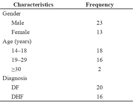
As shown in Table 1, more male patients enrolled
in the study. About half of the patients were ≤18
years of age. Out of thirty six, twenty subjects
had been diagnosed with dengue fever (DF) and
sixteen suffered from dengue hemorrhagic fever
(DHF).
The pattern of CD4+ T-lymphocyte absolute count between DF and DHF based on day of fever is shown in Figure 1. There was a similar pattern of CD4+ T-lymphocyte absolute count between DF and DHF, in which both of them consistently increase from the beginning of the febrile phase
Table 1 Baseline Characteristics of the Patients
MKB, Volume 46 No. 4, Desember 2014 223 to the end of day 7. On the second day of fever,
the average of total CD4+ T-lymphocyte absolute
count was 177 cells/µL. It increased accordingly to 245 cells/µL on the third day, 313 cells/µL on the ifth day, and to 518 cells/µL on the seventh
day.
In contrast, there was no signiicant difference
in relative CD4+ percentage from day to day in both DF and DHF. The average relative CD4+ percentage was relatively constant from time to
time and the mean ranged from 22% to 26%.
When a test was performed to analyze the median number of CD4+ T-lymphocytes, most of
the results were not signiicantly different among
DF and DHF patients, except for the relative
percentage on day 7 (p=0.022).
Discussion
The CD4+ T-lymphocyte count has been known to
be inluenced by age, sex, race, time of specimen
collection, drug administration, viral infection, and physical exercise. Compared to adults, the CD4+T cell count in children is relatively higher. A study conducted among healthy Asian people reported a range of CD4+ cell counts starting from
401to 1,450 cells/uL.6 Clearly, in the beginning
of the disease course, the CD4+ absolute cell count was below the normal level. Our study found that absolute CD4+ T-cells was low in the beginning but gradually increased from day 2 of fever to its normal level at day 7. The absolute
CD4+ T-lymphocyte count differed signiicantly among days of fever (p<0.05). Unlike our study,
Azeredo et al.7 reported that the mean percentage
of CD4+ T-cells was reduced during acute phase of dengue infection and its absolute cell counts decreased when compared to controls. This did not return to normal level until the convalesence
period. Meanwhile, a study that was conducted by Mabalirajan et al8 showed that around the time
of fever defervescence, the CD4+: CD8+ ratio was signiicantly lower in patients with dengue
infection than in healthy people. Another study also reported that after the CD4+ T-lymphocyte decreased in the early course of disease, it would gradually increase and returned to its normal absolute level on the second day after the time of fever defervescence or after shock.9 Another
suggestion have been raised recently that the CD4+ T-lymphocytes increase gradually until the 14th day post-infection, deducted by the
incubation period, that it would be apparent on day 7 after the clinical manifestation.10
The mean relative CD4+ percentage in
this study was ranging from 22% to 25% and was statistically insigniicant (p=0.573). The
percentage of CD4+ T cells, unlike the absolute CD4+ T cells count, is relatively stable in terms of the time of the day, reagents and biological
factors inluencing the absolute CD4+T cells count.6 Several other studies have found that those
patients with relatively high absolute CD4+ T lymphocytes count but low CD4+ T lymphocyte percentage have faster disease progression when they are compared to persons with low absolute CD4+T lymphocytes count but high percentage of CD4+T lymphocytes.11,12 The results showed
that absolute CD4+ T-lymphocytes counts have
no signiicant difference in term of the severity
of the cases. Several studies have pointed out Flavivirus-cross-reactive CD4+ T-cell activation in heterologous infection and have suggested its contribution to the severity of disease and to a comparable difference in absolute CD4+ count among mild and severe dengue infection cases.13 This may probably be due to the lower threshold of memory T-lymphocytes when compared to the naive cellsor the low afinity towards the second serotype which leads to delay in viral clearance
Figure 1 CD4+ T-lymphocyte Absolute Count for Dengue Fever (DF) and Dengue
Hemorrhagic Fever (DHF)
Figure 2 Relative CD4+ Percentage of Dengue Fever (DF) and Dengue Hemorrhagic Fever (DHF)
MKB, Volume 46 No. 4, Desember 2014 224
and immunopathology.14 The increasing amount
of CD4+ T-lymphocyte may be correlated with
the response of the secondary infection when the cells were exposed to the heterologus antigens,
which then produced signiicantly higher amount
of cytokines. A study by Yauch et al.3 in an animal model found that although CD4+T-lymphocyte does not have any contribution in controling DENV2 primary infection, by using epitope
identiication, there was a signiicant role of CD4+
T-lymphocyte in the secondary homologous and heterologous infections.
In conclusion, absolute CD4+ T-lymphocyte count is low in the beginning of the disease course and increases from day 2 of fever to the normal level on day 7 of fever. The relative CD4+
T-lymphocyte percentage shows no signiicant differences in increment. No signiicant difference
in the absolute CD4+ T-lymphocyte count and relative CD4+ T-lymphocyte percentage is found between dengue fever and dengue hemorrhagic fever.
Further studies involving longer follow up period and other lymphocytes are required to have a more integrated perspective on the kinetics of CD4+ T-lymphocytes in dengue infection.
References
1. World Health Organization; 2010 (accessed
10 June 2014). Available from: http://www.
searo.who.int/entity/vector_borne_tropical_ diseases/data/data_factsheet/en/
2. World Health Organization; July 2007
(accessed 24 February 2014). Available from: www. searo. who. int/en/Section10/ Section332_1104. htm
3. Yauch LE, Prestwood TR, May MM, Morar
MM, Zellweger RM, Peters B, et al. CD4+
T cells are not reqiured for the induction of
dengue virus-speciic CD8+ T cells or antibody
responses but contribute to protection after
vaccination. J Immunol. 2010;185:5405–16.
4. Mongkolsapaya J, Duangchinda T, Dejnirattisai W, Vasanawathana S, Avirutnan P, Jairungsri A, et al. T Cell responses in dengue hemorrhagic fever. Are cross-reactive T cells suboptimal. J Immunol.
2006;176:3821– 9.
5. Simmons CP, Dong T, Vin Chau N, Phuong
Dung NT, Bich Chau TN, Thu Thao LT,
et al. Early T-cell responses to dengue virus epitopes in Vietnamese adults with secondary dengue virus infections. J Virol.
2005;79:5667–75.
6. Shete A, Thakar M, Abraham PR, Paranjape R. A review on peripheral blood CD4+ T
lymphocyte counts in healthy adult Indians.
Indian J Med Res. 2010;132(6):667–75.
7. Azeredo EL, Zagne SM, Alvarenga AR, Nogueira RM, Kubelka CF, de
Oliveira-Pinto LM. Activated peripheral lymphocytes
with increased expression of cell adhesion molecules and cytotoxin markers are associated with dengue fever disease. Mem
Inst Oswaldo Cruz. 2006;101(4):437–49. 8. Mabalirajan U, Kadhiravan T, Sharma SK,
Banga A, Ghosh B. Th(2) immune response
in patients with dengue during defervescence:
preliminary evidence. Am J Trop Med Hyg.
2005;72(6):783–5.
9. Mladinich KM, Piaskowski SM, Rudersdorf
R, Eernisee CM, Weisgrau KL, Martins MA, et al. Dengue virus-speciic CD4+ and CD8+ T-lymphocytes target NS1, NS3 and NS5 in infected Indian rhesus macaques. Immunogenetics. 2012; 64(2):111–21.
10. Nielsen DG. The relationship of interacting immunological components in dengue
pathogenesis. Virol J. 2009;6:211.
11. Campbell PJ, Aurelius S, Blowes G, Harvey D. Decrease in CD4 lymphocyte counts with rest; implications for the monitoring of HIV infection. Int J STD AIDS.1997;8:423–6. 12. Hulgan T, Shepherd BE, Raffanti SP,
Fusco JS, Beckerman R, Barkanic G, et al. Absolute count and percentage of CD4+ lymphocytes are independent predictors of disease progression in HIV-infected persons initiating highly active antiretroviral therapy.
J Infect Dis. 2007;195:425–31.
13. Lühn K, Simmons CP, Moran E, Dung
NT, Chau TN, Quyen NT, et al. Increased frequencies of CD4+CD25high regulatory T
cells in acute dengue infection. J Exp Med.
2007;204(5):979–85.
14. Mongkolsapaya J, Dejnirattisai W, Xu XN, Vasanawathana S, Thangthawornchaikul N, Chairunsri A, et al. Original antigenic sin and apoptosis in the pathogenesis of dengue
hemorrhagic fever. Nat Med. 2003;9(7):921–
7.