Association Between Degree of Gastritis and
Malondialdehyde Level of Gastritis Patients
at Adam Malik General Hospital Medan
Darmadi, Gontar Alamsyah Siregar, Leonardo Basa Dairi
Division of Gastroentero-hepatology, Department of Internal Medicine, Faculty of
Medicine, University of Sumatera Utara/Adam Malik General Hospital Medan, Indonesia
Corresponding author:
Gontar Alamsyah Siregar. Division of Gastroentero-hepatology, Department of Internal Medicine, Adam Malik General Hospital, Jl. Bunga Lau No.17 Medan Indonesia. Phone/ facsimile: +62-61-8365742. Email: gontarsir@gmail.com
ABSTRACT
Background: The main pathogenesis of gastritis is inflammation which process can not be separated from the role of free radicals. Malondialdehyde (MDA) is a free radical biomarker and which increases in gastritis patients. However, studies on MDA were generally performed on experimental animals or examined MDA in
gastric mucosa. The aim of this study is to determine the association of histopathological degrees of gastritis (the
degree of lymphocyte infiltration, neutrophil activity, atrophy, and intestinal metaplasia) with plasma MDA level. Method: Cross-sectional study of 40 consecutive gastritis patients who came to endoscopic unit of Adam Malik General Hospital Medan, from January to May 2017. Assessment for the severity of chronic inflammation,
neutrophil activity, atrophy, and intestinal metaplasia refers to Updated Sydney System. Plasma MDA levels
were examined using an HPLC MDA kit. Univariate and bivariate (Chi-square and fisher exact test) analysis were performed with SPSS version 22.
Results: A total of 26 patients (65%) were men with an average age of 49.25 years. Lymphocyte infiltration was observed in 100% of specimens, neutrophil infiltration in 37.5%, atrophy in 22.5%, and intestinal metaplasia in 22.5%. There was a significant association between degree of lymphocyte infiltration with MDA level (p = 0.014; PR = 8.667; 95% CI: 1.52-89.52). There was a significant association between degree of neutrophil activity with high MDA level (p = 0.002; PR = 11.33; 95% CI: 2.64-48.74). There was a significant association between degree of atrophy with high level of MDA (p < 0.001; PR = 14; 95% CI: 3.4–57.648). There was a significant association between degree of intestinal metaplasia with high MDA level (p = 0.001; PR = 12.5; 95% CI: 3.76-24).
Conclusion:There were significant associations between degree of lymphocyte infiltration, neutrophil activity, atrophy, and intestinal metaplasia with high level of MDA.
Keywords:gastritis, Malondialdehyde, Updated Sydney System
ABSTRAK
Latar belakang: Patogenesis utama kasus gastritis adalah inflamasi yang tidak terlepas dari peranan radikal bebas. Malondialdehyde (MDA) merupakan biomarker radikal bebas dan dijumpai meningkat pada pasien gastritis. Namun, penelitian-penelitian tersebut umumnya dilakukan pada hewan coba maupun pemeriksaan MDA di mukosa gaster. Tujuan penelitian ini adalah mengetahui hubungan derajat keparahan gastritis secara histopatologi (derajat infiltrasi limfosit, aktivitas neutrofil, atrofi, dan metaplasia intestinal) dengan kadar MDA
Metode: Studi potong lintang terhadap 40 pasien gastritis secara konsekutif yang datang ke unit endoskopi
Rumah Sakit Umum Adam Malik Medan dari Januari – Mei 2017. Dinilai derajat keparahan inflamasi kronik, aktivitas neutrofil, atrofi, dan metaplasia intestinal mengacu pada Updated Sydney System. Dilakukan pemeriksaan kadar MDA plasma dengan menggunakan kit MDA HPLC. Analisis univariat, bivariat (uji Chi-square dan fisher exact) dengan SPSS versi 22.
Hasil: Sebanyak 26 orang pasien (65%) adalah laki-laki dengan rerata umur 49,25 tahun. Dijumpai infiltrasi limfosit pada 100% spesimen, infiltrasi neutrofil 37,5%, atrofi 22,5%, dan metaplasia intestinal 22,5%. Terdapat hubungan yang signifikan antara derajat infiltrasi limfosit dengan kadar MDA (p = 0,014; PR = 8,667; 95% CI: 1,52-89,52). Terdapat hubungan yang signifikan antara derajat aktivitas neutrofil dengan kadar MDA yang tinggi (p = 0,002; PR = 11,33; 95% CI: 2,64-48,74). Terdapat hubungan yang signifikan antara derajat atrofi dengan kadar MDA yang tinggi (p < 0,001; PR = 14; 95% CI: 3,4–57,648). Terdapat hubungan yang signifikan antara derajat metaplasia intestinal dengan kadar MDA yang tinggi (p = 0,001; PR = 12,5; 95% CI: 3,76-24).
Simpulan: Terdapat hubungan yang signifikan antara derajat infiltrasi limfosit, aktivitas neutrofil, atrofi,
dan metaplasia intestinal dengan kadar MDA yang tinggi.
Kata kunci:gastritis, Malondialdehyde, Updated Sydney System
INTRODUCTION
Gastritis is an inflammatory process in the mucosa and submucosa of the gaster in response to injuries that may be acute or chronic.1 The precise prevalence of gastritis is difficult to obtain because most chronic gastritis patients are asymptomatic. In worldwide, the incidence of gastritis was about 1.8 to 2.1 million of the total population per year.2 Gastritis can be caused
by Helicobacter pylori infection (H. pylori), bile reflux, non-steroidal anti-inflammatory, autoimmunity, or allergic response.3 Chronic gastritis progression may lead to complication of chronic atrophic gastritis, intestinal metaplasia, dysplasia, and gastric cancer.4
Zhang et al reported that there was currently an increased prevalence of chronic inflammation, neutrophil activity, glandular atrophy, and intestinal metaplasia compared in cases of superficial gastritis, erosive gastritis, gastric ulcers, and early gastric cancer both in cases accompanied by H. pylori infection or not. The extent and severity of gastritis objectively can be seen through an approved histopathological test using the Updated Sydney System (USS) that assesses lymphocyte infiltration, neutrophil activity, atrophy, and intestinal metaplasia, where atrophic gastritis and intestinal metaplasia are classified as gastric premalignant lesions.5
The main pathogenesis of gastritis is inflammation that can not be separated from the role of free radicals. Free radicals cause mucosal damage by causing degradation of the epithelial basement membrane, disruption of cell metabolism, and impairment of DNA.6 Anion superoxide radicals (O
2-) are produced
by neutrophil infiltration reactions to cellular lipid
membranes that lead to lipid peroxidation formation metabolized into malondialdehyde (MDA). Cytotoxic MDA is reported to play role in tumorigenesis/ carcinogenesis. Damage to lipid tissue due to free radicals can be evaluated by measuring the MDA compound which is a lipid peroxidation product. The production of free radicals is indirectly assessed with lipid peroxidation levels.7 The aim of this study was to determine the association of histopathological degrees of gastritis (degree of lymphocyte infiltration, neutrophil activity, atrophy, and intestinal metaplasia) with plasma MDA level.
METHOD
The present study was a cross sectional study on 40 consecutive gastritis patients who were admitted to endoscopy unit at Adam Malik General Hospital Medan, Indonesia from January - May 2017. Inclusion criteria included all adult patients with gastritis on endoscopy and histopathological examination. Patient with evidence of malignancy, systemic disease (pulmonary, liver, kidney, metabolic disorders, etc), gastrointestinal haemorrhage, and patients who had history of gastric surgery were excluded. All patients gave informed consent and the study was approved by the local ethics committee.
referring to visual analogue scale of the updated Sydney System. The higher degree was used if the differences of degree were found between the body and antrum. The degree of chronic inflammation, neutrophil infiltration, atrophy, and intestinal metaplasia were determined.
High performance liquid chromatographic (HPLC) analysis was performed with the isocratic method using an Agilent 1200 HPLC system (San Jose, CA, USA) with a commercial MDA kit (Immundiagnostik AG, Germany). The first step in determining MDA was a sample preparation with a derivatization reagent that transformeds MDA into a fluorescent product. Afterwards, the pH was optimized and the reaction mixture (20 mL) was chromatographed on a reversed phase C18 column (18.5 mm, 125 × 4 mm) at 30°C. The flow rate was 0.8 mL/min. Fluorimetric detection was performed with excitation at 515 nm and emission at 553 nm. The detection limit was 0.15 µmol/L.8 The
level above the mean were categorized as high level and the level below the mean were categorized as low level.
SPSS version 22 (SPSS Inc., Chicago) was used for the analysis. The data were analysed using univariate and bivariate analysis with 95% confidence interval. Bivariate analysis was carried out using fisher’s exact test, Chi-square test with a significance levels set at p < 0.05.
RESULTS
From the histopathological results of gastric mucosal biopsy specimens, there was infiltration of lymphocytes in all subjects (100%), most of whom had a mild degree of lymphocyte infiltration (70%), followed by moderate lymphocyte infiltration (22.5%), and severe lymphocyte infiltration (7.5%). In a total of 37.5% of specimens were with neutrophil infiltration, 22.5% were included in mild infiltration and 15% in moderate degrees. From 22.5% of specimens with atrophy, moderate and mild severity were 12.5% and 10%, respectively. While from 22.5% of specimens with intestinal metaplasia, mild and moderate percentages were 17.5% and 5%, respectively.
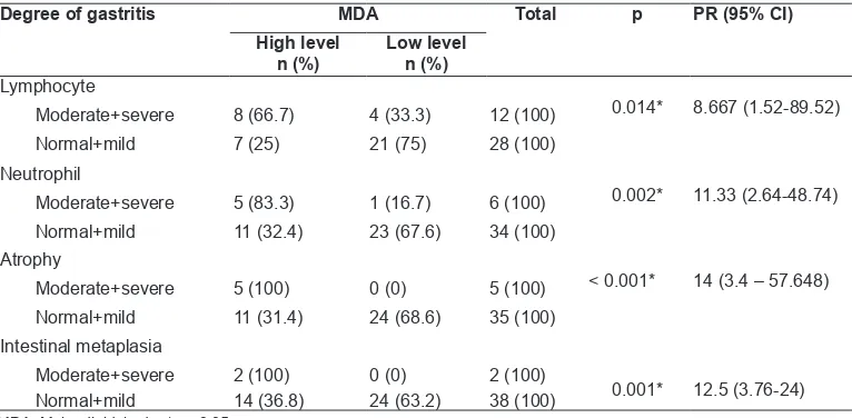
With Chi-square test, there was a significant association between degree of lymphocyte infiltration with MDA level (p = 0.014), where moderate + severe lymphocyte infiltration at risk of 8.667 fold of having higher MDA level compared to normal + low grade lymphocyte infiltration. Fisher exact tests were used to assess the associations between the degree of neutrophil activity, degree of atrophy, and degree of intestinal metaplasia with MDA levels. There was a significant association between degree of neutrophil activity and MDA levels (p = 0.002), where moderate and severe neutrophil activity at risk of 11.33 fold of having higher MDA level compared to normal + low grade neutrophil activity. There was a significant association between the degree of atrophy and MDA level (p < 0.001), where moderate and severe atrophy at risk of 14 times of having higher MDA level compared to normal + low-grade atrophy. There was a significant association between degree of intestinal metaplasia and MDA level (p = 0.001), where moderate + severe intestinal metaplasia at risk of 12.5 times of having higher MDA level compared to normal + low-grade intestinal metaplasia.
DISCUSSION
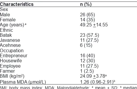
From the results of this study, it was found that the number of male suffering from gastritis was greater than female (65% vs.35%). This result was in accordance with previous studies. Elsawaf et al reported more men Table 1. Basic characteristics of the subjects (n = 40)
Characteristics n (%)
A total of 26 patients (65%) were men with an average age of 49.25 years old. The majority of subjects’ ethnic was Batak (57.5%). Two major occupations of subjects were entrepreneur (40%), followed by housewives (30%). The mean of subject's BMI was 24.09 kg/m2. The median value of plasma
MDA was 1.26 with a minimum value of 0.96 and a maximum value of 2.91.
Table 2. Degree of lymphocyte infiltration, neutrophil activity, atrophy, and intestinal metaplasia
(normal) 1(Mild) 2(Moderate) 3(Severe)
compared to women who had chronic gastritis (54.1%) in Saudi Arabia.9 Takao et al stated that the majority
of chronic gastritis in Japan were males (62.7%).10 Choudhury et al reported 65% of chronic gastritis subjects in India were male.11 While the study by Siregar GA in Medan in 2015 exhibited that 51.25% of subjects with chronic gastritis were male.12
The mean of chronic gastritis patients in this study was 49.25 years old, which was classified as productive age. The mean age in this study also did not differ from previous studies. Studies by Elsawaf et al in Saudi Arabia, Du et al in China, Takao et al in Japan, Choudhury et al in India and Siregar et al in Medan reported that the mean age of chronic gastritis patients were 43 + 10.75 years, 49.4 + 13.2 years, 56.5 years, 54.17 years, and 49.3 + 13.4 years, respectively.9-13
All subjects had chronic inflammation where all specimens were found mild, moderate, and severe lymphocyte infiltration. Most of them had mild lymphocyte infiltration (70%), followed by moderate lymphocyte infiltration (22.5%), and severe lymphocyte infiltration (7.5%), while neutrophil infiltration, atrophy, and intestinal metaplasia were found in 37.5%, 22.5%, and 22.5%, respectively. Previous studies have shown different results. Garg et al reported that there was chronic inflammation in 100% of subjects, neutrophil infiltration in 33.33%, atrophy in 12.33%, and intestinal metaplasia in 7%.14 Whereas Zhang et al reported chronic inflammation in 90.3%, neutrophil infiltration in 56.2%, atrophy in 36.8%, and intestinal metaplasia in 37%.5 Choudhury et al found chronic inflammation and neutrophil infiltration in 100% of specimens, atrophy and intestinal metaplasia in 19.23% of specimens. 11
Under normal circumstances, free radicals will be formed in a small quantity. However, this is not pathological because the amount of free radicals will be offset by the increased endogenous antioxidants (reduced glutathione, superoxide dismutase, and catalase) as a compensatory mechanism to prevent further tissue damage. Recruitment of phagocytes in gastritis will induce an increase in free radicals. Anion superoxide radicals (O2-) are generated by neutrophil
infiltration reactions to cellular lipid membranes that lead to lipid peroxidation formation that is metabolized into MDA.15 These lipid peroxidation reactions damage the cell membranes that cause the release of intracellular components such as lysosomal enzymes, cause further tissue damage, and will cause degradation of epithelial basement membrane, disrupt cell metabolism, and MDA reactions with DNA forming the mutagenic malondialdehyde deoxyguanosine (Mi-dG).16-19
Mahmood et al reported that there was a significant increase of MDA level in gastritis patients, but did not further analysed the relationship between MDA level with histopathologic degrees of gastritis.20 This study
evaluated the association of histopathological degree of gastritis with plasma MDA level. From previous studies, MDA level in the gastric mucosa were generally investigated in regard to its association with the degree of gastritis. Studies that evaluated plasma MDA concentrations focused on gastric malignancy patients.
Turkkan et al conducted a study of 35 dyspeptic patients undergoing endoscopy proving that MDA levels of gastric mucosa were significantly higher in chronic inflammatory degrees (p = 0.04).21 The result
of Turkkan et al was supported from the result of this study. There was a significant association between Table 3. Association between degree of gastritis and MDA level
Degree of gastritis MDA Total p PR (95% CI)
High level
n (%) Low leveln (%) Lymphocyte
0.014* 8.667 (1.52-89.52)
Moderate+severe 8 (66.7) 4 (33.3) 12 (100)
Normal+mild 7 (25) 21 (75) 28 (100)
Neutrophil
0.002* 11.33 (2.64-48.74)
Moderate+severe 5 (83.3) 1 (16.7) 6 (100)
Normal+mild 11 (32.4) 23 (67.6) 34 (100)
Atrophy
< 0.001* 14 (3.4 – 57.648)
Moderate+severe 5 (100) 0 (0) 5 (100)
Normal+mild 11 (31.4) 24 (68.6) 35 (100)
Intestinal metaplasia
0.001* 12.5 (3.76-24)
Moderate+severe 2 (100) 0 (0) 2 (100)
Normal+mild 14 (36.8) 24 (63.2) 38 (100)
MDA level and lymphocyte infiltration degrees, where moderate + severe lymphocyte infiltration at risk of 8.667 times fold of having higher MDA level compared to those with normal + low grade lymphocyte infiltration.
There was a significant association between degree of neutrophil and MDA level. Patients with gastritis with moderate or severe neutrophil activity at risk of 11.33 times higher level of MDA compared to those with normal + low grade neutrophil activity. This result supported previous theories. The presence of neutrophil infiltration reactions to cellular lipid membranes leads to the formation of lipid peroxidation metabolized into MDA. Everett et al who analyzed MDA levels in gastric mucosa also obtained that MDA concentrations in gastric mucosa were positively correlated with neutrophil infiltration in gastric antrum, where the higher mucosal gastric MDA level, the greater the infiltration of neutrophils in gastric antrum (r = 0.32; p = 0.05).22 Demir et al reported that high levels of gastric
mucosal MDA might indicate increased neutrophil activity in patients with peptic ulcer and gastritis.6
There was a significant association between degree of atrophy and MDA level, where moderate and severe atrophy were at risk of 14 times of higher MDA level compared to normal + low-grade atrophy. There was a significant association between degree of intestinal metaplasia and MDA level, where moderate + severe intestinal metaplasia had 12.5 times higher risk of MDA level compared to normal + low-grade intestinal metaplasia. Chronic atrophic gastritis, intestinal metaplasia, and dysplasia are included in premalignant gastric lesions, but only atrophy and intestinal metaplasia are included in the assessment of gastritic degrees according to the Updated Sydney System, which is the degree of gastritis accepted universally by the Anatomical Pathologist in the world.23 The risk
of having higher MDA levels was greater in patients with both moderate + severe atrophy and intestinal metaplasia compared to those with moderate + severe neutrophil activity. This indicated that elevation of MDA level was an important parameter for detecting severe tissue damage, and it has been reported from previous studies that MDA level were associated with mutagenesis and carcinogenesis.24
CONCLUSION
There were significant associations between degree of lymphocyte infiltration, neutrophil activity, atrophy, and intestinal metaplasia with high level of MDA.
REFERENCES
1. El-Zimaity HMT. Recent advances in the histopathology of gastritis. Current diagnostic pathology 2007;13:340-8. 2. Sonnenberg A, Lash RH, Genta RM. A national study of
Helicobacter pylori infection in gastric biopsy specimens. Gastroenterology 2010;139:1894-901.
3. Rugge M, Genta RM. Staging and grading of chronic gastritis. Human pathology 2005;36:228-33.
4. Suerbaum S, Michetti P. Helicobacter pylori infection. N Engl J Med2002;347:1175-86.
5. Zhang C, Yamada N, Wu Y. Helicobacter pylori infection,
glandular atrophy and intestinal metaplasia in superficial gastritis, gastric erosion, erosive gastritis, gastric ulcer and early gastric cancer. World J Gastroenterol 2005;11:791-6. 6. Demir S, Yilmaz M, Koseoglu M, Akalin N, Aslan D, Aydin
A. Role of free radicals in peptic ulcer and gastritis. Turk J Gastroenterol 2003;14:39-43.
7. Tiwari SK, Manoj G, Sharma V, Sivaram G, Saikant R, Bardia A, et al. Relevance of Helicobacter pylori genotypes in gastric pathology and its association with plasma malondialdehyde and nitric oxide levels. Inflammopharmacol 2010;18:59-64. 8. Immundiagnostik AG. Malondialdehyde HPLC Kit. 2010
[serial online] [cited 2016 December 10]. Available from: http://immundiagnostik.com/fileadmin/pdf/Malondialdehyd_ KC1900. pdf
9. Elsawaf ZM, Albasri AM, Hussainy AS, Alhujaily AS. Histopathological pattern of benign endoscopic gastric biopsies in Western Saudi Arabia: a review of 1236 cases. J Pak Med Assoc 2017;67:252-5.
10. Takao T, Ishikawa T, Ando T, Takao M, Matsumoto T, Isozaki Y, et al. Multifaceted assessment of chronic gastritis: a study of correlations between serological, endoscopic, and histological diagnostics. Gastroenterol Res Pract 2011;2011:ID631461. 11. Choudhury S, Laishram RS, Punyabati P, Moirangthem
GS, Debnath K. Histopathological study of gastric mucosal biopsies in chronic gastritis patients with special correlation to
Helicobacter pylori infection at Rims Hospital. J Evid Based Med Health 2016;3:2829-35.
12. Siregar GA, Halim S, Sitepu RR. Serum TNF-α, IL-8, VEGF levels in Helicobacter pylori infection and their association with degree of gastritis. Acta Med Indones 2015;47:120-6. 13. Du Y, Bai Y, Xie P, Fang J, Wang X, Hou X, et al. Chronic
gastritis in China: a national multi-center survey. BMC Gastroenterol 2014;14:21-30.
14. Garg B, Sandhu V, Sood N, Sood A, Malhotra V. Histopathological analysis of chronic gastritis and correlation of pathological features with each other and with endoscopic findings. Pol J Pathol 2012;3:172-8.
15. Li J, Tang HL, Chen Y, Fan Q, Shao YT, Jia M, et al. Malondialdehyde and SOD-induced changes of gastric tissues in acute gastric mucosal injury under positive acceleration. Genetics and Molecular Research 2015;14:4361-8.
16. Choi MA, Kim BS, Yu R. Serum antioxidative vitamin levels and lipid peroxidation in gastric carcinoma patients. Cancer Lett 1999;136:89-93.
17. Drake IM, Mapstone NP, Schorah CJ, White KL, Chalmers DM, Dixon MF, et al. Reactive oxygen species activity and lipid peroxidation in Helicobacter pylori associated gastritis: relation to gastric mucosal ascorbic acid concentrations and effect of H pylori eradication.Gut 1998;42:768-71.
mucosal injury: evidence for a dual mechanism involving lipid peroxidation and nitric oxide. Aliment Pharmacol Ther 1999;13:203-8.
19. Santra A, Chowdhury A, Chaudhuri S, Das Gupta J, Banerjee PK, Mazumder DN. Oxidative stress in gastric mucosa
in Helicobacter pylori infection. Indian J Gastroenterol
2000;19:21-3.
20. Mahmood NA. Measurement of malondialdehyde and thiol level in Iraqi patients with gastroduodenal diseases. Iraqi J Biotech2009;8:465-72.
21. Turkkan E, Uslan I, Acarturk G, Topak N, Kahraman A, Dilek
FH, et al. Does Helicobacter pylori-induced inflammation
of gastric mucosa determine the severity of symptoms in functional dyspepsia? J Gastroenterol 2009;44:66-70. 22. Everett SM, Singh R, Leuratti C, White KL, Neville
P, Greenwood D, et al. Levels of Malondialdehyde-Deoxyguanosine in the gastric mucosa: relationship with lipid peroxidation, ascorbic acid, and Helicobacter pylori. Cancer Epidemiology, Biomarkers, & Prevention 2001;10:369-76. 23. Rugge M, Pennelli G, Pilozzi E, Fassan M, Ingravallo G,
Russo VM, et al. Gastritis: the histology report. Dig Liver Dis 2011;43S:S373–84.