The Effect of Ethanolic Extract of Black and White Rice Bran
(
Oryza sativa
L.) on Cancer Cells
Rizal Maarif Rukmana
1,4, Nyoman Puniawati Soesilo
2, Rumiyati
3,
Rarastoeti Pratiwi
2,*1Student of Doctoral Program, Departement of Biology, Faculty of Biology, Universitas
Gadjah Mada, Jl.Teknika Selatan, Sekip Utara,Yogyakarta 55281, Indonesia
2Faculty of Biology, Universitas Gadjah Mada, Jl.Teknika Selatan, Sekip Utara,Yogyakarta
55281, Indonesia
3Faculty of Pharmacy, Universitas Gadjah Mada, Sekip Utara Yogyakarta 55281, Indonesia 4Departement of Medical Laboratory Technology, Faculty of Health Science, Setia Budi
University, Jl. Letjend Sutoyo, Mojosongo, Surakarta, Indonesia
Abstract
Indonesia has a wide range of rice cultivars and pigments. This rice can be used as a source of phytochemical compounds for cancer prevention. This research aims to analyze the cytotoxic activities of the ethanolic extract of black rice bran of 4 local cultivars i.e. ‘Cempo Ireng’, ‘Woja Laka’, ‘Toraja’ and ‘IR64’ (white rice) on cancer cells and to determine the compounds groups of those extracts. First step, rice bran was extracted with ethanol. This extract was applied to Raji (a human Burkitt Lymphoma cancer), HepG2 (a human liver cancer), and Vero (a nonhuman cell line) cells in order to measure the cytotoxic activities by using MTT assay. To determine descriptively the compounds groups of phenolics, flavonoids, terpenoids, steroids, and alkaloids the thin layer chromatography method was performed. The IC50value was analyzed quantitatively by using probit analysis. Results showed that the IC50values of ethanolic extract of rice bran ‘Woja Laka’, ‘Toraja’, ‘Cempo Ireng’ and ‘IR 64’ on HepG2 cells were 857.23±99.19; 1,896.55±83,8; 1,494.47±87.81 and 727.89±145,97 µg/ml respectively. The IC50on Raji cells were 816.61±85.31; 1,079.93±28.31; 1,627.82; ±119.82, and 769.33±61.43 µg/ml respectively. The IC50on Vero cells were 1,295.2±37; 1,232.07±165.51; 1,874.14±169.56, and 724.4±122.79 µg/ml respectively. The ethanolic extracts of rice bran from four cultivars contain phenolics, flavonoids, terpenoids, and steroids. However, alkaloids could not be detected. The variety of rice cultivars indicates the variation of cytotoxic activities on cancer cells. The ethanolic extracts of rice bran from those four rice cultivars contain similar kinds of organic compounds groups but vary in the Rf values.
Keywords: Cancer cells, organic compound group, cytotoxicity, ethanolic extract, rice bran
Introduction
Rice is the staple food that dominantly consumed by Indonesian people rather than other commodities (Mokoet al., 2013). Rice is a source of energy and can supply more than 70% of human caloric intake (Kanawape et al., 2011). The most common rice consumed by humans is white rice, followed by brown rice, red rice and black rice (Sutharut dan Sudarat, 2012).
Indonesian rice is not only has a high variety of cultivars but also in colors. Rice milling yields 70% of rice (endosperm) as the major product and byproducts consist of 20% rice husk, 8% rice bran and 2% rice germ (Shettharaksaet al., 2008). Aleurone layer of rice has a variety of phytochemical compounds. Mokoet al., (2013) reported that ethanolic extract of white rice bran cultivars ‘Superwin’ and ‘Cigeulis’ from Minahasa, North Sulawesi determined the phytochemical content such as phenolics, flavonoids, triterpenoids, steroids, saponins, and alkaloids. Methanolic extract of white rice bran contains tocopherol, tocotrienol, and γorisanol (Chen and Bergman, 2005).
*Corresponding author:
Rarastoeti Pratiwi
Faculty of Biology, Universitas Gadjah Mada, Jl.Teknika Selatan, Sekip Utara,Yogyakarta 55281, Indonesia, Phone/Fax: +62(274) 580839
Black rice contains anthocyanin pigment that found predominantly in the aleurone layer, pericarp and seed coat (Chaudhary, 2003). Many studies have indicated that black rice bran contains phytochemicals compounds such as polyphenols, γoryzanol, ferulic acid, caffeic acids, flavones, flavonols, carotenoids and flavonoid dominated by anthocyanin (cyanidin, peonidin, and malvidin). These compounds have been used as an antioxidant, anticancer, antiinflammatory, antibacterial, antidiabetic, anticholesterol and antiallergenic (Forsteret al., 2013; Phetpornpaisanet al., 2014).
Cancer is a group of diseases involving abnormal cell growth with the uncontrolled proliferation (Hanahan and Weinberg, 2011). Development of anticancer from natural compounds and particularly on foodstuffs is still being conducted. Previous studies have shown that anthocyanin compounds isolated from the ethanolic extract of black rice bran cultivar ‘Chinese’ have cytotoxic activities on breast cancer cells MCF7, MDAMB231, and MDAMB453. The IC50 values of anthocyanin extract of black rice bran on MCF7, MDA MB231, dan MDAMB453 cells were 374.7, 209.9 and 179.5 µg/mL respectively (Huiet al., 2010). Previous studies also reported that methanolic extract of white rice bran cultivar ‘Hommali 105’ inhibit the growth of prostate cancer cells, followed by cervical and breast cancer cells. The present study identified the composition of compounds groups and cytotoxic activities of ethanolic extract of black rice bran from local cultivars ‘Cempo Ireng’, ‘Woja Laka’, ‘Toraja’ and ‘IR64’ (white rice) on HepG2 cells (Human liver cancer), Raji cells (Burkitt lymphoma cancer) and Vero cells (derived from a normal adult African green monkey).
Materials and Methods
Chemicals
IMDM medium, DMEM medium, M199 medium, FBS (Fetal Bovine Serum) 10% v/v, MTT (3(4,5dimethylthiazol2yl) 2,5 diphenyltetrazolium bromide) were obtained from Gibco®, Grand Island, USA. Penicillinstreptomycin 2% and phosphate
buffer saline (PBS) were purchased from Sigma/Aldrich, St. Louis, MO, USA. Dimethyl sulfoxide (DMSO); stopper reagent SDS 10% in 1 N HCl, ethanol, HCl, ethyl acetate, formic acid, toluene, glacial acetic acid, nhexane, nbutanol, gallic acid, rutin, thymol, stigmasterol, sitroborat, FeCl3, anisaldehydesulfuric acid, diethylamine, quinine, dragendorf (these reagents mostly from Merck®, Darmstadt, Germany).
Rice Samples
Samples of white rice bran cultivars ‘IR 64' were collected from the organic farmer in Minggir, Sleman, Yogyakarta, Indonesia. Black rice bran cultivar 'Cempo Ireng' was obtained from the organic farmer in Sayegan, Sleman, Yogyakarta, Indonesia. Another cultivar of black rice bran ‘Woja Laka' was obtained from the farmer in Kepanjen, Malang, East Java Province, Indonesia. Meanwhile, black rice bran cultivar ‘Toraja' was obtained from the farmer in Bandar Lampung, Lampung Province, Indonesia. All rice samples planted during July to December in 2015.
Preparation of Rice Bran Ethanolic Extract
The extraction of rice bran was carried out according to the method of Pranatami, (2016) with a slight modification. Rice bran four cultivars ‘IR 64’, ‘Cempo Ireng’, ‘Woja Laka’, and ‘Toraja’ sieved with 60mesh sieve and extracted with ethanol acidified with HCl 1 N. The powder of rice bran samples 10 mg were macerated with 100 ml of solvent (ethanol: HCl 1N = 85: 15) for 48 hours and stir occasionally. The extract was filtered using Whatman No. 1. The residue was macerated with 50 ml of a mixture of solvent containing ethanol: HCl 1 N = 85:15 two times and each for overnight at room temperature. The extract was evaporated by using a fan.
Identification of Compounds groups Composition
were applied for this TLC. The using of solvent system in accordance with Table 1. The movement of the active compound was expressed by its retention factor (Rf), then the Rfvalues were calculated for each sample.
Cell cultures and treatment conditions
HepG2 cells (ATCC@HB8065), Raji cell (ATCC@CCL86TM) and Vero cells (ATCC@CCL81) were obtained from the Laboratory of Parasitology, Faculty of Medicine Universitas Gadjah Mada, Yogyakarta, Indonesia. The HepG2 cells were cultured in DMEM medium. The Raji cells were cultured in the IMDM medium. The normal Vero cells were cultured in the M199 medium. The ethanolic extract of rice bran four cultivars was dissolved in dimethyl sulfoxide (DMSO) as a solute and the maximal volume used did not exceed to 10 µl/ml of media. The cell lines were grown at 37°C in a 5% CO2incubator. Each HepG2, Raji, and Vero cell cultures were treated with the extracts with a serial concentration of 125, 250, 500, 750, 1000, 2000 µg/ml respectively and incubated for 48 h.
Cytotoxic Test by MTT assay
Each cell cultures were plated at a density of 104 cells/mL in 96well plates
(each well 100 µl) and allowed incubated in a 5% CO2incubator, at 37°C overnight. After that, the culture medium was replaced with media that containing rice bran extracts and then incubated for 48 h. Treatment medium was removed after 48 h and replaced with an MTT solution (sterile stock solution of 5 mg/ml) was added to cell media at the final concentration of 100μg/ml and this mixture was incubated at 37°C for 4–6 h in 5% CO2 incubator. MTT reaction was stopped with
stopper reagent (SDS 10% in 0.01N HCl) and then incubated overnight at room temperature. Fluorescence was measured at 595 nm (microplate ELISA reader) and the cell viability was expressed as percent of fluorescence relative to the media control. Cell experiments were replicated three times and conducted in a triplicate test.
Results and Discussion
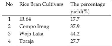
Percentage of yield of ethanolic extract of rice bran
The yield of sequential extracts is shown in Table 2. A number of rendements from the black rice (Cempo Ireng’, ‘Woja Laka’, and ‘Toraja’) was higher than from the white rice (‘IR 64’).
Cytotoxicity of Rice Bran Extracts
Plant biodiversity is not only about plant species and subspecies but also could be about plant secondary metabolites. This variety can be observed between species or even within species. This diversity of secondary metabolites and its cytotoxic activities within species are presented in this study. Four different local cultivars of ethanolic extract of rice bran showed different cytotoxicities on HepG2, Raji and Vero cell lines.
Ethanolic extract of black rice bran (Cempo Ireng, Woja Laka, Toraja) did not show the cytotoxic effect on Vero cell lines. Table 1.Solvent system and spray reagent for detecting secondary metabolites group.
However, the ethanolic extract of white rice bran (IR 64) has the effect to Vero cells. The lowest IC50value of black rice bran extract was observed on ethanolic extract of ‘Woja Laka’ with the IC50 value of 816.61±85.31
μg/ml against Raji cells, followed with the IC50 value of 857.23±99.19μg/ml against HepG2 cells (Table 3).
As Meyeret al., (1982) stated that extracts and fractions showed cytotoxicity against cancer cells if the IC50value of less than 1000 µg/ml. This current result showed that the ethanolic extract of cultivar ‘IR 64’ and ‘Woja Laka’ have IC50values of less than 1000 µg/ml on HepG2 and Raji cells. Ethanolic extract of cultivar ‘IR 64’ have IC50values of less than 1000 µg/ml on Vero cells. Meanwhile, the ethanolic extract of cultivar ‘Toraja’ and ‘Cempo Ireng’ have IC50values of more than 1000 µg/ml on HepG2 and Raji cells. This evidence seems that those extracts need to be separated by using a fractionation method to
get the more active secondary metabolites compounds. However, ethanolic extract of black rice bran ‘Cempo Ireng’, ‘Woja Laka’, ‘Toraja’ do not show the cytotoxic activity on Vero cells. Therefore, recent results showed that black rice bran extracts might safe for food material because there was no cytotoxicity observed in Vero cells. Whereas, ‘IR 64’ is not only has a cytotoxic activity on the Raji cells but also has cytotoxic activity on Vero cells. In contrast to ‘Woja Laka’ which showed more cytotoxic on HepG2 and Raji cells but less cytotoxic activity on Vero cells. This means that white rice extract rice bran less selectivity on those cells compares to the black rice bran extracts.
Previous studies of the methanolic extract on black rice bran ‘Payao’ (Banjerpongchaiet al., 2013) have shown a cytotoxic effect and inhibited cancer cell growth of HepG2. The IC50 value of methanolic extract 'Payao' on HepG2 cells Table 3.The IC50values of ethanolic extract of rice bran on HepG2, Raji, and Vero cells.
was 175.95±8.02 µg/ml. Meiyanto et al., (2005) stated that phytochemical compounds isolated from plants which less cytotoxic activity can be called as chemopreventive compounds. Further, a chemoprevention could be determined as a compound that prevents, inhibits, and normalizes carcinogenesis or prevents invasive cancer development.
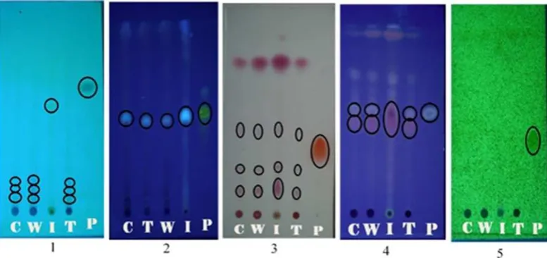
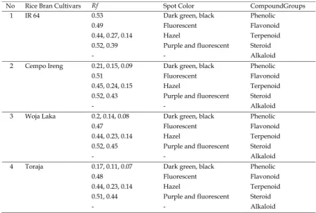
Phytochemical Screening
Thin Layer Chromatography profiles were used to identify the organic compounds groups containing rice bran. Results showed that ethanolic extracts of rice bran from four cultivars contain phenolics, flavonoids, terpenoids, and steroids groups of organic compounds however an alkaloid group could not be detected. Thin Layer Chromatography profiles of ethanolic extract of rice bran from four cultivars and theRfvalues of spots are presented in Figure 1 and Table 4.
In the present study, phytochemical screening for all four extracts showed significant indication about the presence of metabolites. Phenolics, steroids, flavonoids,
and terpenoids, were found to be present in the all the ethanolic extracts of rice bran. However, the alkaloid could not be detected in the all of the ethanolic extracts of rice bran from four cultivars. This result similar with the previous study reported that methanolic extract of black rice bran ‘Cempo Ireng’ showed the TLC profile of phytochemicals compounds such as phenolic, flavonoid, steroid, and terpenoid (Pratiwiet al., 2015).
that found in this present study are similar to the previous study that reported by Moko et al. (2013), except for alkaloids. This evidence could be due to the cultivars and environments differences in both studies.
Conclusions
Results of this study suggested that among 3 cultivars of black rice and one of white rice bran have the variation of cytotoxic activities on cancer cells. The ethanolic extracts of rice bran from those four cultivars contain phenolic, flavonoid, terpenoid, and steroid compound groups, however, the alkaloid could not be detected.
Acknowledgements
We thank the Ministry of Research, Technology, and Higher Education, Republic of Indonesia, which has given The Postgraduate Team Grant.
References
Banjerdpongchai, R., Wudtiwai. B. and Sringarm, K. 2013. The cytotoxic effect of purple rice extracts on human cancer cells related to their active compounds. Research. Gate. Conference Paper. ASEAN Food Conference.
Chaudhary, R.C. 2003. Specialty rices of the world: Effect of WTIO and IPR on its production trend and marketing. J. Food. Agric. Env., 1(2), 34 41.
Chen, M.H., and Bergman, C.J. 2005. A rapid procedure for analysing rice bran tocopherol, tocotrienol and goryzanol contents. J. Food. Comp. Anal., 18, 139–151.
Forster, G.M., Raina, K., Kumar, A., Kumar, S., Agarwal, R., Chen. M., Bauer. J. E., McClung, A. M., and Ryan, E.P. 2013. Rice Varietal Differences in Bioactive Bran Components for Inhibition of Colorectal Cancer Cell Growth. J. Food. Chem., 141, 15451552.
Gujjeti, R.P., and Mamidala, E. 2013. Phytochemical Screening and Thin Layer Chromatographic Studies of Aerva lanataRoot Extract. Inter. J. Innov. Res. Sci. Eng. Tech., 2, 57255730.
Hanahan, D., and Weinberg, R.A. 2011. The Hallmarks of Cancer. Cell, 100, 5770. Hui, C., Bin, Y., Xiaoping, Y., Long, Y.,
Chunye, C., and Mantian. 2010. Anticancer Activities of an AnthocyaninRich Extract From Black Rice Against Breast Cancer Cells In Vitro and In Vivo. Nutr. Cancer, 62(8), 11281136.
Kanawapee, J., Sanitchon, J., Srihaban, P., and Theerakulpisut, P. 2011. Genetic diversity analysis of rice cultivars (Oryza sativa L.) differing in salinity tolerance based on RAPD and SSR markers. Elec. J. Biotech., 14, 07173458. Meiyanto, E., Jenie, R.I., Rahmi, F., and Septisetyani, E.P. 2005. Aktivitas Antikanker Minyak Buah Merah terhadap Sel Kanker Plasma Darah, Sel Kanker Payudara, dan Sel Kanker Leher Rahim. Final Report. Gadjah Mada University, Bernard T. Wahyu Wiryanta.
Meyer, B. N., Ferrigni, N.R., Putnam, J.E., Jacobsen, L.B., Nichols, D.E., McLauglin, J.L. 1982. Brine Shrimp: A Convenient General Bioassay for Active Plant Constituent. Planta Med., 45, 31 34.
Moko, E.M., Purnomo, H., Kusnadi, J. and Ijong, F.G. 2014. Phytochemical content and antioxidant properties of colored and non colored varieties of rice bran from Minahasa, North Sulawesi, Indonesia. Inter. Food. Res. J., 21(3), 10531059.
Phetpornpaisan, P., Tippayawat, P., Jay, M., Sutthanut, K. 2014. A local Thai Cultivar Glutinous Black Rice Bran: A Source of Functional Compounds in immunomodulation, Cell Viability and Collagen synthesis, and Matrix metalloproteinase2 and 9 Inhibition. J. Funct. Foods, 7, 650661.
Pratiwi, R., Tunjung, W.A.S., Rumiyati, and Amalia, A.R. 2015. Apoptosis Induction in Human cervical Cancer Cells by Pigmented Rice Bran Fractions Containing Cyanidin 3glucoside and Peonidin 3glucoside. Indones. J. Biotechnol., 20(1), 1926.
Settharaksa, S., Madaka, F., Sueree, L., Chankana, N., Chakree, K., and Charoenchai, L. 2008. Cytotoxic activity and Antioxidant Potentials of Cold Pressed Rice Bran Oil. Inter. J. Pharm. Tech. Res., 6(2), 686691.