Soebagjo et al. European Journal of Biomedical and Pharmaceutical Sciences
THE ROLE OF p53 PROTEIN EXPRESSION IN RETINOBLASTOMA
Hendrian Dwikoloso Soebagjo1 *, Susy Fatmariyanti2, Delfitri Lutfi3, Aulanni’am4 , Sutiman Bambang Sumitro5 1,2,3
Department of Ophthalmology, Faculty of Medicine Airlangga University, Dr. Soetomo Hospital Surabaya,
East Java, Indonesia.
4
Biochemistry Laboratory, Department of Chemistry, Mathematics and Natural Sciences Faculty of Brawijaya
University, Malang, East Java, Indonesia.
5
Department of Biology, Mathematic and Natural Sciences Faculty of Brawijaya University, Malang, East Java, Indonesia.
Article Received on 23/04/2016 Article Revised on 13/05/2016 Article Accepted on 02/06/2016
INTRODUCTION
Retinoblastoma is the most common primary intraocular malignancy in children. The incidence of retinoblastoma reported in the literature ranges from 1 in 10.000 to 1 in 34.000 live birth (Meel et al., 2012; Moll et al. 1997). Retinoblastoma arise within the retina to form soft white tumor masses that expand within the posterior chamber of the eye and if untreated commonly invade through the sclera or along the optic nerve with poor prognosis (Divan et al., 2001).
Several approaches have been undertaken for detection, diagnosis and screening of retinoblastoma. Protein expression of retinoblastoma has been reported in few studies. Combination of markers such as RB1, E2F, p53 and polycomb group has resulted in improved diagnostic of retinoblastoma. Divan et al. (2001) reported that p53 might mediate cell cycle arrest in differentiating retinoblasts, in which a differentiation program could be initiated by induction of p21. p53 is a tumor suppressor gene, located on the short arm of chromosome 17, producing a 53 kDa nuclear protein. p53 is associated with many human cancers and p53 suppressor gene pathway are present in more than 50% of all human
tumors (Holstein et al., 1991; Kumamoto H et al., 2004). p53 gene is a well known tumor suppressor gene and its product is a transcriptional factor that plays an important role in response to DNA cellular damage. It induces G1/S cell cycle arrest in order to proceed to DNA repair or apoptosis, the latter in case of irreparable damage. DNA damage, hypoxia, oncogene activation and senescence can activate p53-mediated response (Martinez J-C et al., 2005).
Retinoblastoma is one of the few tumors in which the initial genetic mutation is known. Laurie et al., (2006) suggested that retinoblastoma bypasses p53 tumor suppressor pathway because it arises from intrinsically death-resistant cells. That study proposed that inactivation of the p53 pathway promotes the transition from differentiated retinoblastoma cells with amacrine/horizontal cell features to a more immature cell with retinal progenitor cell features.
Most of the reports cited concern in various cancer. Thus very little information is available on retinoblastoma and differential type of retinoblastoma. In present study, we collected samples from tumor tissue of intratumoral
European Journal of Biomedical
AND Pharmaceutical sciences
http://www.ejbps.com
Volume: 3 Issue: 7
01-07 Year: 2016
* Corresponding Author: Hendrian Dwikoloso Soebagjo
Department of Ophthalmology, Faculty of Medicine Airlangga University, Dr. Soetomo Hospital Surabaya, East Java, Indonesia.
ABSTRACT
This study was undertaken to demonstrate the expression of p53 in retinoblastoma. Thirty three retinoblastoma tissue samples of intratumoral retinoblastoma were tested with SDS-PAGE to find the expression of retinoblastoma protein, followed by Western blotting and immunohistochemistry to confirm p53 expression. The electrophoresis examination identified 10 major protein fractions with molecular weight from 103 – 14 kDa. Poorly differentiated retinoblastoma samples revealed 10 protein fractions (molecular weight 103 – 14 kDa) and well differentiated retinoblastoma revealed 9 protein fractions (molecular weight from 97 – 14 kDa). Western blot and immunohistochemistry assay using monoclonal antibody p53 showed that samples are positively expressed p53. No significant correlation was found between p53 expression and differentiation of retinoblastoma (p<0.429, C = 0,214).
retinoblastoma then tested by electrophoresis with SDSPAGE (sodium deodecyl polyacrylamide gel electrophoresis) to find protein expression of retinoblastoma and compare with the differentiation of retinoblastoma then confirmed the characteristic of protein by using Western blotting and immunohistochemistry assay.
MATERIAL AND METHODS Tissue samples.
We selected 33 retinoblastoma tissue samples from Dr. Soetomo Hospital –Faculty of Medicine Airlangga University, Surabaya, Indonesia. The tissues were collected after getting informed consent from the patients. Fresh surgical specimens were immediately frozen and transported to the pathology laboratory. Samples of adjacent soft tissue i i i - C This samples were useful for immunohistochemistry test and SDS-PAGE then confirmed to Western Blotting. The presence of gross tumour tissue was accessed by histological evaluation of tissue adjacent to the fragmen homogenized b h h pi ’ p h gi
SDS-PAGE
SDS-PAGE is a useful tool to determine the protein profiles of tissues. Basically, the proteins of tissue can be extracted into a buffer. The gel itself was made two parts an upper "stacking gel” and a lower "separating gel." 100V. The gel was stained by using Coomassie Brilliant Blue R-250. The molecular weight of each protein fraction was determined by using molecular weight markers as standard. The raw volume was calculated the Rf (Retardation factor) value of band which was a
Western Blotting was performed as immunological analysis for specificity test of protein. Proteins were transferred to nitricellulose PVDF membrane on <35 mA for 90 minutes. Protein result was stained with Ponceau for 5-10 minutes. Then nitricellulose membrane
containing antigen were cut into strips and blocked for 1 hour in 5% skim milk containing PBS at room temperature. The strips were washed in PBS-tween 3 times for 5 minutes. Membrane nitrocellulose incubated with primary antibodies (Mouse Anti-P53(wt-p53) (D2F7) Monoclonal Antibody-Bioss) 1:600 i i
PBS- i i 5 igh 4 C h 3
times for 5 minutes with TBST. Then membrane nitrocellulose was incubated with secondary antibody
Goat anti mouse IgG Biotin labeled (1:1000) for 1 hour at room temperature and then washed with TBST for 3 times for 5 minutes. Then membrane nitrocellulose PVDF was incubated with SA-HRP (1:2000) peroxidase-conjugated streptavidin for 40 minutes at room temperature and then washed with TBST for 2 times for 5 minutes, added by substrat AEC or western blue substrat for 30 minutes and stopped by ddH2O until band appear. Protein result was confirmed with nacalai marker stained.
Immunohistochemistry Test
The immunohistochemistry test was performed on formalin-fixed, paraffin embedded retinoblastoma tissue samples. Three-micrometer tissue sections were placed on coated slides and allowed to dry overnight. After deparaffinization and rehydration, antigen unmasking was performed by using citrate buffer for 25 minutes. Endogenous peroxidase activity was quenched by using 15 minutes incubation in 3% diluted hydrogen peroxide (H2O2). For blocking nonspecific binding, Dakocytomation (Peroxidase blocking reagen S200/30-2) was applied to the sections and then they were incubated at room temperature, with primary antibodies against p53 (CME298 AK,BK; BIOCARE®) at a dilution of 1:50 for 1 hour. After washing with phosphate-buffered saline (PBS), slides were incubated with biotin-labeled secondary antibody (Trekkte Universal Link) for 20 minutes.
Primary antibody binding was localized in tissues using peroxidase-conjugated streptavidin (Trekavidin-HRP Label) and 3,3-diaminobenzidine tetrahydrochloride (DAB) was used as the chromogen, according to
’ i uctions. The slides were
counterstained with hematoxylin, dehydrated and mounted. In parallel, known positive and negative controls were used. As positive control for p53, colorectal cancer sections were used. The immunohistochemistry slides were evaluated by an experienced pathologist, who was blind to the clinical and pathological features of the patients. The intensity of the staining was graded with proportion of percentage of positive staining in two categories: <1 ≥ 1
Statistical Analysis
variables were analyzed by the Chi-Square Test. Data were presented as percentage and mean ± SD and differences between groups were analyzed by using SPSS 15 software. A p value <0.05 was statistically significant.
RESULTS
The current study was undertaken on 33 patients with retinoblastoma. Retinoblastoma patients were at the age of 1-154 months with mean of 45.70 ±32,165 months and consisted of 57.6% (19) male and 42.4% (14) female patients (table.1).
Table 1. Clinical data, histopathology grading, and molecular weight of protein (kDa) in the 33 patients with retinoblastoma.
Patient no.
Age
(months)/ Sex Differentiated
Molecular Weight (kDa)
103 98 85 72 66 60 53 36 33 14
1 63/M Poorly ND ND D D ND D D D ND D
2 105/F Poorly ND ND D ND ND ND D ND ND D
3 19/M Poorly ND ND D D ND D D D ND D
4 98/M Poorly ND ND D D ND D D ND D D
5 24/F Poorly ND D D D ND ND D ND ND D
6 62/F Poorly ND D D D ND ND D ND ND D
7 30/M Well ND ND D ND ND ND D ND ND D
8 36/F Poorly ND D D D ND ND D ND D D
9 18/M Poorly D D D D D D D D ND D
10 45/M Well ND ND D D ND D D D ND D
11 84/F Poorly ND D D ND D D D ND ND D
12 36/M Well ND ND D ND ND ND D ND ND D
13 40/F Well ND ND D D ND ND D ND ND D
14 54/M Poorly ND D D D D D D ND D D
15 18/M Well ND D D D D D D D D
16 3/F Poorly ND ND D D ND ND D D ND D
17 34/F Poorly ND D D D ND ND D ND ND D
18 1/F Well ND ND D D ND ND D ND ND D
19 42/M Poorly ND D D D ND ND D ND ND D
20 54/M Poorly ND D D D ND D D ND D D
21 60/M Well ND ND D ND ND ND D ND ND D
22 12/M Poorly ND D D D D D D D ND D
23 30/F Poorly ND ND D D ND D D ND ND D
24 55/F Poorly ND ND D D ND ND D ND D D
25 35/F Poorly ND D D D ND ND D D ND D
26 154/F Poorly ND ND D D D D D D ND D
27 36/M Poorly ND ND D ND ND ND D ND ND D
28 29/M Well ND ND D D ND D D ND ND D
29 36/F Well ND ND D D ND ND D ND ND D
30 30/M Poorly ND ND D D D D D ND ND D
31 33/M Poorly ND D D ND ND ND D ND ND D
32 32/M Well ND D D D ND ND D ND ND D
33 100/M Poorly ND ND D D ND ND D ND ND D
D = done ND = not done
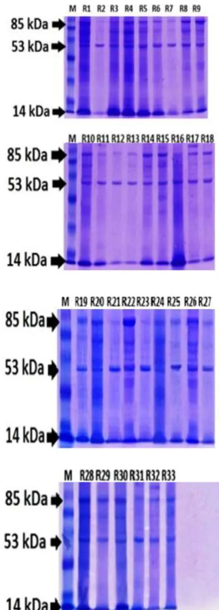
Figure 1. Protein Profile of Retinoblastoma by SDS-PAGE.
M: Protein size markers (from bottom to top): 21, 28, 37, 47, 61, 59, 116, and 200 kDa; 1-33: sample number
(poorly differentiated retinoblastoma: R1-6, R8-9, R11, R14, R16-17, R19-20, 22-27; well differentiated retinoblastoma: R7, R10, R12, R13, R15, R18, R21, R28, R29, R32).
Electrophoresis examination of 23 poorly differentiated samples revealed 10 protein fractions with molecular weight from 103 – 14 kDa. Protein fractions of molecular weight 103 kDa appeared in sample R9, R22 and R26. Protein fractions of minimal molecular weight 14 kDa were expressed in all poorly differentiated retinoblastoma samples.
Well differentiated retinoblastoma had 9 protein fractions with molecular weight from 97 – 14 kDa. Protein fractions of maximal molecular weight was expressed in sample R15 and R32. Meanwhile the minimal molecular weight was expressed in all well differentiated retinoblastoma samples.
The expression of protein fractions from all samples revealed an equal pattern of electrophoresis result. Electrophoresis examination from all samples showed that 3 protein fractions which were always expressed were 85 kDa, 53 kDa and 14 kDa. The third protein was expressed both on poorly differentiated retinoblastoma and well differentiated retinoblastoma (Figure 1). Statistical analysis of molecular weight was not significantly different in poorly differentiated retinoblastoma and well differentiated retinoblastoma (p=0 169,α<0.05).
Three protein fractions which were always expressed from electrophoresis examination, it was followed by Western Blotting only 53 kDa to confirm the molecular weight of the p53.
Figure 2. Result of Confirmation of p53 antibody by Western Blotting
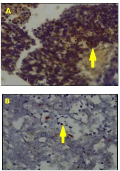
Figure 3. Immunohistochemical determination of p53 in a well differentiated (A) and a poor differentiated (B) retinoblastoma sample. Shown are positive p53 cells (arrow) with brown nucleoplasm (Magnification 400x).
This was in accordance with immunohistochemistry result. From 33 retinoblastoma samples being tested, 21% (5/23) poor differentiated retinoblastoma samples express positive p53 and 21% (2/10) well differentiated retinoblastoma samples express positive p53. No significant correlation was found between p53 and differentiated retinoblastoma (p < 0.911, C = 0,020).
Table 3: Correlation of p53 immunohistochemistry expression with differentiated Retinoblastoma in Western Blotting and Immunohistochemistry
Methods Poor
Differentiated Well Differentiated p-value
Western Blotting 3/23(13%) 3/10 (30%)
0.429 Immunohistochemistry 5/23 (21%) 2/10 (20%)
No significant correlation was found between p53 and differentiated retinoblastoma using both methods (p < 0.429, C = 0,214).
DISCUSSION
Electrophoresis examination from all retinoblastoma samples showed that 10 fraction proteins expressed, 103 kDa, 98 kDa, 85 kDa, 72 kDa, 66 kDa, 60 kDa, 53 kDa, 36 kDa, 33 kDa and 14 kDa. But from all samples, three protein fractions were always seen both in poorly and well differentiated retinoblastoma, which are 85 kDa, 52 kDa and 14 kDa. It has shown that the proteins play a role in retinoblastoma, both poorly differentiated retinoblastoma and well differentiated retinoblastoma.
Poor differentiated and well differentiated retinoblastoma showed insignificant result from SDS-PAGE. All samples express protein with molecular weight 53 kDa. This result confirmed with Western Blot test and showed 18% from all protein expression with molecular weight 53 kDa is p53. It is indicated that protein with molecular weight 53 kDa in retinoblastoma mostly not p53. Western Blott test result reinforced with immunohistochemistry test that showed expression between poor differentiated and well differentiated retinoblastoma has no significant difference (p<0.429, C = 0,214).
While the protein with a molecular weight of 53 might include the p53. p53 is a 53 kDa. p53’ g i functions of eliciting apoptosis, cell cycle arrest and senescence, more recent studies had discovered that limit angiogenesis, regulate autophagy and directly influence survival proteins in the mitochondria, mRNA processing and DNA repair pathways. p53 is frequently mutated in about 50% of human tumours and the remainings seem to have malfunctions in its pathways. This strongly suggests that most cancer cells are defective either in p53 or in its pathways and p53 malfunction is considered one of the most common mechanisms in tumor development. (Kim and Dass, 2011; Teodoro JG et al., 2007; Maiuri MC et al., 2010; Brown CJ et al.,2009).
filament (Sandilands A et al., 1995), ACTL6A actin like 6A that function on cell trancription process and express in cell nucleus (Anagnostopoulos AK et al., 2005). All of these protein have the possibility to express on retinoblastoma and another cancer. Beside that, there is enzym with molecular weight 53 kDa which is EPHX- 1 (epoxide hydrolase-1) that play a role in microsomal epoxide catabolic process that change hydrocarbon policyclic into metabolit carsinogenic, there are lots in lung and kidney cancer (Lin, et al., 2007).
Sitorus (2009) said that retinoblastoma possessed paradox appearance in apoptosis. Retinoblastoma grow because the homeostatic control mechanisms that maintain the appropriate number of cells in normal tissues are defective, leading to an imbalance between cell proliferation and cell death. Generally, retinoblastoma possess an increasing proliferative appearance and so does apoptosis. Divan et al. (2001) said that apoptosis was spatially associated with increased p53 expression and might be p53 mediated in poorly differentiated tumors, but in well-differentiated tumors apoptosis did not colocalize with p53 and maybe p53 independent.
Loss of RB1 gen in retinoblastoma process is the early key of inactivation process of p53 pathway. When Rb pathway inactivated, retina formation and proliferation process begin from activation of Arf-MDM/MDMX-p53 pathway. MDM2 (mouse double minute 2), which encodes a protein capable of binding to the N-terminal region of p53 and negatively regulate its function. Besides, MDM2 protein (Mdm2) functions as an E3 ubiquitin ligase, which promotes degradation of p53, triggering its nuclear exportation and proteosomal destruction (Wallace et al., 2006). MDM2 as p53 suppressor play a role in neoplasia process in retinoblastoma (Ying et al., 2008).
Role of p53 protein in research result showed there is no difference in retinoblastoma differentiated process (well or poor). This is showed that p53 do not play a spesific in both poor and well differentiated retinoblastoma are inactive with low expression p53 on Western Blotting 18% (6/33) and 21% (7/33) on immunohistochemistry test. It showed that p53 do not play significant role in retinoblastoma.
REFERENCES
1. Anagnostopoulos AK, Vougas K, Kolialexi A, et al. (2005). The Protein Profile of the Human Immature T-cell Line CCRF-CEM. Cancer Genomics & Proteomics, 2: 271-300.
2. Brown CJ, Lain S, Verma CS, et al. (2009). Awakening guardian angels: drugging the p53 pathway. Nat Rev Cancer, 9: 862–73.
3. Burkhart DL, Sage J (2008). Cellular mechanisms of tumour suppression by the retinoblastoma gene. Nat Rev Cancer, 8: 671-82.
4. Caganova, M (2011). The Role of Ezh2 in B Cell Development and Adaptive Immunity. Dissertation. European School of Molecular Medicine (SEMM), University of Milan and University of Naples “F i II” F M i i , Divan A, Lawry J, et al. (2001). p53 and p21waf-1 Expression Correlates with Apoptosis or Cell Survival in Poorly Differentiated, but not Well-Differentiated, Retinoblastomas. Cancer Research, 61: 3157-63. 5. Dominguez-Brauer C, Brauer PM, et al. (2010).
Tumor suppression by ARF. Gatekeeper and caretaker. Cell Cycle, 9: 86-9.
6. Eagle, RC (2009). High-risk features and tumor differentiation in retinoblastoma. A retrospective histopathologic study. J.Arch. Pathol Lab Med, 133: 1203–9.
7. Fukuda T, Guo L, et al. (2003). CH-ILKBP regulates cell survival by facilitating the membrane translocation of protein kinase B/Akt. The Journal of Cell Biology, 160: 7.
8. Gupta, A, Cao W, Sadashivaiah K, et al. (2013). Promising Noninvasive Cellular Phenotype in Prostate Cancer Cells Knockdown of Matrix Metalloproteinase 9. J Sci World. Hindawi Pub Co, 1-13.
9. Hollstein M, Sidransky D, et al. (1991). p53 mutations in human cancers. Science, 253: 49. 10. Kim SH, Dass CR (2011). p53-targeted cancer
pharmacotherapy: move towards small molecule compounds. J Pharm and Pharmacol, 63: 603–10. 11. Knudson, AG (2001). Two genetic hits (more or
less) to cancer. Nature Reviews, 1: 157-70.
12. Kumamoto H et al. (2004). p53 gene status and expression of p53, MDM2 and p14ARF proteins in ameloblastomas. Journal of Oral Pathology and Medicine, 33: 292-9.
13. Laurie NA, Donovan SL, Shih CS, et al. (2006). Inactivation of the p53 pathway in retinoblastoma.
Nature, 444(7115): 61-6.
14. Lin TS, Huang HH, Fan YH, et al. (2007). Genetic polymorphism and gene expression of microsomal epoxide hydrolase in non-small cell lung cancer. meningeal haemangiopericytomas. Study by double immunofluorescence and laser scanning confocal microscopy. Histopathology, 46: 184-94.
management of retinoblastoma. Indian J Med Paediatr Oncol, 33(2): 80–8.
18. Moll AC, Kuik DJ, Bouter LM, Otter et al. (1997). Incidence and survival of retinoblastoma in the Netherlands: a register based study 1862–1995.
British J Ophth, 81: 559–62.
19. Sitorus RS, Gumay S, Valk PVD (2009). The apoptosis paradox in retinoblastoma. Natural compounds and their role in apoptotic cell signaling pathways. Ann NY Acad Sci, 1171: 77-86.
20. Teodoro, JG, Evans SK, Green MR (2007). Inhibition of tumor angiogenesis by p53: a new role for the guardian of the genome. J Mol Med, 85: 1175–86.
21. Viatour P, Sage J (2011). Newly identified aspects of tumor suppression by RB. Disease Models & Mechanisms, 4: 581-5.
22. Wallace VA (2006). Second step to retinal tumours.
Nature, 444: 2.
23. Yu JC, Shen CY (2002). Two-hit Hypothesis of Tumor Suppressor Gene and Revisions. J Med Sci,