AMINO ACID SEQUENCE OF B CELL EPITOPE OF
N-TERMINAL REGION OF ESAT-6 MYCOBACTERIUM
LEPRAE ROLE AS SPECIFIC ANTIGEN FOR DIAGNOSIS
OF LEPROSY
Fauziah Nuraini Kurdi, Indro Handojo, Indropo Agoesni and Yoes Priatna Dahlan
Physical and Health Study Program, Teacher Training and Education Faculty and Rehabiltitation Medicine, Medical Faculty, Sriwijaya University, Palembang; Department of Clinical Pathology, Department of Dermatology and Venerology,
Department of Parasitology, Medical Faculty, Airlangga University, Surabaya, Indonesia
Abstract. The objective of this study was to find a specific B-cell epitope of Nterminal region of antigen L-ESAT-6 from leprosy patients, healthy individuals and healthy nurses working for more than 10 years in the leprosy ward of dr.A.Rivai Abdullah Leprosy Hospital, Palembang, Indonesia. Fifty subjects were enrolled in this study, comprising 10 subjects with LL type leprosy, 10 subjects with BB type leprosy , 10 subjects with TT type leprosy, 10 healthy nurses from leprosy ward and 10 healthy individuals as control group. The amino acid sequence of residues 11-36 of the N-terminal region of L-ESAT-6 were divided into a series of 18 peptides each consisting of 9-mer peptides with an overlap of 8-mers and an offset of one amino acid. The series of 18 peptides were synthesized in the form of biotinylated peptides and used to screen sera of 50 subjects using an indirect ELISA method. Our study identified at the N-terminal of L-ESAT-6, LEQCQES, VNELQG and IDALLE as epitope marker for LL and BB type of leprosy, epitope marker for TT type of leprosy and for protective epitope marker) healthy nurses working for more than 10 years in the leprosy ward, respectively. These antigens can be used in immunochromatographic test for the early diagnosis of leprosy.
Key words: L-ESAT-6, B-cell epitope mapping, M. leprae diagnostic tool, protective marker, leprosy marker
INTRODUCTION
Leprosy is still a major public health problem in Indonesia as well as in the world. The number of leprosy cases in Indonesia could be classified as number three in the world after India and Brazil. The world prevalence of leprosy in 2008 was 212,802, while 254,525 new cases were reported in 2007, 15% of which were children (WHO, 2006).
The main problem of leprosy is its potency to cause damage of skin, nerves, eyes and other organs. In the long run it can cause body defects, especially the extremities, which has an impact on patient’s social as well as quality of life. In Indonesia active transmission of the disease still occurs due to the high incidence of multibacillary types of leprosy (Agoesni, 2003; Scollard et al, 2006).
Case finding followed by adequate treatment is the key point in leprosy control program (Scollard et al, 2006; WHO, 2006). For case finding, a reliable and practical diagnostic tool for early diagnosis of leprosy has to be used (Handojo, 2002). However, diagnostic tools currently used for leprosy does not meet the above mentioned criterion (Sengupta, 2001). In the last decade, a low molecular weight protein (L-ESAT-6), secreted extracellularly only by virulent strains of Mycobacterium tuberculosis and
M. leprae). It is therefore assumed to be the virulence factor of Mycobacterium, was
found to play an important role in the immune response against the invading
antibodies against L-ESAT- 6 were significantly higher than the one against phenolic glycolipid-I (PGL-1), which is widely used as the antigen of a serologic test for the diagnosis of leprosy. Besides, the amino acid sequence of Mycobacterium leprae L-EAST-6 differs by 60% from that of M. tuberculosis (Geluk et al, 2002; Spencer et al, 2002). This has led to the conclusion that L-ESAT-6 has a potential to be used as an antigen for early diagnosis of leprosy.
To obtain a very specific test, the antigen used should be very specific and purified. For this purpose, a B-cell epitope mapping of the specific region of L-ESAT- 6 should be performed to search for the epitope marker in leprosy patients and the protective epitope marker of healthy nurses in the leprosy ward. Thus, the aim of this study was to map the B-cell epitopes of L-ESAT-6 of various types of leprosy and those of healthy nurses who are in close contact with leprosy patients for more than 10 years.
MATERIALS AND METHODS
Subjects
This study was carried out on 30 newly found leprosy patients, aged 18-55 years, comprising 20 males and 10 females who attended the leprosy outpatient clinic of Sei Kundur Hospital in Palembang, Indonesia, 10 healthy nurses, aged 28-55 years, comprising 2 males and 8 females who worked for more than 10 years at the leprosy ward; and 10 healthy subjects free from leprosy, aged 18-55 years, comprising 8 males and 2 females who live at least one year in the endemic area.
The leprosy patients were divided into 3 groups as follows: 10 patients with LL type , 10 patients with BB type, and 10 patients with TT type. The diagnosis of leprosy was based on the standard diagnosis of WHO (2000).The leprosy patients enrolled in this study had not received anti-leprosy treatment and were not pregnant.
Healthy nurses enrolled in this study, had positive lymphocyte transformation test (LTT) with a value of IS>1 (Deubner et al, 2001; Klein et al, 2004) and showed elevated concentrations of TNF-〈 of the supernatant of peripheral blood mononuclear cell (PBMC) culture stimulated with the mitogen using Dharmendra lepromins when compared with unstimulated condition. The anti PGL-1 titer of these healthy nurses was within normal limits (Fulya et al, 2006).
Healthy subjects in this study had normal LTT with a value of IS < 1 (Klein et al, 2004) but had elevated concentration of TNF-〈 in the supernatant of PBMC culture stimulated with the mitogen using Dharmendra lepromins when compared with unstimulated condition (Kaur et al, 2001).
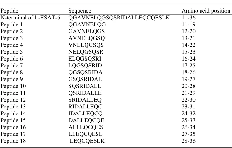
Table 1
Sequence of L-ESAT-6 peptides used in the study
Peptide Sequence Amino acid position
N-terminal of L-ESAT-6 QGAVNELQGSQSRIDALLEQCQESLK 11-36
Peptide 1 QGAVNELQG 11-19
Peptide 2 GAVNELQGS 12-20
Peptide 3 AVNELQGSQ 13-21
Peptide 4 VNELQGSQS 14-22
Peptide 5 NELQGSQSR 15-23
Peptide 6 ELQGSQSRI 16-24
Peptide 7 LQGSQSRID 17-25
Peptide 8 QGSQSRIDA 18-26
Peptide 9 GSQSRIDAL 19-27
Peptide 10 SQSRIDALL 20-28
Peptide 11 QSRIDALLE 21-29
Peptide 12 SRIDALLEQ 22-30
Peptide 13 RIDALLEQC 23-31
Peptide 14 IDALLEQCQ 24-32
Peptide 15 DALLEQCQE 25-33
Peptide 16 ALLEQCQES 26-34
Peptide 17 LLEQCQESL 27-35
Every subject enrolled in this study signed informed consent form, which was approved by the Ethics Committee of Dr A Rivai Abdulah Hospital, Palembang.
Peptide design and screening
The N-terminal region of L-ESAT-6 (residue number 11-36) was the target of this study. Using the GNET computer program (Worthington and Morgan, 1994; Mimotope,2008) the peptide was divided into a series of 18 overlapping peptides, each consisting of 9-mers with an overlapping of 8-mers and an offset of one amino acid (Table 1).
The peptides in biotinylated form were synthesized by Chiron Technologies, Clayton, Victoria, Australia, which also provided streptavidin-coated microtiter plates.
Sera obtained from the 50 subjects of this study were screened using procedures in accordance with the manufacturer’s instructions. Biotinylated peptides were reconstituted in 200 ⎧l of dimethyl sulfoxide or dimethyl formamide. Before starting the test, the reconstituted peptides were diluted 1:1,000 in phosphate-buffered saline solution (PBS) containing 0.1% bovine serum albumin (BSA) and 0.1% sodium azide. The solution was rediluted 1:5 and kept at -20ºC until used. The streptavidin-coated wells were filled with 100 ⎧l of diluted biotinylated peptide and incubated with shaking for 1 hour at 20ºC. Then the wells were washed 4 times with PBS-T20 (Tween 20). Subsequently, 100 ⎧l of diluted serum (1:1,500) were added and the plate was incubated with shaking overnight at 4ºC. After another wash cycle as described above, 100 ⎧l of diluted (1: 500) conjugate solution goat antihuman IgG labeled with horse radish peroxides (HRP) ( KPL cat.number 05-10-06) were added and the plate was incubated for 1 hour at 20ºC. The next step was the wash cycle as described above, followed by washing twice with PBS to remove any traces of Tween 20. Then 100 ⎧l of chromogenic substrate solution (0.03% H2O2 plus ABTS) (Pierce Cat.No.37615) were added and the colorometric reaction was developed in the dark for 45 minutes at 20ºC. The absorbence results of the tests were measured using a micro-ELISA reader at 405 nm or 492 nm. The cut-off value of reactivity of the test is according to Worthington and Morgan (1994), namely, lowest 80 or 90% of the results (absorbance values). Positive values were those 80% above the lowest 20% of all absorbances measured on all 18 peptides reacting with every subject serum. Therefore the cut-off value was different from one subject to another. Absorbance values higher than the cut-off values were considered as reactive (positive) .
The peptides, which were assessed as reactive and significant with the antibody in the sera of the subjects enrolled in this study, were grouped and evaluated based on the method of Worthington and Morgan (1994). Each group of reactive peptides consists at least of 4 adjacently located reactive peptides. According to Tam (1994) an epitope could only be recognized by Bcells if it has minimally 6-mer amino acid sequence. The prevalence of each epitope in the groups of subjects enrolled in this study was statistically evaluated using the z proportional test with an 〈 = 0.05 and z table = 1.96 (Sudjana, 1996).
Table 2
Results of the study on 30 patients with various types of leprosy, 10 healthy nurses working for more than 10 years at the leprosy ward and 10 healthy controls who
lived for more than one years in endemic area of leprosy.
Amino acid sequence Present in Not present in Z-value of the epitope subject group subject group
RESULTS
As shown in Table 2, the epitope with amino acid sequence LEQCQES (28-34) was found in the groups of type LL and BB leprosy patients but not in the other groups of subjects, such as healthy subjects, healthy nurses as well as in patients with type TT leprosy. The epitope with amino acid sequence VNELQG (14-19) was found only in the group of patients with type TT leprosy and not in the other groups of subjects. On the other hand, the epitope with the amino acid sequence IDALLE (24-29) was found only in healthy nurses and not in the other groups of subjects.
DISCUSSION
From the data obtained in this study, it is very clear that the leprosy epitope markers were located at the N-terminal of L-ESAT-6, ie LEQCQES (for LL and BB type leprosy) and VNELQG (for TT type leprosy), as these epitopes were only found in the patient groups. However, the protective marker epitope IDALLE, which has a very high prevalence rate in healthy nurses, was also present in low percentage in the group of healthy individuals (free from leprosy) who lived more than one year in the leprosy endemic area. It is possible that these apparently healthy individuals who lived in endemic area of leprosy might have been infected with M. leprae with a low degree of virulency. Thus, the immune response of such individuals could have killed the infecting
M. leprae and produced protective antibodies against L-ESAT-6. Further studies should
be performed to prove this hypothesis.
To our knowledge these epitopes are unique to Indonesian subjects because this study was performed using sera from Indonesian people who suffered from leprosy and have not yet been studied in other Southeast Asian regions. Further research should be done to compare the finding of L-ESAT-6 epitope with other races in Southeast Asian region or other parts of the world.
Some infectious diseases such as leprosy, for which no vaccines are available and this can have an impact on the social and the individual quality of life of the patients. Early case finding followed by adequate treatment is mandatory. For this purpose, a reliable, practical and inexpensive diagnostic tool for the early diagnosis of the disease is the key point for a control program. This study has shown that the specific epitope marker of type LL and BB leprosy was the amino acid sequence LEQCQES of L-ESAT-6, and that of type TT leprosy was VNELQC, while the specific protective epitope marker was IDALLE. It is worth mentioning that these marker epitopes are not found in T-ESAT- 6 and thus are very specific markers of leprosy. If epitopes are used as antigens for the detection of antibodies in the sera of leprosy patients and contact subjects, it is expected that these antigens will be very specific antigens for the serologic test of leprosy. To increase the sensitivity of the diagnostic test, these epitopes may have to be polymerized into antigens with higher molecular weights. To get a high degree of practicability, the above mentioned antigens can be used in lateral flow immunochromatographic test (ICT) (Handojo, 2002). Besides, ICT is a relatively inexpensive test and its high degree of detectability makes this test very suitable for use in the early diagnosis of leprosy.
In summary, we have identified at the N-terminal of L-ESAT-6, LEQCQES, VNELQG and IDALLE as epitope marker for LL and BB type of leprosy, epitope marker for TT type of leprosy and protective epitope marker for healthy nurses working for more than 10 years in the leprosy ward, respectively. These antigens can be used in ICT for the early diagnosis of leprosy.
ACKNOWLEDGEMENTS
REFFERENCES
Agoesni I. Leprosy, the ancient disease with an abundance of mysteries (Penyakit Kusta Penyakit Tua Dengan Segudang Misteri). Oration as Professor at Airlangga University. Surabaya: Airlangga University Press, 2003.
Deubner DC, Goodman M, Iannuzzi J. Variability, predictive value, and uses of the Beryllium Blood Lymphocyte Proliferation Test (BLPT): Preliminary analysis of the ongoing workforce survey. App Occup Envirm Hyg 2001; 16: 521-6.
Fulya I, Mehmet O, Handan O, Vedat B. Cytokine measurement in lymphocyte culture supernatant of inactive lepromatous leprosy patients. Indian J Med Microbiol 2006; 24: 121-3.
Geluk A, van Meijgaarden KE, Franken KL, et al. Identification and characterization of the ESAT-6 homologue of Mycobacterium leprae and T-cell cross-reactivity with
Mycobacterium tuberculosis. Infect Immun 2002; 70: 2544-8.
Geysen HM, Rodda SJ, Mason TJ, Tribbick G. Strategies for epitope analysis using peptide synthesis. J Imunol Methods 1987; 102: 259-74
Handojo I. Introduction to basic immunoassay. Surabaya: Airlangga University Press, 2002: 109-90.
Handojo I. Development of diagnosis and prevention of tuberculosis through epitope mapping of ESAT-6 Mycobacterium tuberculosis. Oration as Professor at Airlangga University. Surabaya: Airlangga University Press, 2003.
Kaur I, Agnihotri N, Mehta M, Dogra S, Ganguly NK. Tumor necrosis factor (TNF) production in leprosy patients. Int J Lepr Other Mycobact Dis 2001; 69: 249-50. Klein R, Schwenk M, Ramm RH, Templeton DM. Diagnostic relevance of the
lymphocyte transformatiom test for sensitization to beryllium and other metals.
Pure Appl Chem 2004; 76: 1269-81.
Mimotopes. Mimotope peptides and immunology. Mimotope Library, Clayton, Victoria: Australia, 2008.
Parkash O, Pandey R, Kumar A, Kumar A. Performance of recombinant ESAT-6 antigen (ML0049) for detection of leprosy patients. Lett Appl Microbiol 2007; 44: 524-30. Scollard DM, Adams LB, Gillis TP, Krahenbuhl JL, Truman RW, Williams DL. The
continuing challenges of leprosy. Clin Microbiol Rev 2006; 19: 338-81.
Sengupta U. Difficulties in the early serodiagnosis of leprosy. ICMR Bull 2001; 31(11). Spencer JS, Marques MA, Lima MCBS, et al. Antigenic specificity of the
Mycobacterium leprae homologue of ESAT-6. Infect Immun 2002; 70: 1010-3.
Sudjana. Statistic method. Bandung:Tarsito Press, 1996: 246-7.
Tam JP. Immunization with peptide-carrier complexes : traditional and multiple-antigen peptide systems. In: Wisdom GB, ed. Peptide antigen: a practical approach. Oxford, New York, Tokyo: IRL Press at Oxford University Press, 1994: 111-5. World Health Organization (WHO). Guide to eliminate leprosy as a public health
problem. Geneva: WHO, 2000: 1-5.
World Health Organization (WHO). Global leprosy situation. Wkly Epidemiol Res 2006; 81: 309-16.