See discussions, stats, and author profiles for this publication at: https://www.researchgate.net/publication/51752749
Evaluation of myelin sheath and collagen
reorganization pattern in a model of peripheral
nerve regeneration using an...
Article in Histochemie · December 2011
DOI: 10.1007/s00418-011-0874-3 · Source: PubMed
CITATIONS
10
READS
95
4 authors, including:
Some of the authors of this publication are also working on these related projects:
“ALIGNED PLASMA BIO-ACTIVATED NANOSTRUCTURED CONDUITS FOR PERIPHERAL NERVE REGENERATION”. Referencia: 24J2014.0007.01.View project
Co-Investigador Principal en el proyecto “ELABORACIÓN Y EVALUACIÓN PRECLÍNICA DE UN NUEVO MODELO DE NERVIO PERIFÉRICO ARTIFICIAL GENERADO POR INGENIERÍA TISULAR” Referencia: FIS PI14-1343View project
Víctor Carriel
University of Granada
62PUBLICATIONS 248CITATIONS
SEE PROFILE
Ingrid Garzon
University of Granada
82PUBLICATIONS 494CITATIONS
SEE PROFILE
All content following this page was uploaded by Víctor Carriel on 22 January 2017.
1 23
Histochemistry and Cell Biology
ISSN 0948-6143
Volume 136
Number 6
Histochem Cell Biol (2011) 136:709-717
DOI 10.1007/s00418-011-0874-3
Evaluation of myelin sheath and collagen
reorganization pattern in a model of
peripheral nerve regeneration using an
integrated histochemical approach
1 23
S H O R T C O M M U N I C A T I O N
Evaluation of myelin sheath and collagen reorganization pattern
in a model of peripheral nerve regeneration using an integrated
histochemical approach
Vı´ctor Carriel•Ingrid Garzo´n• Miguel Alaminos•
Antonio Campos
Accepted: 9 October 2011 / Published online: 29 October 2011
ÓSpringer-Verlag 2011
Abstract Peripheral nerves are complex histological structures that can be affected by a variety of conditions with different degree of axonal degeneration and demye-lination. For the study of peripheral nerve regeneration in pathology and tissue engineering, it is necessary to evaluate the regeneration, remyelination and extracellular matrix reorganization of the neural tissue. Currently, different histochemical techniques must be used in parallel, and a correlation among their findings should be further per-formed. In this work, we describe a new histochemical method for myelin and collagen fibers based on luxol fast blue and picrosirius methods, for the evaluation of the morphology, the myelin sheath and the collagen fiber reorganization using a model of peripheral nerve regener-ation. Whole brain, normal sciatic nerve and regenerating peripheral nerve samples were fixed in 10% neutral buf-fered formalin and paraffin-embedded, for the performance of the hematoxylin-eosin stain, the Luxol fast blue method and the new histochemical method for myelin and colla-gen. The results of this technique revealed that this new histochemical method allowed us to properly evaluate histological patterns, and simultaneously observe the his-tochemical reaction for myelin sheath and collagen fibers in normal tissue, and during the regeneration process. In conclusion, this new method combines morphological and histochemical properties that allowed us to determine with high accuracy the degree of remyelination and collagen fibers reorganization. For all these reasons, we hypothesize
that this new histochemical method could be useful in pathology and tissue engineering.
Keywords Myelin sheathCollagen fibers
Peripheral nerve regeneration Histochemistry
Tissue engineering
Introduction
Peripheral nerves are complex histological structures whose main components are neuron axons, myelin sheaths syn-thesized by Schwann cells and a collagen-rich extracellular matrix (ECM) (Mills2007). These structures can be affected by a variety of conditions and neuropathies with different degrees of axonal degeneration and demyelination (Oh
2001). The study of the regeneration processes that occur during peripheral nerve reparation is one of the goals of current biomedical research (Chalfoun et al.2006; Jiao et al.
2009); and assessment of peripheral nerve morphology is a pillar in the investigation of nerve damage and regeneration in tissue engineering (Vleggeert-Lankamp2007). The use of light microscopy plays an essential role in the evaluation of morphological features of the pathologic and regenerative processes that occur in the peripheral nerve structure. The use of morphological, histochemical and immunohisto-chemical techniques provides information about patho-physiological conditions in peripheral nerves.
Light microscopy allows observing the complex histo-logical structure of the peripheral nerve with several lim-itations. In many cases, the histological image that can be observed using routine nerve staining methods is poor. For most researchers, the gold standard in peripheral nerve histology is toluidine blue staining of resin-embedded semi thin sections, which allows the accurate identification of
V. Carriel (&)I. Garzo´nM. AlaminosA. Campos
Department of Histology (Tissue Engineering Group), University of Granada, Avenida de Madrid 11, 18012 Granada, Spain
most myelinated fibers (Mills 2007; Hirano 2005). How-ever, this methodology is time-consuming, expensive and requires special equipment such as ultramicrotomes.
Regarding the histochemical techniques, there are several reagents that specifically stain myelin, and these techniques have been an important part of the histopathologic exami-nation of the nervous system (Bancroft and Gamble2008; Kiernan 2008; Mills 2007). The Luxol fast blue (LFB) method described by Klu¨ver and Barrera in 1953 is most often used in histopathology for the evaluation of myelin in central and peripheral nervous system. Myelin can also be detected by immunohistochemistry and immunofluores-cence using antibodies that specifically recognize the myelin basic protein (Mills2007; Taylor and Cote2005).
For the study of peripheral nerve injuries and the regeneration process that is associated to these injuries, it is necessary to evaluate the presence of axonal sprouting, remyelination and ECM remodeling in these tissues (Mills
2007; Oh 2001; Jiao et al. 2009). Several techniques are available, but most of these must be performed and inter-preted in parallel, and a correlation among their findings should be further carried out. For the evaluation of these parameters, the development of techniques that allow a comprehensive assessment of the main histochemical properties using a single histological slide would be useful. In this work, we describe a new (MCOLL) histochem-ical method based on conventional luxol fast blue and picrosirius histochemical methods, for the simultaneous staining of the morphology pattern, myelin sheath and stromal collagen fibers in different nerve tissues.
Materials and methods
Animal tissues
All animals used in this study were obtained from the Service of Production and Animal Experimentation, Uni-versity of Granada, with the approval of the Ethics Com-mittee of the University of Granada, Spain. The animals were housed in a temperature controlled environment (21±1°C), maintained on a 12 h light/dark cycle, and
given free access to tap water and standard rat chow. To evaluate this new histochemical method in normal native neural tissues, we used normal brains and sciatic nerves from 12 female Wistar rats of 12 weeks old weighing 250–300 g.
For the evaluation of different degrees of axonal degeneration, demyelination and remyelination process, we used a peripheral nerve regeneration model. In this case, animals were anesthetized by an intraperitoneal injection of a mixture of acepromazine (Calmo-NeosanÒ0.001 mg per gram of weight of the animal) and ketamine (Imalgene
1000Ò 0.15 mg per gram of weight of the animal). Then, 10 mm of the left sciatic nerve were surgically removed from each animal, and a commercially available type I-collagen conduit (CC) (NeuraGenÒ) was microsurgically implanted between both nerve ends to induce nerve regeneration as clinically used in human patients with a peripheral nerve lesion (Wangensteen and Kalliainen
2010). The right sciatic nerve was used as control in each animal. After 12 weeks of the implant of the CC, the ani-mals were euthanatized under general anesthesia and both sciatic nerves were harvested (the normal right sciatic nerve and the regenerating left sciatic nerve).
Histological analysis
Normal and regenerating peripheral nerve samples were fixed in 10% formalin in 0.1 M PBS for 8–12 h and embedded in paraffin. Whole brains were sagittally sec-tioned and fixed in 10% formalin in 0.1 M PBS for 48 h and embedded in paraffin. Samples were cut in 5lm thick
sections for staining with conventional hematoxylin-eosin (HE) staining, conventional LFB (Klu¨ver and Barrera
1953; Kiernan 2008) and the new MCOLL histochemical method described in this work.
Conventional LFB staining procedure was carried out as follows:
1. De-wax paraffin sections in xylene and hydrate in 100 and 95% ethanol.
2. Stain sections in 0.1% LFB (Color Index C.I. 74180, BDH Chemicals) diluted in 95% ethanol with 2.5 ml of 10% acetic acid at 56°C 16–24 h (overnight).
3. Rinse in 95% ethanol, and then in distilled water. 4. Differentiate in 0.05% lithium carbonate until gray and
white matter can be distinguished. 5. Rinse in 70% ethanol, two changes.
6. Counterstain in 0.1% aqueous solution of crystal violet with 5 mg oxalic acid for 10 min at 56°C.
7. Differentiate in 70% ethanol until there is no back-ground color.
8. Dehydrate in 99% ethanol (three changes).
9. Clear using two changes of xylene and mount using a hydrophobic medium.
The MCOLL procedure was performed as follows: 1. Perform the steps 1–5 as described above. 2. Rinse in distilled water for 5 min.
3. Stain sections in 0.2% sirius red F3B (C.I. 35780, Sigma-Aldrich) in a saturated solution of picric acid for 30 min at room temperature.
4. Rinse in distilled water, two changes.
5. Counterstain in Harris hematoxylin (Panreac, Barce-lona, Spain) for 3 min.
710 Histochem Cell Biol (2011) 136:709–717
123
6. Rinse in tap water for 3–5 min.
7. Dehydrate in increasing concentrations of ethanol. 8. Clear using two changes of xylene and mount using a
hydrophobic medium.
Stained tissue sections were examined under a Nikon Eclipse 90i light microscope, and images were captured with a Nikon Digital Camera DXM 1200c and NIS Ele-ments software (Nikon, Tokyo, Japan) for light micros-copy. To analyze the three-dimensional collagen fiber organization, a polarized light microscopy study was per-formed using an Olympus BX 51 microscope, and images were captured with an Olympus digital camera DP70 and DP manager software (Olympus Optical, Tokyo, Japan).
Results
Analysis of the normal tissues
In the brain sections stained with HE, we observed the tissue pattern, which allowed us to recognize gray matter (GM) and white matter (WM), but the limits between both areas were imprecise and difficult to identify (Fig.1a, e, h, k). With the LFB method, it was possible to observe the myelinated structures of the WM (in blue) and the meta-chromatic reaction of the Nissl bodies in the GM (data not shown), but the contrast was weak, and it was difficult to recognize all the histological structures of the central ner-vous system (CNS) (Fig.1b, f, i, l).
With the new MCOLL histochemical method, an intense histochemical reaction for myelin was observed in blue, while the WM was clearly distinguishable from the GM with high contrast in brain and cerebellum (Fig.1c, g). With this new method it was possible to accurately identify the cortex layers based on morphological and histochemi-cal parameters (Fig.1d, g). The histochemical evaluation of the cerebellum nuclei and the multiform layer of the cerebral cortex allowed us to simultaneously observe the soma of the neurons and individual myelinated fibers (in blue) with high contrast. (Fig.1j, m). Regarding the CNS stroma, with this new histochemical method it was possible to identify the collagen fibers network on the blood vessels wall, the meninges (Fig.1d, g, j) and the choroid plexus (data not shown).
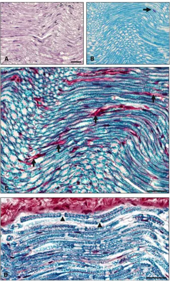
The analysis of the normal sciatic nerve with HE stain allowed us to observe the tissue pattern with the typical undulated and parallel organization of the nerve fibers. The myelin sheath stain was weak and unspecific, with poor contrast between the different histological structures (Fig.2a). With the LFB method it was only possible to identify the myelin sheath of the myelinated nerve fibers and the metachromatic granules of the mast cells (Fig.2b).
The analysis of the normal sciatic nerve with the new MCOLL histochemical method allowed us to observe tis-sue pattern as HE stain did (Fig.2c, d). The specific identification of the myelin sheath (in blue) and collagen fibers (in red) allowed us to simultaneously observe and correlate the distribution pattern and organization of both components with high contrast and specificity (Fig. 2c, d). With MCOLL histochemical method it was possible to accurately identify the Nodes of Ranvier and the organi-zation of the collagen fibers in the connective tissue sur-rounding the nerve fibers and blood vessels (Fig.2c, d). Analysis of the nerve regeneration model
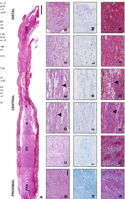
The morphologic analysis with HE stain of the CC after 12 weeks of in vivo implantation allowed us to observe the axonal regeneration along the graft. This axonal regener-ation was abundant only in the proximal anastomosis. We observed the formation of a thick regeneration cone, accompanied by an increased axonal sprouting (Fig.3b–e). It was observed that the thickness of the cone regeneration progressively declined and we did not observe any nerve regeneration at the distal nerve (Fig.3f, g). In relation to the ECM, we could identify some acidophilic components, but it was not possible to determine and observe any changes in the remodeling of the ECM during the process of regeneration.
The analysis with LFB method allowed us to confirm the presence of a myelin sheath at the proximal anastomosis (Fig.3h). Remyelination process was observed at the regeneration cone and axonal sprouting (Fig.3i–k). This histochemical reaction progressively decreased, and was negative at the distal anastomosis (Fig. 3l, m). However, it was not possible to properly observe the morphologic characteristics of the regenerating nerve, because the con-trast was low and the resolution quality was poor.
The analysis with the new MCOLL histochemical method allowed us to observe the axonal regeneration process in the grafted CC (Fig.3a), and correlate all the morphologic parameters with the histochemical reaction for myelin sheath and collagen fibers simultaneously, with high contrast, specificity and sensibility (Fig.3n–s).
In the proximal sciatic nerve, we observed the typical undulated tissue pattern of the peripheral nerve with signs of Wallerian degeneration and a significant degree of demye-lination. The histochemical reaction for collagen fibers was intense and the fibers tended to remain properly oriented (Fig.3n). The structural disorganization of the proximal anastomosis, with the formation of a thick regeneration cone, was accompanied by remyelination of the axonal sprouting and reorganization of the collagen fibers. These collagen fibers were oriented around the axonal sprouting forming fascicles (Fig.3o–q). We observed a marked
M
I
GM GM GM
II
III
WM WM WM
III
IV
A
B
C
WM GM
V
E
G
F
VI
H
J
I
WM
K
D
M
L
712 Histochem Cell Biol (2011) 136:709–717
123
process of fibrosis with disorganized collagen fibers and demyelination process at the distal anastomosis (Fig.3r, s).
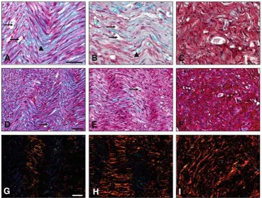
With the polarized light microscopy (Fig.4a–h), we observed the specific increase of the birefringence of the collagen fibers and their reorganization during the periph-eral nerve regeneration process. Birefringence was weak and fibers were well organized at the proximal sciatic nerve (Fig.4b), but became strong and disorganized at the central portion, with high levels of fibrosis at the distal anasto-mosis (Fig.4f, h).
Discussion
Histopathologic analysis and tissue engineering of periph-eral nerves should be accompanied by the application of histological quality controls. The observation of normal CNS, PNS and peripheral nerve regeneration using CC with HE allowed us to evaluate the main morphologic features and regeneration process. However, with this method it was not possible to evaluate the integrity of the myelin sheath and the changes that occur in the fibers of the ECM. The identification of myelin sheaths and collagen fibers acquires an important role in the evaluation of the peripheral nerve in pathology and tissue engineering (Mills
2007; Jiao et al.2009; Oh2001). Currently, it is necessary to carry out different specific histochemical methods for the identification of these elements and these results should be interpreted in parallel (Mills2007). Recently, Rutschow et al. (2010) described the use of both LFB and picrosirius histological methods for the evaluation of myocytolysis and fibrosis in myocardial tissue. However, the method
reported by these authors required the use of two inde-pendent staining processes using two different samples, since both staining procedures were carried out separately. One of the main advantages of the novel MCOLL method is the use of one single staining process.
In paraffin-embedded tissues the LFB method is most often used in histopathology for the study of the myelin in CNS and PNS (Mills 2007; Klu¨ver and Barrera 1953). In substitution of the previously described method, several techniques have been developed for the identification of myelin using light and electron microscopy (Savaskan et al.
2009; Di Scipio et al.2008; Larsen et al.2003; Xiang et al.
2005; Schmued and Slikker 1999; McNally and Peters
1998; Tolivia et al. 1994; Schmued 1990; Schmued et al.
1982; Stilwell 1957). However, all these methodologies only allow the specific identification of myelin, and it is not possible to make a comprehensive assessment of the morphologic parameters and the status of the ECM. The osmium-tetroxide has been described for myelin sheath impregnation in paraffin-embedded samples with different staining contrast with promising results (Di Scipio et al.
2008). But this method provides a permanent coloration of the myelin sheath. Therefore, it may limit the application of other histochemical and immunohistochemical tech-niques in these samples.
The HE staining is commonly non-specific for many tissue elements, due to its electropolar nature. Using this staining, we were not able to accurately identify the myelin sheath. In relation to the ECM, HE staining allowed us to recognize some acidophilic elements such as the collagen fibers. However, the staining pattern was non-specific because all these elements became stained with similar tones and intensities in comparison with other histological structures.
The new MCOLL histochemical method based on con-ventional luxol fast blue and picrosirius histochemical methods allowed us to evaluate the morphological features of the nerve tissue as HE did. However, the MCOLL method had the advantage of simultaneously identifying the myelin sheath with the same staining pattern, speci-ficity and accuracy of the conventional LFB method, and the collagen fibers with the same sensibility and specificity of picrosirius techniques, using a single analysis.
On the other hand, the analysis of the CNS, with this MCOLL histochemical method allowed us to observe the histological structure and analyze a specific histochemical reaction for myelin in the white matter. The evaluation of the CC grafted for nerve regeneration allowed us to eval-uate with high contrast all major morphologic parameters, the specific histochemical identification of the myelin sheath in different stages of remyelination or demyelina-tion, and the collagen fibers reorganization during the regeneration process. In the new-formed nerve fascicles we Fig. 1 Morphologic and histochemical study of the normal CNS.
Sagittal section of the cerebral cortex at low magnification stained
with HE (a), the LFB histochemical method for myelin inblue(b),
and the new MCOLL method, where it is possible to distinguish the
gray matter (GM) from the white matter (WM) stained inbluewith
high contrast (c). Scale bar=500lm. Overview of the cerebral
cortex with the MCOLL method, where it is possible to observe the
WM, and the different layers of the cerebral cortex (Mmeninges,
Imolecular layer,II external granular layer,IIIexternal pyramidal
layer, IV internal granular layer, V internal pyramidal layer, VI
polymorphic or multiform layer). Bar=100lm (d). Section of
cerebellum stained with HE (e), the LFB stain (f), and the new
MCOLL method with a positive histochemical reaction for myelin in
WM and collagen fibers (arrows) at the meninges (g). Scale
bar=100lm. Section of the cerebellum nucleus stained with HE
(e), the LFB method (f), and the new MCOLL method where the
neuronal soma is clearly distinguished (arrowheads) between
mye-linated fibers stained in blue(j). Scale bar=50lm. Polymorphic
layer of the cerebral cortex stained with HE (k), LFB (l) and the
MCOLL method with a positive histochemical reaction for individual
myelinated nerve fibers inblue(arrows) between the bodies of the
neurons (arrowhead) with high contrast (m).Scale bar=50lm
b
could clearly identify and evaluate the presence of axonal sprouting and nerve regeneration. All this allowed us to establish a more precise diagnosis of the structures that were present in during the regeneration process in a single sample of tissue.
The identification of collagen fibers using the MCOLL method was specific, because it uses a strong anionic
tetrakisazo dye called sirius red. This dye interacts with cationic groups on the surface of the collagen molecules in parallel, giving an intense red color to the collagen fibers in light microscopy. In addition, this dye specifically increa-ses the natural birefringence of collagen fibers, and allows us to selectively identify them by polarizing microscopy. This phenomenon is particularly induced by Picrosirius
A
B
C
D
Fig. 2 Histochemical analyses of the normal sciatic nerve. Longitudinal sections of the sciatic nerve stained with HE, where it is possible to observe the typical undulated tissue pattern (a). Positive histochemical reaction for myelin sheath and
metachromatic reaction for mast
cells (arrow) stained with the
LFB stain (b). The same
sections stained with the new MCOLL method, where it is possible to observe the typical tissue pattern, the intense histochemical reaction for
myelin sheath inblue, and the
histochemical identification of
the collagen fibers (arrows) in
red(c). The axons and the
Nodes of Ranvier are clearly
visualized (arrowheads) along
the myelinated nerve fibers; observe the thick collagen fibers
of the epineurium stained inred
(d).Scale bar=50lm
714 Histochem Cell Biol (2011) 136:709–717
123
CENTRAL
Fig. 3 Morphologic analysis of the collagen conduit (CC) graft. Histochemical composition of the entire collagen conduit graft
(a) stained with the new
MCOLL method. The morphological study with HE shows the nerve regeneration process with axonal sprouting along the graft (b–g). The histochemical evaluation of the CC graft with the LFB method, shows a progressive decrease of the myelin histochemical reaction along the graft (h–m). The histochemical evaluation with the MCOLL method, shows the regeneration process accompanied by axonal sprouting, a progressive decrease of the myelin histochemical reaction, and the progressive increase of the histochemical reaction for
collagen fibers stained inred
(n–s).CCcollagen conduit wall,
PANproximal nerve
anastomosis,RCregeneration
cone,DANdistal anastomosis
and distal nerve.Arrowheads
label illustrative examples of axonal sprouting and
new-formed nerve fascicles.Scale
barin panela=1000lm.
Scale barin panels b–s=100lm
staining (Junqueira et al. 1979; Montes and Junqueira
1991; Trau et al. 1991; Carriel et al. 2011). Interestingly, the histochemical reaction of the myelin with LFB in this new MCOLL method was not affected by subsequent procedures, maintaining the sensibility and specificity, with a considerable improvement in the contrast without back-ground color. In addition, the histochemical reaction for collagen fibers was not affected by treatment of the section with high temperature, which has been previously descri-bed (Carriel et al.2011), and provides a high contrast that allowed us to identify the different components separately and specifically.
Currently, there are several methods for the evaluation of the myelin; however, a histochemical method with these histochemical and morphologic characteristics does not exist. Furthermore, this new MCOLL histochemical method is carried out in about 18 h, being a specific, simple, and inexpensive procedure for paraffin-embedded tissue. In addition, the picrosirius and LFB solutions are very stable at room temperature for at least 5 years, and can be used repeatedly (Kiernan2008).
Anyway, this is not a method that aims to replace the conventional procedures used routinely; this new method could be useful not only to help in a better diagnosis of the different degrees of axonal degeneration and demyelination in CNS and PNS injuries, but also to evaluate the
simultaneous correlation between the morphologic param-eters, the myelin integrity and the collagen fibers reorga-nization during peripheral nerve regeneration carried out with different protocols of tissue engineering.
In conclusion, the use of this new MCOLL histochem-ical method allowed us to simultaneously evaluate and correlate the histological pattern, the specific histochemical reaction for myelin sheath, and the histochemical reaction for the stromal collagen fibers in a single slide with high contrast. This method combines the properties of sensi-bility and specificity of the used reagents, providing a high intensity and specificity of the histochemical reaction in the identification of the histological structures described in this work. For all these reasons, we hypothesize that this new histochemical method could be useful in pathology and tissue engineering.
Acknowledgments The authors are grateful to Ms. Ariane Ruyff-elaert from Ghent University, Belgium for revising and correcting the English manuscript. This work was supported by grant SAS PI-135/ 2007 from Junta de Andalucı´a, Spain.
References
Bancroft JD, Gamble M (2008) Theory and practice of histological techniques, 6th edn. Churchill Livingstone, UK
B
C
D
F
H
E
G
I
A
Fig. 4 Light (a–f) and polarized light (g–i) microscopy images of the CC graft stained with the new MCOLL histochemical method. Proximal sciatic nerve (a,d, g). Proximal nerve anastomosis (b,e,h).
Distal nerve anastomosis (c, f, i). Arrows illustrative examples of
axons showing Wallerian degeneration.Arrowheadsmyelin sheaths.
Scale bar=50lm
716 Histochem Cell Biol (2011) 136:709–717
123
Carriel VS, Aneiros-Fernandez J, Arias-Santiago S, Garzo´n IJ, Alaminos M, Campos A (2011) A novel histochemical method for a simultaneous staining of melanin and collagen fibers. J Histochem Cytochem 59(3):270–277
Chalfoun CT, Wirth GA, Evans GRJ (2006) Tissue engineered nerve constructs: where do we stand? J Cell Mol Med 10:309–317 Di Scipio F, Raimondo S, Tos P, Geuna S (2008) A simple protocol
for paraffin-embedded myelin sheath staining with osmium
tetroxide for light microscope observation. Microsc Res
Tech 71(7):497–502
Hirano A (2005) The role of electron microscopy in neuropathology: a personal historical note. Acta Neuropathol 109:115–123 Jiao H, Yao J, Yang Y, Chen X, Lin W, Li Y, Gu X, Wang X (2009)
Chitosan/polyglycolic acid nerve grafts for axon regeneration from prolonged axotomized neurons to chronically denervated segments. Biomaterials 30:5004–5018
Junqueira LC, Bignolas G, Brentani RR (1979) Picrosirius staining plus polarization microscopy, a specific method for collagen detection in tissue sections. Histochem J 11:447–455
Kiernan JA (2008) Histological and histochemical methods. Theory and practice, 4th edn. Scion, Oxfordshire
Klu¨ver H, Barrera E (1953) A method for the combined staining of cells and fibers in the central nervous system. J Neuropathol Exp Neurol 12:400–403
Larsen M, Bjarkam CR, Stoltenberg M, Sørensen JC, Danscher G (2003) An autometallographic technique for myelin staining in formaldehyde-fixed tissue. Histol Histopathol 18:1125–1130 McNally KJ, Peters A (1998) A new method for intense staining of
myelin. J Histochem Cytochem 46(4):541–545
Mills SE (2007) Histology for pathologists, 3rd edn. Lippincott Williams and Wilkins, Philadelphia
Montes GS, Junqueira LC (1991) The use of the picrosirius-polarization method for the study of the biopathology of collagen. Mem Inst Oswaldo Cruz 3:3–11
Oh SJ (2001) Color atlas of nerve biopsy pathology. CRC press, Rutschow S, Leschka S, Westermann D, Puhl K, Weitz A, Ladyszenskij
L, Jaeger S, Zeichhardt H, Noutsias M, Schultheiss HP (2010) Left
ventricular enlargement in coxsackievirus-B3 induced chronic myocarditis: ongoing inflammation and imbalance of the matrix degrading system. Eur J Pharmacol 630(1–3):145–151
Savaskan NE, Weinmann O, Heimrich B, Eyupoglu IY (2009) High resolution neurochemical gold staining method for myelin in peripheral and central nervous system at the light and electron microscopic level. Cell Tissue Res 337:213–221
Schmued LC (1990) A rapid, sensitive histochemical stain for myelin in frozen brain sections. J Histochem Cytochem 38:717–720 Schmued L, Slikker W Jr (1999) Black-gold: a simple,
high-resolution histochemical label for normal and pathological myelin in brain tissue sections. Brain Res 837:289–297 Schmued LC, Swanson LW, Sawchenko PE (1982) Some fluorescent
counterstains for neuroanatomical studies. J Histochem Cyto-chem 30:123–128
Stilwell DL (1957) A Sudan black B myelin stain for peripheral nerves. Stain Technol 32:19–23
Taylor CR, Cote RJ (2005) Immunomicroscopy: a diagnostic tool for the surgical pathologist. W. B. Saunders, Philadelphia Tolivia J, Navarro A, Tolivia D (1994) Differential staining of nerve
cells and fibers for sections of paraffin-embedded material in mammalian central system. Histochemistry 102:101–104 Trau H, Dayan D, Hirschberg A, Hiss Y, Bubis JJ, Wolman M (1991)
Connective tissue nevi collagens. Study with picrosirius red and polarizing microscopy. Am J Dermatopathol 4:374–377 Vleggeert-Lankamp CL (2007) The role of evaluation methods in the
assessment of peripheral nerve regeneration through synthetic conduits: a systematic review. J Neurosurg 107:1168–1189 Wangensteen KJ, Kalliainen LK (2010) Collagen tube conduits in
peripheral nerve repair: A retrospective analysis. HAND 5:273–277
Xiang Z, Nesterov EE, Skoch J, Lin T, Hyman BT, Swager TM, Bacskai BJ, Reeves SA (2005) Detection of myelination using a novel histological probe. J Histochem Cytochem 53(12):1511– 1516
Histochem Cell Biol (2011) 136:709–717 717