Non-invasive detection of Spatio-temporal activation of SBE and
NFAT5 Promoters in transgenic reporter mice following Stroke
Ashkaun Shaterian, B.S., Alexandra Borboa, B.S., Raul Coimbra, MD, PhD, Andrew Baird, PhD, and Brian P. Eliceiri, PhD
Author Institutional Affiliations: Dept of Surgery, University of California of San Diego, San Diego, CA
Summary
The characterization of molecular responses following cerebral ischemia-induced changes in animal models capable of undergoing real-time analysis is an important goal for stroke research. In this study, we use transgenic mice to examine the activation of two different promoters in a firefly luciferase reporter mouse analyzable through a non-invasive bioluminescent imaging system. In the first model, we examine the middle cerebral artery occlusion (MCAO)-induced activation of Smad-binding elements (SBE), a downstream target of Smad 1/2/3 transcription factors, in which SBEs regulate the expression of the fluc reporter. We observed that MCAO induces a bilateral activation (i.e. both ipsilateral and contralateral brain hemispheres) of the SBE-luc reporter with a peak at 24 hours. In the second model, we examined MCAO-induced activation of the osmolarity-sensitive promoter Nuclear factor of activated T-cells 5 (NFAT5) and identified a peak reporter expression 72 hours post-MCAO in the ipsilateral but not contralateral hemisphere. In each of these models, the assessment of post-MCAO fluc-expression provided both a
quantitative measure (i.e. radiance in photons/sec/cm2/steradian) as well as qualitative localization of the molecular response following focal ischemic injury.
Keywords
Nuclear factor of activated T-cells 5 (NFAT5); Smad-binding elements (SBE); non-invasive; stroke/ischemic wound
INTRODUCTION
The molecular pathophysiology of stroke is a dynamic and spatially localized process that leads to the activation of various injury cascades and host responses (i.e. cytotoxic and vasogenic edema) 1. Local and early changes (i.e. initial hypoxia and cell death) are followed by cascades of more global and sustained cellular and molecular responses resulting in vascular and neurological damage 1,2. Stroke-induced stress signaling,
neurovascular damage, and inflammation all further aggravate the initial ischemic insult 3,4, yet the cellular and molecular mechanisms governing these responses remain poorly understood. In this study, we assess two pathways of brain injury with a focus on the kinetics of stroke-induced activation of the Nuclear factor of activated T-cells 5 (NFAT5) and Smad binding elements (SBEs) promoters. The NFAT5 promoter is the only promoter known to date that is responsive to changes in osmolarity, therefore we examined NFAT5 promoter activation as a non-invasive in-vivo readout for stroke-induced changes in sodium
NIH Public Access
Author Manuscript
Neuropathology. Author manuscript; available in PMC 2013 April 1.
Published in final edited form as:
Neuropathology. 2012 April ; 32(2): 118–123. doi:10.1111/j.1440-1789.2011.01242.x.
NIH-PA Author Manuscript
NIH-PA Author Manuscript
homeostasis. Likewise, SBEs, the downstream targets of TGFβ-receptor mediated CNS scarring 5, were examined as a non-invasive in-vivo readout for stroke-induced CNS scarring.
Specific members of the Smad transcription factor family (Smads 2 and 3) have been shown to mediate TGFβ signaling and ischemic scarring 6,7, however the relationship between the two has yet to be explored in a non-invasive reporter model. Ischemia-induced CNS scarring is a dynamic process that results in the inhibition of neuronal regeneration 8. Smad2/3 and its upstream mediator, TGFβ, mediate CNS scarring as well as wound healing outside the CNS 9–11. TGFβ is a potent fibrogenic mediator that stimulates excessive deposition of extracellular matrix leading to subsequent fibrosis and scarring12. In this study, we have used transgenic mice expressing fluc under the regulation of multiple Smad 2/3 binding elements (SBE-luc), and follow the activation profile following a timecourse after MCAO.
Nuclear Factor of Activated T-cells 5 (NFAT5) is the only transcription factor known to be regulated by sodium homeostasis and therefore its promoter offers a unique reagent to assess stroke–induced changes in osmolarity 13,14. Recent studies have elucidated NFAT5 as a responsive mediator providing mammalian cell adaptation to hypertonic environments 15. NFAT5 is a transcription factor that increases the expression of osmo-protective proteins, such as salt channels, reducing hyperosmolar gradients that would otherwise diminish cell survival 16. Considering the role of NFAT5 in osmoregulation, sodium homeostasis and fluid balance, we used transgenic mice expressing fluc under the regulation of the NFAT5 promoter to explore the potential of NFAT5 as a surrogate marker of stroke-induced changes in sodium homeostasis/fluid balance. These studies define the distinct stroke-induced expression profiles of SBE-luc vs. NFAT5-luc transgenic models to provide a better understanding of the molecular pathophysiology of stroke.
MATERIAL AND METHODS
Ethics statement
To ensure animal welfare and to ameliorate suffering, mice were anesthetized with isoflurane and verified to be anesthetized before operating. Post surgical analgesia was provided with topical lidocaine and systemic buprenorphine. All procedures were done according to the UCSD Institutional Animal Care and Use Committee guidelines.
Generation of Transgenic Mice
The generation of NFAT-luc transgenic mice has been previously described20 and were the generous gift of Dr. J. Molkentin. In short, the NFAT-luciferase transgene was constructed from nine copies of an NFAT binding site from the IL-4 promoter, while SBE-Luc transgenic mice were generated by Dr. T. Wyss-Coray17 and are commercially available from Jackson Laboratory. The SBE-luc transgenic mice contain the firefly luciferase gene under the control of 12 Smad-binding element repeats in a C57BL/6J background.
Permanent Middle Cerebral Artery Occlusion (MCAO)
16 week-old NFAT5-luciferase transgenic mice and Smad2/3 Binding Element-luciferase transgenic mice were used in experiments. Mice were anesthetized with isoflurane, the surgical site removed of hair using a shaver and Nair® (Church & Dwight Co. Inc.; Princeton, New Jersey, USA) and prepared in a routine aseptic fashion. After verifying adequate anesthesia, skulls were exposed by a surgical incision and the skin and underlying temporal muscle were retracted. Craniotomy was then performed using a hand held drill equipped with a 1/32- inch high-speed cutting bit (Dremel, Racine, WI), producing a small
NIH-PA Author Manuscript
NIH-PA Author Manuscript
anterior branch of the right middle cerebral artery was occluded by coagulation with a heating filament producing a permanent artery occlusion. The incision was closed with four 3-0 silk interrupted sutures, followed by topical lidocaine. Animals were housed in separate cages with a 12 hour light/dark cycle and given access to feed and water. At various time points, 1, 3, 5 days after surgery, animals were imaged and tissue was harvested.
Tissue Collection, Immunohistochemistry and Image Anaysis
To corroborate our non-invasive analysis of Smad2/3 and to identify a cellular source for stroke-induced Smad2/3 signaling, an immunohistochemical analysis was performed on ischemic brain tissue and compared to controls. Specifically, animals were sacrificed, subjected to systemic intracardiac perfusion with heparin-saline, and brain tissue was harvested either pre-operatively or post-MCAO. For 2,3,5-triphenyltetrazolium chloride (TTC) staining, mice brain was dissected, cut into 1mm serial sections, and incubated for 5– 10 minutes in TTC, a mitochondrial viability stain to visualize infarction. For
immunohistochemistry, tissue samples were fixed overnight in 3.7% paraformaldehyde and then cryopreserved in phosphate-buffered 30% sucrose. Indirect immunofluorescence was performed on cryosections using anti-Phospho-Smad2 antibody (Cell Signaling
Technologies; Beverly, MA, USA) at 1:500 dilution and detected with TSA kit #12 (Invitrogen; Carlsbad, CA, USA). Phosphorylated Smad 2 in brain tissue was used as an indicator of Smad activation, as both Smad 2 and 3 undergo phosphorylation upon TGFβ receptor binding. Samples were stained according to manufacturer’s TSA kit #12 protocol. Fluorescent images of immunostained tissue sections were acquired with an Olympus Fluoview 1000 confocal microscope using exposure-matched settings (Advanced Software V1.6, Olympus, Center Valley, PA, USA).
Non-invasive imaging of Host Responses following MCAO
To characterize NFAT5 and SBE-mediated fluc expression following ischemic injury, transgenic mice were subjected to MCAO and monitored non-invasively for MCAO-induced fluc expression. At various timepoints, mice were anesthetized and given an intraperitoneal injection with the substrate D-luciferin (1.5 mg in a volume of 150 µL in saline) (Caliper Life Sciences; Hopkinton, MA, USA). Five minutes after injection, mice were imaged as whole live animals or dissected brain tissue on a Lumina CCD Imaging System (Caliper Life Sciences; Hopkinton, MA, USA). Exposure time for imaging was 5 minutes taken at field of view “A” and “D” for whole animals and dissected brain sections, respectively. Exposure-matched images for each animal was acquired using the Living Image Version 3 software, where mice were evaluated post MCAO and compared to pre-operative baseline fluc activity levels. Pre-pre-operative mice, as opposed to sham-operated mice, were chosen for analysis due to the heightened inter-animal variability inherent in using such animal models. Using pre-operative images for data analysis provides an internal control, reduces inter-animal variability, and provides greater statistical power21,22. Using a fixed region of interest for data analysis, the bioluminescence was measured and quantified at each timepoint. The regions of interest used for quantitation are shown in the
representative images in Figures 1 and 2. Bioluminescence emission was normalized and displayed in physical units of surface radiance (photons · s−1 · cm−2 · steradian–1 [sr]). The Wilcoxon Rank Sign test was used for statistical analysis. The animal images shown in this study are represented as pseudocolor images indicating light intensity (red being most intense), and are superimposed over gray-scale reference photographs. Separate groups of mice were assessed for invasive imaging and TTC staining to allow for continued non-invasive analysis at future time points; thus we analyzed animals in parallel at each time point to validate the model.
NIH-PA Author Manuscript
NIH-PA Author Manuscript
RESULTS
Non-invasive imaging and quantitation of SBE-mediated fluc expression following stroke
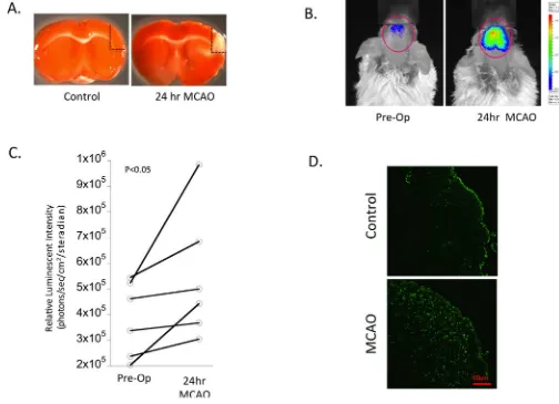
To characterize stroke-induced SBE-mediated luciferase expression, we monitored SBE-luc activity levels following MCAO over an extended timecourse. Analysis of bioluminescence in the head revealed both qualitative (Figure 1B) and quantitative (Figure 1C) changes following MCAO. A significant increase in signal was detected 24 hours post-MCAO in SBE-luc animals when compared to pre-operative background levels (i.e. a matched region of interest), with the signal originating almost exclusively from brain tissue. While the ischemic injury was performed on the right middle cerebral artery, we found stroke-induced up-regulation of SBE-luc expression that was interestingly generalized to both hemispheres, indicating a global up-regulation of Smad2/3-mediated activity in the brain post-MCAO (Figure 1B). Pre-operative quantification of fluc signal compared with post-operative fluc signal from the same animal revealed a statistically significant increase in SBE-mediated fluc activity at 24 hours post-MCAO (Figure 1C, P<0.05). In contrast there was no detectable difference in SBE-luc signal at later timepoints (i.e. 72–120 hours) post-MCAO (data not shown).
Analysis of stroke-induced Smad activation using immunohistochemical analysis To support the fluc imaging of the SBE reporter model, we performed an immunohistochemical analysis of brain sections from parallel animals with a phosphorylation state-specific anti-Smad antibody. Similar to our non-invasive data, immunohistochemical analysis revealed a global increase in phosphorylated Smad 2 vs. control (non-MCAO) brains (Figure 1D). These findings support the non-invasive SBE-luc study in that MCAO activation of the Smad pathway was detectable within 24 hours and originated from a cellular source within the brain. Importantly, we see an ischemia-induced increase in the phosphorylation of Smad using immunohistochemistry. This increase in signal intensity was not restricted to the ipsilateral hemisphere, further supporting our bioluminescent studies showing bilateral SBE-luc activation.
Non-invasive imaging and quantitation of NFAT5-mediated fluc expression following stroke
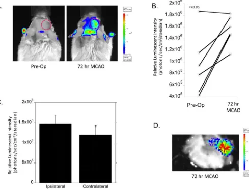
To characterize the kinetics of NFAT5-mediated fluc expression, NFAT5-luc transgenic mice were subjected to MCAO and subjected to bioluminescent imaging. Non-invasive imaging post-MCAO revealed both qualitative (Figure 2A) and quantitative (Figure 2B) changes 72 hours post-MCAO when compared to pre-operative activity levels from the same animal (P< 0.05). In contrast to the SBE-luc model, increased fluc activity in the NFAT5-luc model was localized to the ipsilateral hemisphere (Figures 2A,C), along with a notable increase in the caudal regions of the brain. In contrast to the SBE-luc model with a peak fluc reporter signal at 24 hours post-MCAO, the NFAT5-luc model did not reveal a statistically significant difference 24 or 120 hours post-MCAO (data not shown). Next, we imaged brain slab sections to verify the source of the NFAT5-mediated luc activity. We confirmed that the fluc signal activity originated from a cellular source within the brain and corroborated our non-invasive data showing NFAT5-mediated bioluminescence localizing to the ipsilateral hemisphere (Figure 2D). Taken together, the NFAT5-fluc and SBE-fluc reporter models provide complementary non-invasive readouts for the molecular pathophysiology following stroke.
Discussion
In this study, we have identified distinct patterns of SBE-mediated fluc activation vs.
NIH-PA Author Manuscript
NIH-PA Author Manuscript
24 hours for SBE-luc vs. 72 hours for NFAT5-luc) and localization (i.e. both hemispheres for SBE-luc vs. ipsilateral for NFAT5-luc) was distinct in the SBE-luc vs. NFAT5-luc models. Consistent with previous studies demonstrating the molecular basis of SBE as a readout for TGFβ receptor signaling 9,14,15,17, and the NFAT5 promoter for transcriptional assessment of changes in osmolarity 14,15, these studies demonstrate a novel approach for characterizing the molecular physiology of the stroke response. In both examples, the non-invasive and quantitative monitoring of MCAO-mediated remodeling of the brain provides an important tool to assess the biological activity of specific genes following brain injury (i.e. through assessing promoter activity in vivo).
A transgenic mouse expressing fluc under the regulation of the glial fibrillary acidic protein (GFAP) promoter has been previously validated as a pathophysiologically relevant model for astrogliosis 18. In this model, kainic acid induced a peak GFAP-mediated fluc signal at 24 hours, which correlated with neuronal damage. This study demonstrated the GFAP-luc model as a potentially useful surrogate marker and tool for monitoring neurochemistry non-invasively.
Similar to the use of GFAP-mediated fluc activity as readout for astrogliosis, and with extensive studies linking TGFβ-Smad2/3 signaling to scarring and NFAT5 to changes in osmolarity, we propose the SBE-luc and NFAT5-luc reporters as potentially useful surrogate markers for stroke-induced scarring and edema, respectively. The distinct patterns of activation of the SBE and NFAT5 promoters following stroke can be exploited both to better understand the molecular physiology of the brain19 and validate the potential for
therapeutic-targeting windows in drug development. Used in combination, the expression profiles of injury and treatment may not only elucidate the molecular pathology, but could also provide evidence-based specificity for treatment modalities based on molecular analysis19. Treatments that have been shown to specifically target a particular molecular mechanism can then be individualized and tailored for specific disease processes based on a better understanding of the spatiotemporal expression profiles in vivo.
Currently, most non-MRI animal models for stroke research involve cranial dissection for data analysis with loss of the animal. However, the use of transgenic reporter animals expressing luciferase under the transcriptional control of a responsive promoter provides investigators a flexible and less expensive model to analyze host gene expression levels. Here, we provide evidence for distinct spatio-temporal activation of the SBE and NFAT5 promoters that can be the basis for further characterization of stroke-induced changes in TGFβ signaling and osmolarity.
Acknowledgments
Sources of Support: NIH/NHLBI (BE) and the American Heart Association (AS)
References
1. Dirnagl U, Iadecola C, Moskowitz MA. Pathobiology of ischaemic stroke: an integrated view. Trends Neurosci. 1999; 22:391–397. [PubMed: 10441299]
2. Lo EH, Dalkara T, Moskowitz MA. Mechanisms, challenges and opportunities in stroke. Nat Rev Neurosci. 2003; 4:399–415. [PubMed: 12728267]
3. Mehta SL, Manhas N, Raghubir R. Molecular targets in cerebral ischemia for developing novel therapeutics. Brain Res Rev. 2007; 54:34–66. [PubMed: 17222914]
4. Nilupul Perera M, Ma HK, Arakawa S, et al. Inflammation following stroke. J Clin Neurosci. 2006; 13:1–8. [PubMed: 16410192]
NIH-PA Author Manuscript
NIH-PA Author Manuscript
5. Kirkbride KC, Townsend TA, Bruinsma MW, Barnett JV, Blobe GC. Bone morphogenetic proteins signal through the transforming growth factor-beta type III receptor. J Biol Chem. 2008; 283:7628– 7637. [PubMed: 18184661]
6. Krupinski J, Kumar P, Kumar S, Kaluza J. Increased expression of TGF-beta 1 in brain tissue after ischemic stroke in humans. Stroke. 1996; 27:852–857. [PubMed: 8623105]
7. Logan A, Green J, Hunter A, Jackson R, Berry M. Inhibition of glial scarring in the injured rat brain by a recombinant human monoclonal antibody to transforming growth factor-beta2. Eur J Neurosci. 1999; 11:2367–2374. [PubMed: 10383626]
8. Fawcett JW, Asher RA. The glial scar and central nervous system repair. Brain Res Bull. 1999; 49:377–391. [PubMed: 10483914]
9. Luo J, Lin AH, Masliah E, Wyss-Coray T. Bioluminescence imaging of Smad signaling in living mice shows correlation with excitotoxic neurodegeneration. Proc Natl Acad Sci U S A. 2006; 103:18326–18331. [PubMed: 17110447]
10. Mustoe TA, Pierce GF, Thomason A, Gramates P, Sporn MB, Deuel TF. Accelerated healing of incisional wounds in rats induced by transforming growth factor-beta. Science. 1987; 237:1333– 1336. [PubMed: 2442813]
11. Shah M, Foreman DM, Ferguson MW. Control of scarring in adult wounds by neutralising antibody to transforming growth factor beta. Lancet. 1992; 339:213–214. [PubMed: 1346175] 12. Cui X, Shimizu I, Lu G, et al. Inhibitory effect of a soluble transforming growth factor beta type II
receptor on the activation of rat hepatic stellate cells in primary culture. J Hepatol. 2003; 39:731– 737. [PubMed: 14568254]
13. Ho SN. The role of NFAT5/TonEBP in establishing an optimal intracellular environment. Arch Biochem Biophys. 2003; 413:151–157. [PubMed: 12729611]
14. Lopez-Rodriguez C, Antos CL, Shelton JM, et al. Loss of NFAT5 results in renal atrophy and lack of tonicity-responsive gene expression. Proc Natl Acad Sci U S A. 2004; 101:2392–2397. [PubMed: 14983020]
15. Lee JH, Kim M, Im YS, Choi W, Byeon SH, Lee HK. NFAT5 induction and its role in
hyperosmolar stressed human limbal epithelial cells. Invest Ophthalmol Vis Sci. 2008; 49:1827– 1835. [PubMed: 18436816]
16. Trama J, Go WY, Ho SN. The osmoprotective function of the NFAT5 transcription factor in T cell development and activation. J Immunol. 2002; 169:5477–5488. [PubMed: 12421923]
17. Lin AH, Luo J, Mondshein LH, et al. Global analysis of Smad2/3-dependent TGF-beta signaling in living mice reveals prominent tissue-specific responses to injury. J Immunol. 2005; 175:547–554. [PubMed: 15972691]
18. Zhu L, Ramboz S, Hewitt D, Boring L, Grass DS, Purchio AF. Non-invasive imaging of GFAP expression after neuronal damage in mice. Neurosci Lett. 2004; 367:210–212. [PubMed: 15331155]
19. Contag CH, Ross BD. It's not just about anatomy: in vivo bioluminescence imaging as an eyepiece into biology. J Magn Reson Imaging. 2002; 16:378–387. [PubMed: 12353253]
20. Wilkins BJ, Dai YS, Bueno OF, et al. Calcineurin/NFAT coupling participates in pathological, but not physiological, cardiac hypertrophy. Circ Res. 2004; 94:110–118. [PubMed: 14656927] 21. Peterson CY, Shaterian A, Borboa AK, et al. The noninvasive, quantitative, in vivo assessment of
adenoviral-mediated gene delivery in skin wound biomaterials. Biomaterials. 2009; 30:6788–6793. [PubMed: 19781761]
22. Lee J, Borboa AK, Chun HB, Baird A, Eliceiri BP. Conditional deletion of the focal adhesion kinase FAK alters remodeling of the blood-brain barrier in glioma. Cancer Res. 2010; 70:10131– 10140. [PubMed: 21159635]
NIH-PA Author Manuscript
NIH-PA Author Manuscript
Figure 1. Non-invasive imaging of Smad2/3 response following MCAO in Smad-Binding Element (SBE)-Luciferase mice
(A) Mice brain was dissected, cut into 1mm serial sections, and incubated in 2,3,5-triphenyltetrazolium chloride (TTC), a mitochondrial viability stain used to provide qualitative visualization of infarction and to validate the model of ischemic injury. The red dotted line orients the anatomical location for the images used in figure 1D. (B)
Representative images of real time Smad2/3 expression were acquired at 0 hours (pre-operative) and 24 hours post-MCAO by intraperitoneal injection with substrate luciferin and imaging with a cooled CCD imaging system. Regions of interest are delineated with the red outline. (C) Quantification of luciferase expression kinetics following MCAO. Data is presented as individual animal pairings showing the change in signal intensity between pre-operative imaging and post-pre-operative imaging (i.e. 24 hours post-MCAO). Each time point is taken from non-invasive images with matched acquisition settings as described in the Materials and Methods (n=6 per time point, *P < 0.05 compared to pre-operative/
background luminescence). (D) Indirect immunofluorescence staining of brain cryosections with anti-phosphorylated Smad 2 antibody in control vs. 24 hours after MCAO surgery. The regions shown are delineated in Figure 1A with a red dotted line for orientation.
Representative images of the infracted cortex are shown and were acquired using a laser scanning confocal microscope at magnification of 20×. Bar = 100 µm.
NIH-PA Author Manuscript
NIH-PA Author Manuscript
Figure 2. Non-invasive imaging of NFAT5 response following MCAO in NFAT5-Luciferase mice
(A) Representative images of real time NFAT5 expression was determined pre-operatively and 72 hours following MCAO surgery by intraperitoneal injection with substrate luciferin using a cooled CCD imaging system. Regions of interest are delineated with the red outline. (B) Quantification of luciferase expression kinetics following MCAO. Data is presented as individual animal pairings showing the pre-operative vs.post-operative signal (i.e. 72 hours post-MCAO) intensity of each animal. Each time point is taken from non-invasive images with matched acquisition settings as described in the Materials and Methods (n=6 per time point, *P < 0.05 compared to pre-operative/background luminescence). (C) Comparative quantification of luciferase expression following MCAO. Luciferase expression was quantitated over the contralateral vs. ipsilateral hemispheres relative to the sight of ischemic insult. Each value represents the averaged signal intensity obtained from matched images as described in the Materials and Methods. (D) Representative image of a 1mm dissected brain sections showing NFAT5-mediated fluc expression 72 hours following MCAO surgery.