DAFTAR ISI
1. Cover jurnal
2. Halaman pengesahan
3. Artikel final yang sudah dipublikasi 4. Detail jurnal
5. Submission Acknowledgement 6. Hasil Review
7. Published
2.
Halaman Pengesahan
3.
Artikel Final Yang
Sudah Dipublikasi
INSIST Vol. 4 No. 2, October 2019 (246–249) http://insist.unila.ac.id/
DOI:10.23960/ins.v4i2.246
Received: 03/07/2019 Accepted: 05/09/2019 Published online: 20/10/2019
INSIST Vol. 4 No. 2, October 2019 (246 - 249) 246
Abstract—Odontoglossum ringspot virus (ORSV) is one of the viruses that are reported to infect the most orchids in Indonesia.
Viral infections cause economic losses. This research was conducted to study the introduction of symptoms of infection that appear as the initial stages of disease control. The source of the virus is orchid leaves Phalaenopsis amabilis positively infected by ORSV collected from Magelang. Virus isolation was carried out by mechanical inoculation to indicator plants (Chenopodium amaranticolor and Nicotiana tabaccum). Furthermore, the results of inoculation were multiplied using various types of orchids (Dendrobium, Cymbidium, Chattleya, Phalaenopsis, Spatthoglotis, Liparis, and Pecteilis). Virus detection on orchid tissue was done trough the DAS-ELISA and observation of virus structure using TEM. The results showed that Pectelis sussanae (L.) Raf. was the only orchid plant that was immune against ORSV approached.
Dendrobium sp., Cymbidium sp., Chattleya sp., Phalaenopsis sp., and Spathoglotis sp. showed a response of susceptible of ORSV severe infection symptoms, while Liparis sp. were resistant. The variation of resistance from several orchid plant against infection ORSV showed severe symptoms with the incubation periode was seen earlier. The observation of virus structure using TEM showed rigid road shape particle, 300 x 18 nm in size, which is general characteristic of Tobamovirus. This indicate an infection of ORSV is a dangerous disease and require serious control.
Keywords—DAS-ELISA, Orchidaceae, ORSV, TEM.
I. INTRODUCTION
RCHID is one type of ornamental plant that has a high aesthetic value and is most in demand by the community. Disease infections are still the main obstacle in the cultivation and development of the potential of natural orchids [4]. The Odontoglossum ringspot virus (ORSV) is one of the most infectious orchid viruses. This virus was first discovered in the United States [1] and has spread to other countries including Indonesia. ORSV has a wide spread in Java, Ujung Pandang, Kalimantan, Bali, and Papua [3, 4-10]. In general, ORSV infection can reduce photosynthetic ability because of damage to chloroplasts [9, 10], inhibiting plant growth and resilience, and decreasing aesthetic value and selling power on a regular basis.
To facilitate disease control, it is better to do an inventory of data regarding the infection. Observation of variations in symptoms in response to disease infection is the first data needed for virus identification. This information is a very important aspect to determine disease management and
1 Department of Biology, Faculty of Mathematics and Natural Sciences, Lampung University, Bandar Lampung, Indonesia
2 Faculty of Biology, Gadjah Mada University, Yogyakarta, Indonesia
* Correspondence to F. Author, email: [email protected].
Tel.:+62-721-704947; fax.:+62-721-704948.
control measures in the field. In line with the knowledge of the response of various types of orchid plants can also be the basis for selecting orchids that will be developed in areas endemic to ORSV so that possible diseases can be avoided.
II. MATERIALS AND METHODS
A. Propagation Test
This method aims to purify and multiply the virus through mechanical inoculation of the test plants. The source of the virus is orchid leaves Phalaenopsis amabilis positively infected by ORSV collected from Borobudur Orchids Center, Magelang. Virus isolation was carried out by transmission to indicator plants (Chenopodium amaranticolor and Nicotiana tabaccum). Furthermore, the results of inoculation were multiplied using various types of orchids (Dendrobium, Cymbidium, Chattleya, Phalaenopsis, Spatthoglotis, Liparis, and Pecteilis). Inoculation was carried out at the top of the leaf mechanically using a 600 mesh carborondum with the addition of a 0.05 M buffer solution pH 7.0. Inoculated plants are maintained in a greenhouse with conditions 25-30º C and carried out observations every day. Observations were made on the variation of symptoms that arise, the incubation period, and the number of symptomatic plants or the percentage of disease occurrences.
B. Virus Detection on Orchid Plants
Orchid plant samples that have been inoculated by ORSV were detected by the DAS-ELISA method. Enzyme-linked immunosorbent assay (ELISA) is a virus detection method using specific antibodies. This method commonly used to detect plant viruses is direct ELISA (direct-ELISA) such as double antibody sandwich (DAS) where antigens are flanked by one type of antibody. The advantage of the ELISA technique is that it can identify multiple samples at once with relatively low costs and relatively short time.
Variation Symptoms and Resistance Response of Different Types on Orchids (Orchidaceae) Against Odontoglossum ringspot virus (ORSV) Infection
Mahfut1, Budi Setiadi Daryono2
O
eSSN: 2502-8588
A B
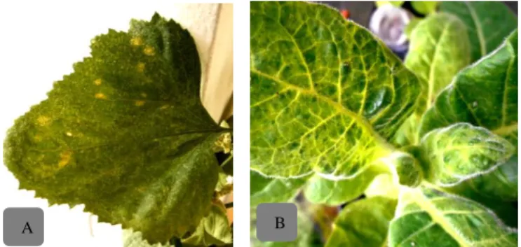
Fig. 1. Symptoms of ORSV infection: (A) Necrotic local lesion in C.
amaranticolor, (B) mosaic and vein clearing on N. tabacum
INSIST Vol. 4 No. 2, October 2019 (246–249) http://insist.unila.ac.id/
DOI:10.23960/ins.v4i2.246
Received: 03/07/2019 Accepted: 05/09/2019 Published online: 20/10/2019
INSIST Vol. 4 No. 2, October 2019 (246 - 249) 247
C. Determination of Plant Resistance
Determination of response criteria for various types of orchids against ORSV infection is based on several factors, including the percentage of occurrence of diseases and viral infections. The response of orchid plants is grouped to be close to immune, tolerant, somewhat resistant and vulnerable.
D. Virus Particle Analysis
Leaf samples of infected indicator plants were cut into small pieces and crushed for viral extraction. Prepared petri dishes which have been placed in the base by parafilm. Then a drop of sap is extracted on the surface of the parafilm, then the petri dish is closed to avoid evaporation. Samples were incubated for 30 minutes at 25°C and then washed using 30 drops of aquades. The sample is dried with filter paper for a few seconds and drops of a negative 2% phoshotungstenic acid dye solution of 7 drops. The sample is dried again using filter paper. The next sample was observed under the JEOL JEM-1400 (TEM) electron microscope at a magnification of 20000. The presence of a rigid rod-shaped morphology was an indication of the presence of ORSV.
III. RESULTS AND DISCUSSION
A. Propagation Test
The response of host plants to the transmission test shows that ORSV can infect almost all plants tested with different variations in symptoms and incubation time (Table 1). The response generally starts to appear around 2-3 weeks after inoculation in all host plants.
Specific symptoms commonly caused by indicator plants are symptoms of necrotic local lesions on Chenopodium amaraticolor on the 5th days and become clearer until the beginning of the second week, and chlorotic symptoms in the Nicotiana tabacum plant which after some time (first week) turn into a systemic mosaic (Fig. 1). Another symptom in the two plants is the leaf edge that rolls down on the young leaves as the initial symptom of ORSV infection. Chenopodiaceae is the fastest symptomatic plant with a short incubation period and a high percentage of events reaches 80%. Necrotic local lesions that appear are a symptom of the results of a hypersensitive response that is responsible for limiting pathogens so that the plant becomes more resistant to disease.
Symptoms of viral disease in host plants can occur due to the use of metabolic results of plants for viral synthesis, buildup of virions or parts of viruses and the effects of typical polypeptides that are encoded by viral genes. The response to ORSV infection shows that variations in symptoms between orchids are necrotic, necrotic, mosaic, chlorotic, and symptomless. Based on the symptoms shown, the ORSV inoculum used in the study was very infective. For necrotic symptoms appear in Phalaenopsis sp. namely day 23 and Dendrobium sp on day 15 after inoculation (Fig. 2 A & B).
Dendrobium sp. besides showing necrotic symptoms, another symptom that appears is mosaic on the 23rd day.
Mosaic symptoms are also seen in Cattleya sp. Orchids. on day 35 and Spathoglotis sp. on day 65 (Fig. 2 C, D, & E).
Mosaic symptoms are marked in the form of "green islands"
where there is a mixture of yellow or light green leaves in green. Another symptom is chlorotic in Spathoglotis sp.
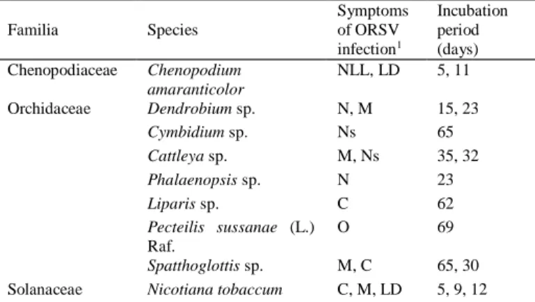
Table 1. Variation in symptoms of ORSV infection in test plants
Familia Species
Symptoms of ORSV infection1
Incubation period (days) Chenopodiaceae Chenopodium
amaranticolor
NLL, LD 5, 11
Orchidaceae Dendrobium sp. N, M 15, 23
Cymbidium sp. Ns 65
Cattleya sp. M, Ns 35, 32
Phalaenopsis sp. N 23
Liparis sp. C 62
Pecteilis sussanae (L.) Raf.
O 69
Spatthoglottis sp. M, C 65, 30 Solanaceae Nicotiana tobaccum C, M, LD 5, 9, 12
1M: Mosaic; NLL: Necrotic local lesions; N: Necrotic; Ns: Necrocis; LD:
Leaf Demalformation; C: Chlorotic; O: No symptom.
Fig. 2. Variations in symptoms of ORSV infection in several types of orchids: Necrotic in (A) Phalaenopsis sp. and (B) Dendrobium sp.; Mosaic on (C) Spathoglotis sp., (D) Dendrobium sp., and (E) Cattleya sp.; Chlorotics in (F) Spathoglottis sp., and (G) Liparis sp.; and Necrosis in (H) Cattleya sp.
and (I) Cymbidium sp.
Orchids. appears earlier than the symptoms of mosaic which is on the 30th day. Other orchids that show chlorotic symptoms are Liparis sp. on day 62 (Fig. 2 F & G). Chlorotic is a type of symptom caused by damage to chloroplasts which causes yellow parts of plants that are normally normal to turn yellow. Chloroplast damage can be caused by lack or absence of chlorophyll due to pathogenic poisons, mineral deficiencies, air pollution, lack of water, or due to chemicals.
Chlorotic symptoms often precede necrotic symptoms so that they gradually turn brown. Sometimes chlorotic symptoms are often associated with necrotics where chlorotic surrounds the necrotic called "halo".
Symptoms of necrosis are black spots where cell death and leaf tissue occur only in Cattleya sp. and Cymbidium sp. (Fig.
2 H & I). The difference is necrosis in Cattleya sp. bigger, clearer, and shorter incubation period of 32 days compared to Cymbidium sp, which is 65 days after inoculation. This is
D D
A B C D
E F G H I
INSIST Vol. 4 No. 2, October 2019 (246–249) http://insist.unila.ac.id/
DOI:10.23960/ins.v4i2.246
Received: 03/07/2019 Accepted: 05/09/2019 Published online: 20/10/2019
INSIST Vol. 4 No. 2, October 2019 (246 - 249) 248
because the leaves of Cymbidium sp. have thicker and harder leaves because of the layers of lignin and wax. Necrosis is a type of symptom caused by physical damage or death to cells, cell parts, or tissues. Some symptoms including necrotic types are specks (necrose), rot (rot), die-back (die back), and cancer (dead bark dries with clear boundaries).
Pectelis sussanae L. (Raff.) was the only test plant that showed no symptoms after being inoculated until the 69th day. This shows that this type of orchid has a resistant response to ORSV. Latent symptoms (symtompless) are systemic symptoms of infection. Viral propagation of test plants that are resistant to ORSV attacks produces latent symptoms (symtompless) caused by environmental factors such as the state of the environment in which they grow and temperature treatment. In addition, other factors that also play a role are the test plant is not an ORSV host, the amount of virus in the inoculum is insufficient, and the content of inhibitors in the test plant can eliminate the stability of the virus in sap.
B. Virus Detection on Orchid Plants
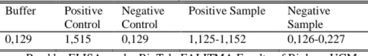
The results of the DAS-ELISA serological test (Table 2) indicate the overall sample of plant leaves from inoculation, except for the sample of Pectelis sussanae leaves, showed positive infection with ORSV with an average absorbance value of 1.125-1.152. [2] suggested that a test sample is said to be positively infected based on DAS-ELISA if the absorbance value at 405 nm wavelength approaches the absorbance value of positive control, or has a value 2-3 times the absorbance value of the control buffer.
This serological detection result reinforces evidence that the method can be used to detect ORSV which causes mosaic, chlorotic, necrotic, necrotic, and malformed orchid plants in plants infected with ORSV.
C. Determination of Plant Resistance
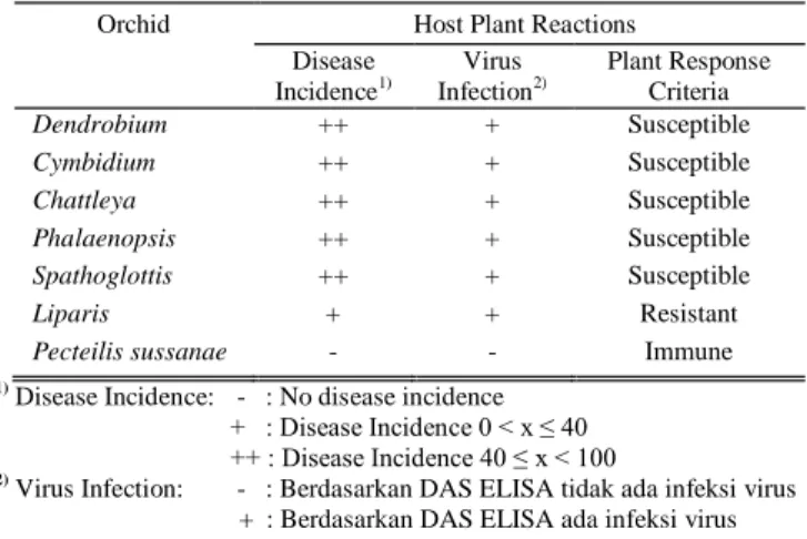
Plant responses to pathogens can be classified as immune, resistant, tolerant, and vulnerable. Based on observations of orchid plants' responses to single ORSV infections by observing the occurrence of diseases and viral infections, the response of orchid plants is grouped to be close to immune, somewhat resistant, and vulnerable. The results showed that Pecteilis sussanae plants were orchids that were close to the immune system against ORSV infection. Dendrobium, Cymbidium, Chattleya, Phalaenopsis, and Spathoglottis are susceptible and Liparis is resistant to ORSV infection (Table 3).
The response of Pecteilis sussanae to ORSV is close to the immune system, presumably because ORSV cannot replicate in plant cells. Orchids that have a resistant response to ORSV are orchid plants that can be infected with viruses, but plant cells do not support the growth and development of viruses, so that no disease occurs. In orchids that have a response.
While orchids that have a susceptible response to ORSV are orchids that support the growth and development of viruses, and viruses can cause damage.
Based on the severity of the symptoms seen and the incubation period, interactions that occur in the majority of orchid plants resulting from inoculation cause severe
Table 2. The range of absorbance mean values is based on DAS-ELISA at wavelength 405 nm
Buffer Positive Control
Negative Control
Positive Sample Negative Sample 0,129 1,515 0,129 1,125-1,152 0,126-0,227
Read by ELISA-reader BioTek, FALITMA Faculty of Biology UGM Table 3. The level of resistance of various types of orchids to ORSV
infections
Orchid Host Plant Reactions
Disease Incidence1)
Virus Infection2)
Plant Response Criteria
Dendrobium ++ + Susceptible
Cymbidium ++ + Susceptible
Chattleya ++ + Susceptible
Phalaenopsis ++ + Susceptible
Spathoglottis ++ + Susceptible
Liparis + + Resistant
Pecteilis sussanae - - Immune
1) Disease Incidence: - : No disease incidence + : Disease Incidence 0 < x ≤ 40 ++ : Disease Incidence 40 ≤ x < 100
2) Virus Infection: - : Berdasarkan DAS ELISA tidak ada infeksi virus
+ + : Berdasarkan DAS ELISA ada infeksi virus
symptoms of infection. The difference in the severity of the symptoms of the disease is related to the process of development and spread of the virus in plant cells. The severity will be higher with the rapid process of virus development and spread on infected plant cells. Viruses move into plant tissue through phloem vessels and interfere with plant's physiological function by utilizing the existing amino acids for the replication process. These physiological disorders cause systemic symptoms that appear on young leaves. So, the faster the process of development and spread of the two viruses in plant cells, then the systemic symptoms appear more quickly and the severity increases.
Unlike other orchid species, Liparis and Pecteilis sussanae exhibit less severe symptoms. In both of these orchids, the interaction of viral inoculation is interference and it is thought that the plant has resistance to viral infection so that the virus cannot cause significant damage. When the virus enters plant cells, the elicitor in the virus will be associated with receptors on plant cells to determine the relationship of subsequent infections. If it occurs incompatible, then all parts of the plant will provide a systemic reaction of resistance when infected with the virus, so that the virus cannot do multiplication and cause symptoms. If the interaction is compatible, the virus can infect host plants.
D. Virus Particle Analysis
The leaves of Nicotiana tabaccum symptomatic infection resulting from inoculation were used in the analysis of virus particles using electron microscopy (TEM) with 1%
ammonium molybdate as a negative dye and distilled water as a buffer. The TEM working principle is the conversion of electrical energy into electrons penetrated into specimens to be emitted into light energy. The electron beam is transmitted and absorbed by all specimens, for the next monitor screen captures the specimen structure.
INSIST Vol. 4 No. 2, October 2019 (246–249) http://insist.unila.ac.id/
DOI:10.23960/ins.v4i2.246
Received: 03/07/2019 Accepted: 05/09/2019 Published online: 20/10/2019
INSIST Vol. 4 No. 2, October 2019 (246 - 249) 249
Based on the results of the observations (Fig. 3) showing the structure of rigid elongated stem particles which is a common character of Tobamovirus. ORSV particles have a length of 300 nm and a width of 18 nm. On further observation, infection with this virus causes form malformations in the chloroplasts, mitochondria, and other cell organelles. In addition, X-body formation and paramural bodies occur between cell membranes and cytoplasm. The stability of a virus is the result of interactions between subunits of proteins and viral genomes.
The results of this study as an initial inventory of ORSV infection in Indonesia. This data is then expected to be used as basic information in the application of the concept of natural orchid conservation in Indonesia through crop protection efforts.
IV. CONCLUSION
The ability of ORSV to infect plants as a result of inoculation is very fast and shows symptoms of a fairly severe infection. Indicator plants show specific symptoms of necrotic local lesions in Chenopodium amaraticolor and chlorotic symptoms and mosaics in Nicotiana tabacum.
Another symptom in both plants is leaf malformation. While the response in the host orchid plants showed a variety of symptoms, namely in the form of necrotic, necrotic, mosaic, chlorotic, and symptomless (symtompless). The response of orchids to a single infection of ORSV based on the incidence of disease and viral infection shows Pecteilis sussanae is an orchid plant that is close to the immune system against ORSV infection. Dendrobium, Cymbidium, Chattleya, Phalaenopsis, and Spathoglottis are vulnerable, and Liparis is somewhat resistant to ORSV infection. The observation of the structure of the virus particles showed a rigid elongated rod measuring 300 nm long and 18 nm wide which is a common character of Tobamovirus.
ACKNOWLEDGMENT
This research was conducted with financial support from Ministry of Research, Technology and High Education,
Republic of Indonesia under project of Doctoral Dissertation Research with grant number 89/UN26/8/LPPM/2016.
REFERENCES
[1] K. N. Corbett, “Some distinguish characteristhic of the orchid strain of Tobacco Mosaic Virus,” in The Handbook on Orchid Pest and Disease:
American Orchid Soc., vol. 4, R. H. Lawson and S. Ali. 1967, pp. 62- 100.
[2] B.S. Daryono, K. T. Natsuaki, “Survei Virus yang Menyerang Labu- Labuan di Yogyakarta dan Jawa Tengah,” Jurnal Perlindungan Tanaman Indonesia, vol. 15, no 2, pp. 83-89, 2009.
[3] N. Inouye, I. W. Gara, “Detection and identification of virus of orchid in Indonesia,” Bull, Res, Inst., vol. 4, pp. 109-118, 1996.
[4] A. D. Kumalawati, S. Abdullah, Mahfut, B. S. Daryono, “Study on genetic diversity and convervation of orchids in Wonosadi forest, Gunung Kidul based on molecular analysis,” in Proc. 1st International Conference on Biological Science, Yogyakarta, 2011, pp. 54.
[5] Mahfut, B. S. Daryono, “Deteksi Odontoglossum ringspot virus (ORSV) Terhadap Anggrek Alam di Hutan Wonosadi, Gunung Kidul,”
Biogenesis, vol. 2, no. 2, 2014, pp. 101-108.
[6] Mahfut, T. Joko, B. S. Daryono, “Molecular Characterization Molecular of Odontoglossum ringspot virus (ORSV) in Jawa and Bali, Indonesia,” Asian Journal of Plant Pathology, vol. 10, no. 1-2, 2016, pp. 9-14.
[7] Mahfut, B. S. Daryono, T. Joko, S. Somowiyarjo, “Survei Odontoglossum ringspot virus (ORSV) yang menginfeksi anggrek alam tropis di Indonesia,” Jurnal Perlindungan Tanaman Indonesia, vol. 20, no. 1, 2016, pp. 1-6.
[8] Mahfut, B. S. Daryono, S. Somowiyarjo, “Deteksi Odontoglossum ringspot virus (ORSV) yang menginfeksi anggrek asli koleksi kebun raya di Indonesia,” Jurnal Fitopatologi Indonesia, vol. 13, no. 1, 2017, pp. 1-8.
[9] Mahfut, B. S. Daryono, S. Somowiyarjo, “Identifikasi Molekuler DNA Kloroplas Pada Anggrek Terinfeksi Odontoglossum ringspot virus (ORSV) di Magelang, Jawa Tengah,” in Prosiding Seminar Nasional Pengendalian Penyakit Pada Tanaman Pertanian Ramah Lingkungan II Perhimpunan Fitopatologi Indonesia Komisariat Daerah Yogyakarta, Solo, dan Semarang,Yogyakarta, 2017, pp. 354-360.
[10] Mahfut, B. S. Daryono, A. Indrianto, S. Somowiyarjo, “Plant-Virus Interaction on Orchids Infected Odontoglossum ringspot virus (ORSV) in Bogor Botanical Garden, Indonesia,” in Proc. 1st International Conference on Science and Technology, Makassar, 2019, pp.
Fig. 3. Particle structure of ORSV with TEM observations; arrow indicates virus particles
4.
Detail Jurnal
5.
Submission
Acknowledgement
6.
Hasil Review
INSIST Vol. 5 No. 1, April 2020 (p1–p4) http://insist.unila.ac.id/index.php/ojs DOI:
Received: 12 April 2020 Accepted: TBA
Published online: TBA
eISSN: 2502-8588
Abstract: Odontoglossum ringspot virus (ORSV) is one of the viruses that are reported to infect the most orchids in Indonesia.
Viral infections cause economic losses. This research was conducted to study the introduction of symptoms of infection that appear as the initial stages of disease control. The source of the virus is orchid leaves Phalaenopsis amabilis positively infected by ORSV collected from Magelang. Virus isolation was carried out by mechanical inoculation to indicator plants (Chenopodium amaranticolor and Nicotiana tabaccum). Furthermore, the results of inoculation were multiplied using various types of orchids (Dendrobium, Cymbidium, Chattleya, Phalaenopsis, Spatthoglotis, Liparis, and Pecteilis). Virus detection on orchid tissue was done trough the DAS-ELISA and observation of virus structure using TEM. The results showed that Pectelis sussanae (L.) Raf. was the only orchid plant that was immune against ORSV approached. Dendrobium sp., Cymbidium sp., Chattleya sp., Phalaenopsis sp., and Spathoglotis sp. showed a response of susceptible of ORSV severe infection symptoms, while Liparis sp. were resistant. The variation of resistance from several orchid plant against infection ORSV showed severe symptoms with the incubation periode was seen earlier. The observation of virus structure using TEM showed rigid road shape particle, 300 x 18 nm in size, which is general characteristic of Tobamovirus. This indicate an infection of ORSV is a dangerous disease and require serious control.
Keywords:DAS-ELISA, Orchidaceae, ORSV, TEM.
I. INTRODUCTION
RCHIDis one type of ornamental plant that has a high aesthetic value and is most in demand by the community. Disease infections are still the main obstacle in the cultivation and development of the potential of natural orchids [4]. The Odontoglossum ringspot virus (ORSV) is one of the most infectious orchid viruses. This virus was first discovered in the United States [1] and has spread to other countries including Indonesia. ORSV has a wide spread in Java, Ujung Pandang, Kalimantan, Bali, and Papua [3, 4-10]. In general, ORSV infection can reduce photosynthetic ability because of damage to chloroplasts [9,10], inhibiting plant growth and resilience, and decreasing aesthetic value and selling power on a regular basis.
To facilitate disease control, it is better to do an inventory of data regarding the infection. Observation of variations in symptoms in response to disease infection is the first data needed for virus identification. This information is a very important aspect to determine disease management and control measures in the field. In line with the knowledge of the response of various types of orchid plants can also be the basis for selecting orchids that will be developed in areas endemic to ORSV so that possible diseases can be avoided.
II. MATERIALSANDMETHODS A. Propagation Test
This method aims to purify and multiply the virus through mechanical inoculation of the test plants. The source of the virus is orchid leaves Phalaenopsis amabilis positively infected by ORSV collected from Borobudur Orchids Center, Magelang. Virus isolation was carried out by transmission to indicator plants (Chenopodium amaranticolor and Nicotiana tabaccum). Furthermore, the results of inoculation were multiplied using various types of orchids (Dendrobium, Cymbidium, Chattleya, Phalaenopsis, Spatthoglotis, Liparis, and Pecteilis). Inoculation was carried out at the top of the leaf mechanically using a 600 mesh carborondum with the addition of a 0.05 M buffer solution pH 7.0. Inoculated plants are maintained in a greenhouse with conditions 25-30º C and carried out observations every day. Observations were made on the variation of symptoms that arise, the incubation period, and the number of symptomatic plants or the percentage of disease occurrences.
B. Virus Detection on Orchid Plants
Orchid plant samples that have been inoculated by ORSV were detected by the DAS-ELISA method. Enzyme-linked immunosorbent assay (ELISA) is a virus detection method using specific antibodies. This method commonly used to detect plant viruses is direct ELISA (direct-ELISA) such as double antibody sandwich (DAS) where antigens are flanked by one type of antibody. The advantage of the ELISA technique is that it can identify multiple samples at once with relatively low costs and relatively short time.
C. Determination of Plant Resistance
Determination of response criteria for various types of orchids against ORSV infection is based on several factors, including the percentage of occurrence of diseases and viral infections. The response of orchid plants is grouped to be close to immune, tolerant, somewhat resistant and vulnerable.
D. Virus Particle Analysis
Leaf samples of infected indicator plants were cut into small pieces and crushed for viral extraction. Prepared petri dishes which have been placed in the base by parafilm. Then a drop of sap is extracted on the surface of the parafilm, then the petri dish is closed to avoid evaporation. Samples were incubated for 30 minutes at 25°C and then washed using 30 drops of aquades. The sample is dried with filter paper for a few seconds and drops of a negative 2% phoshotungstenic acid dye solution of 7 drops. The sample is dried again using filter paper. The next sample was observed under the JEOL JEM-1400 (TEM) electron microscope at a magnification of 20000. The presence of a rigid rod-shaped morphology was
O
Variation Symptoms and Resistance Response of Different Types on Orchids
(Orchidaceae) Against Odontoglossum ringspot virus (ORSV) Infection
INSIST Vol. 5 No. 1, April 2020 (p1–p4) http://insist.unila.ac.id/index.php/ojs DOI:
Received: 12 April 2020 Accepted: TBA
Published online: TBA
eISSN: 2502-8588
an indication of the presence of ORSV.
III. RESULTSANDDISCUSSION A. Propagation Test
The response of host plants to the transmission test shows that ORSV can infect almost all plants tested with different variations in symptoms and incubation time (Table 1). The response generally starts to appear around 2-3 weeks after inoculation in all host plants.
Table 1. Variation in symptoms of ORSV infection in test plants
Familia Species
Symptoms of ORSV infection1
Incubation period (days) Chenopodiaceae Chenopodium
amaranticolor
NLL, LD 5, 11
Orchidaceae Dendrobium sp. N, M 15, 23
Cymbidium sp. Ns 65
Cattleya sp. M, Ns 35, 32
Phalaenopsis sp. N 23
Liparis sp. C 62
Pecteilis sussanae (L.) Raf.
O 69
Spatthoglottis sp. M, C 65, 30 Solanaceae Nicotiana tobaccum C, M, LD 5, 9, 12
1M: Mosaic; NLL: Necrotic local lesions; N: Necrotic; Ns: Necrocis; LD:
Leaf Demalformation; C: Chlorotic; O: No symptom.
Specific symptoms commonly caused by indicator plants are symptoms of necrotic local lesions on Chenopodium amaraticolor on the 5th days and become clearer until the beginning of the second week, and chlorotic symptoms in the Nicotiana tabacum plant which after some time (first week) turn into a systemic mosaic (Fig. 1). Another symptom in the two plants is the leaf edge that rolls down on the young leaves as the initial symptom of ORSV infection. Chenopodiaceae is the fastest symptomatic plant with a short incubation period and a high percentage of events reaches 80%. Necrotic local lesions that appear are a symptom of the results of a hypersensitive response that is responsible for limiting pathogens so that the plant becomes more resistant to disease.
Fig. 1. Symptoms of ORSV infection: (A) Necrotic local lesion in C.
amaranticolor, (B) mosaic and vein clearing on N. tabacum
Symptoms of viral disease in host plants can occur due to the use of metabolic results of plants for viral synthesis, buildup of virions or parts of viruses and the effects of typical polypeptides that are encoded by viral genes. The response to ORSV infection shows that variations in symptoms between
orchids are necrotic, necrotic, mosaic, chlorotic, and symptomless. Based on the symptoms shown, the ORSV inoculum used in the study was very infective. For necrotic symptoms appear in Phalaenopsis sp. namely day 23 and Dendrobium sp on day 15 after inoculation (Fig. 2 A & B).
Fig. 2. Variations in symptoms of ORSV infection in several types of orchids: Necrotic in (A) Phalaenopsis sp. and (B) Dendrobium sp.; Mosaic on (C) Spathoglotis sp., (D) Dendrobium sp., and (E) Cattleya sp.; Chlorotics in (F) Spathoglottis sp., and (G) Liparis sp.; and Necrosis in (H) Cattleya sp.
and (I) Cymbidium sp.
Dendrobium sp. besides showing necrotic symptoms, another symptom that appears is mosaic on the 23rd day.
Mosaic symptoms are also seen in Cattleya sp. Orchids. on day 35 and Spathoglotis sp. on day 65 (Fig. 2 C, D, & E).
Mosaic symptoms are marked in the form of "green islands"
where there is a mixture of yellow or light green leaves in green. Another symptom is chlorotic in Spathoglotis sp.
Orchids. appears earlier than the symptoms of mosaic which is on the 30th day. Other orchids that show chlorotic symptoms are Liparis sp. on day 62 (Fig. 2 F & G). Chlorotic is a type of symptom caused by damage to chloroplasts which causes yellow parts of plants that are normally normal to turn yellow. Chloroplast damage can be caused by lack or absence of chlorophyll due to pathogenic poisons, mineral deficiencies, air pollution, lack of water, or due to chemicals.
Chlorotic symptoms often precede necrotic symptoms so that they gradually turn brown. Sometimes chlorotic symptoms are often associated with necrotics where chlorotic surrounds the necrotic called "halo".
Symptoms of necrosis are black spots where cell death and leaf tissue occur only in Cattleya sp. and Cymbidium sp. (Fig.
2 H & I). The difference is necrosis in Cattleya sp. bigger, clearer, and shorter incubation period of 32 days compared to Cymbidium sp, which is 65 days after inoculation. This is because the leaves of Cymbidium sp. have thicker and harder leaves because of the layers of lignin and wax. Necrosis is a
D
A B
I
A B C D D
E F G H I
INSIST Vol. 5 No. 1, April 2020 (p1–p4) http://insist.unila.ac.id/index.php/ojs DOI:
Received: 12 April 2020 Accepted: TBA
Published online: TBA
eISSN: 2502-8588
type of symptom caused by physical damage or death to cells, cell parts, or tissues. Some symptoms including necrotic types are specks (necrose), rot (rot), die-back (die back), and cancer (dead bark dries with clear boundaries).
Pectelis sussanae L. (Raff.) was the only test plant that showed no symptoms after being inoculated until the 69th day. This shows that this type of orchid has a resistant response to ORSV. Latent symptoms (symtompless) are systemic symptoms of infection. Viral propagation of test plants that are resistant to ORSV attacks produces latent symptoms (symtompless) caused by environmental factors such as the state of the environment in which they grow and temperature treatment. In addition, other factors that also play a role are the test plant is not an ORSV host, the amount of virus in the inoculum is insufficient, and the content of inhibitors in the test plant can eliminate the stability of the virus in sap.
B. Virus Detection on Orchid Plants
The results of the DAS-ELISA serological test (Table 2) indicate the overall sample of plant leaves from inoculation, except for the sample of Pectelis sussanae leaves, showed positive infection with ORSV with an average absorbance value of 1.125-1.152. [2] suggested that a test sample is said to be positively infected based on DAS-ELISA if the absorbance value at 405 nm wavelength approaches the absorbance value of positive control, or has a value 2-3 times the absorbance value of the control buffer.
Table 2. The range of absorbance mean values is based on DAS-ELISA at wavelength 405 nm
Buffer Positive Control
Negative Control
Positive Sample Negative Sample 0,129 1,515 0,129 1,125-1,152 0,126-0,227
Read by ELISA-reader BioTek, FALITMA Faculty of Biology UGM
This serological detection result reinforces evidence that the method can be used to detect ORSV which causes mosaic, chlorotic, necrotic, necrotic, and malformed orchid plants in plants infected with ORSV.
C. Determination of Plant Resistance
Plant responses to pathogens can be classified as immune, resistant, tolerant, and vulnerable. Based on observations of orchid plants' responses to single ORSV infections by observing the occurrence of diseases and viral infections, the response of orchid plants is grouped to be close to immune, somewhat resistant, and vulnerable. The results showed that Pecteilis sussanae plants were orchids that were close to the immune system against ORSV infection. Dendrobium, Cymbidium, Chattleya, Phalaenopsis, and Spathoglottis are susceptible and Liparis is resistant to ORSV infection (Table 3).
The response of Pecteilis sussanae to ORSV is close to the immune system, presumably because ORSV cannot replicate in plant cells. Orchids that have a resistant response to ORSV are orchid plants that can be infected with viruses, but plant cells do not support the growth and development of viruses,
so that no disease occurs. In orchids that have a response.
While orchids that have a susceptible response to ORSV are orchids that support the growth and development of viruses, and viruses can cause damage.
Table 3. The level of resistance of various types of orchids to ORSV infections
Orchid Host Plant Reactions
Disease Incidence1)
Virus Infection2)
Plant Response Criteria
Dendrobium ++ + Susceptible
Cymbidium ++ + Susceptible
Chattleya ++ + Susceptible
Phalaenopsis ++ + Susceptible
Spathoglottis ++ + Susceptible
Liparis + + Resistant
Pecteilis sussanae - - Immune
1) Disease Incidence: - : No disease incidence + : Disease Incidence 0 < x ≤ 40 ++ : Disease Incidence 40 ≤ x < 100
2) Virus Infection: - : Berdasarkan DAS ELISA tidak ada infeksi virus + : Berdasarkan D + : Berdasarkan DAS ELISA ada infeksi virus
Based on the severity of the symptoms seen and the incubation period, interactions that occur in the majority of orchid plants resulting from inoculation cause severe symptoms of infection. The difference in the severity of the symptoms of the disease is related to the process of development and spread of the virus in plant cells. The severity will be higher with the rapid process of virus development and spread on infected plant cells. Viruses move into plant tissue through phloem vessels and interfere with plant's physiological function by utilizing the existing amino acids for the replication process. These physiological disorders cause systemic symptoms that appear on young leaves. So, the faster the process of development and spread of the two viruses in plant cells, then the systemic symptoms appear more quickly and the severity increases.
Unlike other orchid species, Liparis and Pecteilis sussanae exhibit less severe symptoms. In both of these orchids, the interaction of viral inoculation is interference and it is thought that the plant has resistance to viral infection so that the virus cannot cause significant damage. When the virus enters plant cells, the elicitor in the virus will be associated with receptors on plant cells to determine the relationship of subsequent infections. If it occurs incompatible, then all parts of the plant will provide a systemic reaction of resistance when infected with the virus, so that the virus cannot do multiplication and cause symptoms. If the interaction is compatible, the virus can infect host plants.
D. Virus Particle Analysis
The leaves of Nicotiana tabaccum symptomatic infection resulting from inoculation were used in the analysis of virus particles using electron microscopy (TEM) with 1%
ammonium molybdate as a negative dye and distilled water as a buffer. The TEM working principle is the conversion of electrical energy into electrons penetrated into specimens to be emitted into light energy. The electron beam is transmitted and absorbed by all specimens, for the next monitor screen
INSIST Vol. 5 No. 1, April 2020 (p1–p4) http://insist.unila.ac.id/index.php/ojs DOI:
Received: 12 April 2020 Accepted: TBA
Published online: TBA
eISSN: 2502-8588
captures the specimen structure.
Based on the results of the observations (Fig. 3) showing the structure of rigid elongated stem particles which is a common character of Tobamovirus. ORSV particles have a length of 300 nm and a width of 18 nm. On further observation, infection with this virus causes form malformations in the chloroplasts, mitochondria, and other cell organelles. In addition, X-body formation and paramural bodies occur between cell membranes and cytoplasm. The stability of a virus is the result of interactions between subunits of proteins and viral genomes.
Fig. 3. Particle structure of ORSV with TEM observations; arrow indicates virus particles
The results of this study as an initial inventory of ORSV infection in Indonesia. This data is then expected to be used as basic information in the application of the concept of natural orchid conservation in Indonesia through crop protection efforts.
IV. CONCLUSION
The ability of ORSV to infect plants as a result of inoculation is very fast and shows symptoms of a fairly severe infection. Indicator plants show specific symptoms of necrotic local lesions in Chenopodium amaraticolor and chlorotic symptoms and mosaics in Nicotiana tabacum.
Another symptom in both plants is leaf malformation. While the response in the host orchid plants showed a variety of symptoms, namely in the form of necrotic, necrotic, mosaic, chlorotic, and symptomless (symtompless). The response of orchids to a single infection of ORSV based on the incidence of disease and viral infection shows Pecteilis sussanae is an orchid plant that is close to the immune system against ORSV infection. Dendrobium, Cymbidium, Chattleya, Phalaenopsis, and Spathoglottis are vulnerable, and Liparis is somewhat resistant to ORSV infection. The observation of the structure of the virus particles showed a rigid elongated rod measuring 300 nm long and 18 nm wide which is a common character of Tobamovirus.
ACKNOWLEDGMENT
This research was conducted with financial support from Ministry of Research, Technology and High Education, Republic of Indonesia under project of Doctoral Dissertation Research with grant number 89/ UN26/ 8/ LPPM/ 2016.
REFERENCES
[1] K. N. Corbett, “Some distinguish characteristhic of the orchid strain of Tobacco Mosaic Virus,” in The Handbook on Orchid Pest and Disease:
American Orchid Soc., vol. 4, R. H. Lawson and S. Ali. 1967, pp. 62- 100.
[2] B.S. Daryono, K. T. Natsuaki, “Survei Virus yang Menyerang Labu- Labuan di Yogyakarta dan Jawa Tengah,” Jurnal Perlindungan Tanaman Indonesia, vol. 15, no 2, pp. 83-89, 2009.
[3] N. Inouye, I. W. Gara, “Detection and identification of virus of orchid in Indonesia,” Bull, Res, Inst., vol. 4, pp. 109-118, 1996.
[4] A. D. Kumalawati, S. Abdullah, Mahfut, B. S. Daryono, “Study on genetic diversity and convervation of orchids in Wonosadi forest, Gunung Kidul based on molecular analysis,” in Proc. 1st International Conference on Biological Science, Yogyakarta, 2011, pp. 54.
[5] Mahfut, B. S. Daryono, “Deteksi Odontoglossum ringspot virus (ORSV) Terhadap Anggrek Alam di Hutan Wonosadi, Gunung Kidul,”
Biogenesis, vol. 2, no. 2, 2014, pp. 101-108.
[6] Mahfut, T. Joko, B. S. Daryono, “Molecular Characterization Molecular of Odontoglossum ringspot virus (ORSV) in Jawa and Bali, Indonesia,” Asian Journal of Plant Pathology, vol. 10, no. 1-2, 2016, pp. 9-14.
[7] Mahfut, B. S. Daryono, T. Joko, S. Somowiyarjo, “Survei Odontoglossum ringspot virus (ORSV) yang menginfeksi anggrek alam tropis di Indonesia,” Jurnal Perlindungan Tanaman Indonesia, vol. 20, no. 1, 2016, pp. 1-6.
[8] Mahfut, B. S. Daryono, S. Somowiyarjo, “Deteksi Odontoglossum ringspot virus (ORSV) yang menginfeksi anggrek asli koleksi kebun raya di Indonesia,” Jurnal Fitopatologi Indonesia, vol. 13, no. 1, 2017, pp. 1-8.
[9] Mahfut, B. S. Daryono, S. Somowiyarjo, “Identifikasi Molekuler DNA Kloroplas Pada Anggrek Terinfeksi Odontoglossum ringspot virus (ORSV) di Magelang, Jawa Tengah,” in Prosiding Seminar Nasional Pengendalian Penyakit Pada Tanaman Pertanian Ramah Lingkungan II Perhimpunan Fitopatologi Indonesia Komisariat Daerah Yogyakarta, Solo, dan Semarang,Yogyakarta, 2017, pp. 354-360.
[10] Mahfut, B. S. Daryono, A. Indrianto, S. Somowiyarjo, “Plant-Virus Interaction on Orchids Infected Odontoglossum ringspot virus (ORSV) in Bogor Botanical Garden, Indonesia,” in Proc. 1st International Conference on Science and Technology, Makassar, 2019, pp.