I
External Morphology of the Genus Aegla (Crustacea:
Anomura: Aeglidae)
JOEL W. MARTIN and
AWRENCE G. ABELE
SMITHSONIAN CONTRIBUTIONS TO ZOOLOGY • NUMBER 453
Emphasis upon publication as a means of "diffusing knowledge" was expressed by the first Secretary of the Smithsonian. In his formal plan for the Institution, Joseph Henry outlined a program that included the following statement: "It is proposed to publish a series of reports, giving an account of the new discoveries in science, and of the changes made from year to year in all branches of knowledge." This theme of basic research has been adhered to through the years by thousands of titles issued in series publications under the Smithsonian imprint, commencing with Smithsonian Contributions to Knowledge in 1848 and continuing with the following active series:
Smithsonian Contributions to Anthropology Smithsonian Contributions to Astrophysics
Smithsonian Contributions to Botany Smithsonian Contributions to the Earth Sciences Smithsonian Contributions to the Marine Sciences
Smithsonian Contributions to Paleobiology Smithsonian Contributions to Zoology
Smithsonian Folklife Studies Smithsonian Studies in Air and Space Smithsonian Studies in History and Technology
In these series, the Institution publishes small papers and full-scale monographs that report the research and collections of its various museums and bureaux or of professional colleagues in the world of science and scholarship. The publications are distributed by mailing lists to libraries, universities, and similar institutions throughout the world.
Papers or monographs submitted for series publication are received by the Smithsonian Institution Press, subject to its own review for format and style, only through departments of the various Smithsonian museums or bureaux, where the manuscripts are given substantive review.
Press requirements for manuscript and art preparation are outlined on the inside back cover.
Robert McC. Adams Secretary
Smithsonian Institution
S M I T H S O N I A N C O N T R I B U T I O N S T O Z O O L O G Y • N U M B E R 4 5 3
External Morphology of the Genus Aegla (Crustacea:
Anomura: Aeglidae)
Joel W. Martin and
Lawrence G. Abele
SMITHSONIAN INSTITUTION PRESS Washington, D.C.
1988
Martin, Joel W., and Lawrence G. Abele. External Morphology of the Genus Aegla (Crustacea: Anomura: Aeglidae). Smithsonian Contributions to Zoology, number 453, 46 pages, 19 figures, 1988.—External morphology of aeglid "crabs," unusual freshwater anomuran decapods endemic to South America, is described in detail for common members of the genus Aegla from the Arroyo San Antonio in southern Uruguay. Comparisons are made with available species descriptions in the literature. General aeglid morphology resembles that of members of the marine family Galatheidae, with which aeglids are traditionally grouped in the superfamily Galatheoidea. Several morphological features distinguish aeglids from marine members of the Galatheoidea. Of special interest are branchial morphology and sutures of the carapace. The occurrence of characters similar to those seen in Aegla in non-galatheoid anomuran families casts doubt upon the presently accepted phylogenetic placement of the Aeglidae. The hypothesis that aeglids may be related to members of the Paguroidea is presented.
A list of all species of the family is included.
OFFICIAL PUBLICATION DATE is handstamped in a limited number of initial copies and is recorded in the Institution's annual report, Smithsonian Year. SERIES COVER DESIGN: The coral Montastrea cavernosa (Linnaeus).
Library of Congress Cataloging in Publication Data Martin, Joel W.
External morphology of the genus Aegla (Crustacea: Anomura: Aeglidae) (Smithsonian contributions to zoology ; no. 453)
Bibliography: p.
1. Aegla—Morphology. 2. Crustacea—Morphology. I. Abele, Lawrence G. II. Title. III. Series QLI.54 no. 453 [QL444M33] 591s [595.3'844] 86-26244
Contents
Page
Introduction 1 Acknowledgments 1 Annotated History of Aeglid Systematics 2 Morphological Studies of Aegla 2 Materials and Methods 4 Results 5 Carapace and Rostrum 5 Carapace 5 Rostrum 8 Protocephalon 11 Eyes 11 First Antenna 11 Second Antenna 13 Epistome 13 Setal Morphology 13 Gnathothorax 14 Gnathal Region 15 Mandibles 15 Paragnaths 15 First Maxilla 15 Second Maxilla 15 Thorax 15 First Maxilliped 15 Second Maxilliped 16 Third Maxilliped 16 Thoracic Sterna 16 Pereiopods 18 First Pereiopod 18 Second through Fourth Pereiopods 20 Fifth Pereiopod 23 Branchiae 23 Gill Formula 23 Gill Morphology 23 Abdomen 25 Abdominal Somites 27 Pleopods 27 Uropods 28 Telson 30 Discussion 30 Grooves, Sutures, and Lineae 32 Origin of Aegla 32 Appendix I: Abbreviations and Lettering Used on the Figures 34 Appendix II: List of the Known Species and Subspecies of the Aeglidae 36 Museum Abbreviations for Type Depositions 36 Annotated List of Species and Subspecies 36 Literature Cited 43
iii
External Morphology of the Genus Aegla (Crustacea:
Anomura: Aeglidae)
Joel W. Martin and Lawrence G. Abele
Introduction
The Recent Aeglidae are freshwater crab-like decapod crustaceans endemic to South America. They occur in lakes, streams, salt marshes, and caves from Argentina, Bolivia, Brazil, Chile, Paraguay, and Uruguay (Bahamonde and L6pez, 1961; Hobbs, 1979; Manning and Hobbs, 1979; Schmitt, 1942b). These decapods are presently considered to belong to a single genus, Aegla, consisting of some 40 currently recognized species and subspecies, several of which have been described recently (Buckup and Rossi, 1977; Hebling and Rodrigues, 1977; Hobbs, 1979; Jara, 1977,1980b, 1982,1986;
Jara and Lopez, 1981). Although widely distributed across temperate South America, they are perhaps the least known of the freshwater decapod crustaceans.
The group is interesting for several reasons. First, with the possible exception of one fossil species (Haumuriaegla glaessneri Feldmann) from New Zealand (see Feldmann, 1984), they are restricted to temperate and subtropical South America, the only anomuran family thus restricted. Schmitt (1942b:431) noted: "There are no freshwater Crustacea at all like Aegla anywhere else in the world." Thus, their distribution poses some interesting biogeographical as well as ecological questions. Second, their evolutionary relationships are un- known. Morphologically the aeglids appear similar to members of the family Galatheidae (infraorder Anomura, superfamily Galatheoidea) and are included with the galatheids, porcel- lanids, and chirostylids in the superfamily Galatheoidea Samouelle, 1819. However, all members of the Galatheidae and even of the Galatheoidea, except the aeglids, are restricted to marine habitats (an exception is the porcellanid Petrolisthes
Joel W. Martin and Lawrence G. Abele, Department of Biological Science, Florida State University, Tallahassee, Florida 32306.
robsonae Glassell; see Gore and Abele, 1976). In addition, there are some important morphological differences between aeglids and galatheids. In Aegla, males have vestigial abdominal appendages, whereas these appendages (pleopods) are usually well developed in male galatheids (e.g., Pike, 1947;
Tirmizi, 1966). Apart from being of systematic importance, this absence of functional pleopods in aeglid males poses the interesting problem of how sperm transfer occurs. Sutures of the aeglid carapace are unlike those found in any galatheids.
The gill structure, traditionally considered an important systematic character in decapod Crustacea (e.g., Huxley, 1878;
Bate, 1888; Glaessner, 1960,1969) is "penicillate" [trichobran- chiate] in the aeglids, yet "foliose" [phyllobranchiate] in the galatheids (terminology after Dana, 1852). Other morphologi- cal peculiarities of aeglids include a border of scales along the cutting edges of the chelae and a spoon-shaped lobe on the palm of the cheliped in some species (e.g., A. schmitti Hobbs;
see Hobbs, 1979). Finally, aeglids constitute an ecologically unique group of decapod crustaceans in that, during the mating season, females of some species leave the water and congregate beneath stones or logs at the water's edge; they are sometimes found associated with spiders, ants, isopods, and scorpions during this spawning period (Bahamonde and Lopez, 1961;
Burns, 1972).
The gaps in our knowledge of aeglid crabs are large. The present study, in anticipation of a re-examination of the phylogenetic placement of aeglids in relation to other decapods, begins to address the question of aeglid origins by examining the external morphology of the genus Aegla.
ACKNOWLEDGMENTS.—We are extremely grateful to D.
Bruce Means for his invaluable help in transporting aeglids within and from Uruguay. We thank D. Bruce Means,
Guillermo J. Wibmer and Alberto and Maria Cola for help in planning the collecting expedition. In Uruguay, assistance was graciously offered by Lucrecia C. de Zolessi, Departamento dc Artropodos, Facultad de Humanidades y Ciencias, Tristan Narvaja, Montevideo, and by the families of G. Wibmer and A. Cola. R.B. Manning kindly loaned us specimens of Aegla jujuyana from the Smithsonian's National Museum of Natural History and reviewed an early draft of the manuscript.
Specimens of A. platensis from Argentina were loaned by Enrique Boschi. We thank Carlos G. Jara and Estela C.
Lopretto for sending us copies of their many excellent papers on aeglids, and Nilton J. Hebling and especially Carlos G. Jara for reviewing the manuscript. Our sincere appreciation is extended to Ardis B. Johnston for allowing the senior author to examine holdings of Aegla at the Museum of Comparative Zoology, Harvard University, and for checking the MCZ type specimens against our checklist. Finally, we gratefully acknowledge Horton H. Hobbs, Jr., and H.H. Hobbs III, for their extensive efforts to improve the manuscript. This work was supported in part by the National Science Foundation, Grants No. BSR-8414347 (Doctoral Dissertation Improvement series) and BSR-8508430. The senior author sincerely thanks Susan B. Martin for years of love, patience, and encourage- ment.
Annotated History of Aeglid Systematics
Latreille (1818, pi. 308: fig. 2) first illustrated, without description, an aeglid under the name Galathea laevis. Schmitt (1942b) suggests that Latreille may have been unaware that the specimen came from freshwater, as the genus in which Latreille placed it is entirely marine. According to Schmitt (1942b), "a crustacean of this type was recognized (but not described) as early as 1782 (pp. 206, 347; 1789, p. 182) by Molina in his 'Saggio sulla Storia Naturale del Chile' as Cancer apancora"
However, Molina's description is vague and may not have been of an aeglid:
The apancora (Cancer apancora) is larger than the ulicuna. The shell is oval and wholly denticulated, the claws are hairy, and the tail of a triangular form and very long. [From the 1808 English translation.]
The specific name selected by Molina may stem from the fact that the common name for these crabs in parts of Chile is pancora (Burns, 1972). Leach (1821) recognized that La- trcille's species represented a distinct genus, and it was he who chose the genus name Aegla, probably after Aegle, one of the Hcspcridcs who guarded the garden of golden apples of the Isles of the Blest at the western end of the earth in Greek Mythology.
A number of early carcinologists reproduced the figure of Latreille, some introducing inaccuracies and some reproducing the figure only crudely. These workers included Desmarest (1825), who introduced the misspelling Aeglea, Griffith and Pidgcon (1833), H. Milne Edwards (1837), who first mentioned
the habitat (the coasts of Chile), and the "Disciples Edition"
of Cuvier's (1837) Le Regne Animal. The contributions of these workers are discussed in more detail by Schmitt (1942b).
Prior to 1849, the genus was still considered monotypic, the sole species being A. laevis (Latreille). Although distinct species were described by Nicolet (1849), Girard (1855), and Muller (1876), Ortmann (1892, 1902) continued to refer to the genus as monotypic, and only after Schmitt's (1942a,b) work was the diversity of the group appreciated. Schmitt's (1942b) monograph on Aegla remains the primary taxonomic reference.
In that paper, he examined specimens of Aegla from Argentina, Brazil, Chile, and Uruguay, described 13 new species and 2 new subspecies, and included a key to the identification of the 20 forms recognized by him.
In the years since Schmitt's monograph several new species have been described (Bahamonde and L6pez, 1963; Buckup and Rossi, 1977; Hebling and Rodrigues, 1977; Hobbs, 1979;
Jara, 1977, 1980a,b, 1982, 1986; Jara and Ldpez, 1981;
Ringuelct, 1948a,b, 1960b; Tiirkay, 1972) so that there are now approximately 40 currently recognized species and subspecies of the genus Aegla (Appendix II). While this is not a small number of species, it is in no way comparable to some other families of freshwater Decapoda. North American crayfishes, for example, comprise well over 300 species in approximately 9 genera and 19 subgenera (Hobbs, 1972, 1974). It is possible that the smaller number of species known in Aegla reflects a lack of adequate sampling in temperate and subtropical South America.
Morphological Studies of Aegla
Most authors agree that the aeglids are a branch of the galatheid crabs that invaded fresh water. Schmitt (1942b:431) stated that "Its nearest relatives are marine and probably to be found somewhere among the galatheids (tribe Galatheidea)."
However, no authors have ventured to guess which group of the galatheids is most closely related to the aeglids. This stems probably from the absence of any detailed morphological examination of the aeglids. Traditional characters illustrated in descriptions of aeglids are those proposed by Schmitt (1942b) for distinguishing species; these are limited to the shape of the rostrum, outline of the carapace, anterolateral angle of the second abdominal somite, ventral margin of the second pereiopod, armature of the fourth thoracic sternum, and several characters of the cheliped (palmar lobes, basal teeth, and spination of the ischium). It is known that many of these characters are extremely variable among individuals (e.g., see Hobbs, 1979; Jara, 1980a; Ringuelet, 1948b, 1949a,b; Vaz- Ferreira, Gary, and Vaz-Ferreira, 1945). An additional problem is that in the literature often only one or two of the above characters are illustrated, with other characters either incom- pletely described in the text or omitted from the description.
Few papers exist in which aeglid characters other than those proposed by Schmitt are described. H. Milne Edwards (1837)
NUMBFR
32°S
• 34°S
5 6 ' 54°
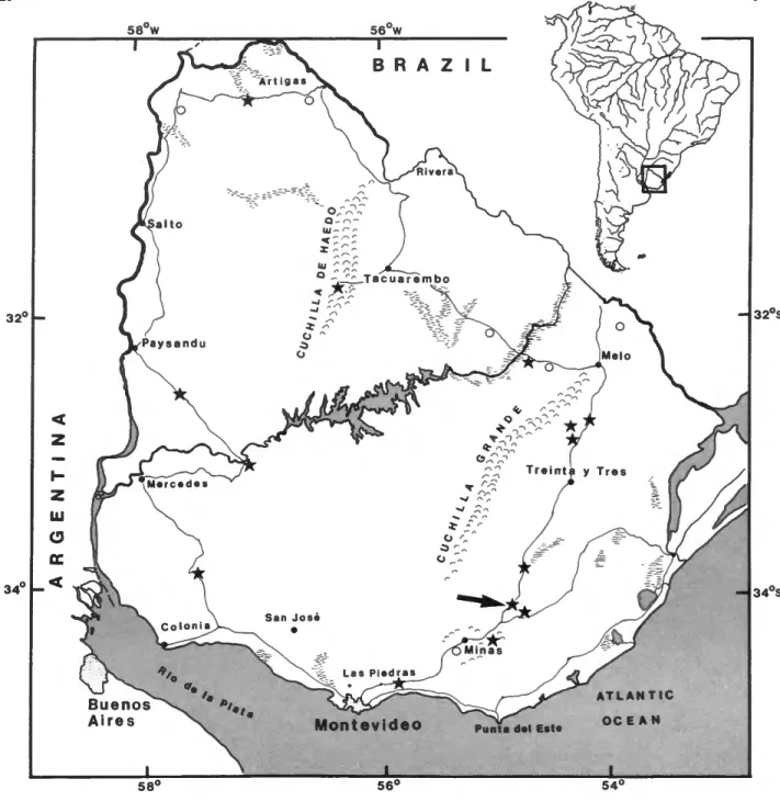
FIGURE 1.—Collecting localities visited by one author (JWM) in Uruguay. Stars indicate collecting sites where aeglids were found; large arrow indicates approximate site of the Arroyo San Antonio from which live aeglids were collected.
presented a brief diagnosis of the genus, Dana (1852) briefly discussed the nature of the carapace sutures and outlined (somewhat erroneously) the characters of the genus, Mocquard (1883, pi. 6: figs. 133-135) published surprisingly detailed figures of the foregut of A. "laevis," and Ortmann (1892) crudely illustrated the mouthparts of A. "laevis." Mouchet (1931a,b, 1932a,b) figured parts of the gills in a study of the
parasites of aeglids. Snodgrass (1950) included a schematic diagram of the protocephalon and gnathal region of A prado.
With the exception of Mocquard's and Dana's work these papers do not provide any basis for comparisons of aeglids with other anomurans at the family or superfamily level;
characters are either insufficiently illustrated (Mouchet) or else do not differ from the same structures in many other anomuran
propodus / —
c a r p a l ridge -x
carpus
m«rus -. ^ epigastric prominence
- - o r b i t a l s i n u s
.» ,---*- vr__extraorbital sinus anterolateral spine
7 hepatic _ _ / ~ lobes
- 3 protogastric lobe - _
gastric area
~ - - A-epibranchlal tooth _ / _ cervical groove
- 1
~ ~ 2 abdominal
— 3 somites 4
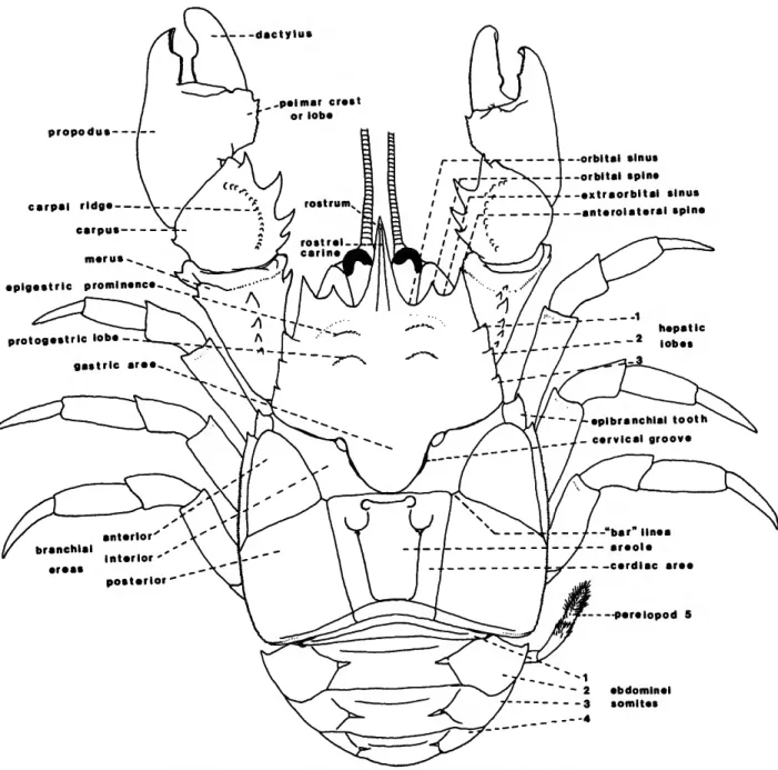
FIGURE 2.—Schematic view of a typical aeglid crab (based on an adult male A. platensis).
groups (Ortmann). Recent work by Lopretto (1978a,b, 1979, 1980a,b, 1981) describes in detail the morphology of the male fifth pcreiopod coxa, a structure that holds promise for clarification of wiihin-family systematics. Below we describe the complete external morphology of aeglids for the first time.
Materials and Methods
Live aeglid crabs were collected by dipnet from the Arroyo
San Antonio, southern Uruguay (Figure 1). The San Antonio, a tributary of the Rio Cebollati, is a shallow (<1.0 m) second order freshwater stream with a bed of loose gravel and stones.
Water temperature at the time of collection (19 April 1984) was 22°C; air temperature was 20°C. The current of the stream varied from 0.8 m/sec in the center to <0.01 m/sec in deeper pools and along the shallow sides of the stream. Density of the crabs, estimated by throwing a 0.5 m2 metal quadrat and
NUMBER 453
FIGURE 3.—Aegla platensis, lateral view of adult male.
removing all stones and crabs from within its confines, varied from 6 to 48 individuals/m2. Crabs were collected three hours before departure from Uruguay and placed in styrofoam chests with crushed ice; towels were added to reduce the risk of damage during shipping and to give the aeglids something to which they could cling. This collection yielded numerous specimens of A. platensis Schmittand A. uruguayana Schmitt.
Additional specimens of both species were preserved in 10%-20% formalin in the field and later transferred to 70%
cthanol. Through the kindness of Dr. R.B. Manning at the National Museum of Natural History, Smithsonian Institution, specimens of the Argentine A. jujuyana were borrowed for comparative purposes, and Dr. Enrique Boschi graciously sent a collection of preserved A. platensis from Argentina.
Fortunately all three of the above named species have been considered to represent "primitive" species of Aegla (see Schmitt, 1942b; Ringuelet, 1949c) so that this examination may serve as a baseline study for further investigations into aeglid morphology.
Illustrations were made from crabs preserved in the field and later transferred to ethanol. In addition, many of the structures were dissected from live crabs in the laboratory; this allowed observation of function as well as form. Other live aeglids were allowed to air dry before they were examined and/or dissected, and a few illustrations were aided by photography of the live crabs (e.g., Figure 3). Illustrations are of A. platensis and A. uruguayana. Comparisons of them with aeglids other than A. jujuyana are made through accounts in the literature.
Terminology follows that of Glaessner (1969), Schmitt
(1942b), and Pike (1947) for characters of the carapace and appendages, Kunze and Anderson (1979) for setal morphology, and Snodgrass (1951, 1952a,b) for characters of the proto- cephalon. Abbreviations used in the figures are explained in Appendix I.
Results
CARAPACE AND ROSTRUM
CARAPACE (Figures 2-4, 16).—Perhaps the single most remarkable feature of the genus Aegla is the carapace; it distinguishes the group not only from other members of the Galatheoidea but from all other decapod crustaceans. As with nearly all features employed in aeglid taxonomy, the carapace displays considerable variation among and within species.
However, some features are consistent and unique to the family. The following description is based upon A. platensis, but applies to all species of the genus except where noted.
The carapace is extremely depressed and gives the animal an overall flattened appearance (Figure Ab,d). The dorsal surface is divided by a distinct cervical groove (Figure 2; eg, Figure 4) into a narrow anterior region and a much wider posterior region. The carapace may be nearly smooth or obviously granulate; small, often setiferous punctations are common on the dorsal surface. Small simple setae are frequent especially along the ventral borders.
Anterior Region: The anterior region is marked by a well-developed rostrum usually with a strong dorsal carina
a
srp lav ept ptr la
brl lad eg
FIGURE 4.—The acglid carapace: a, dorsal view; b, frontal view; c, lateral view of anterior half of carapace; d, lateral view of carapace with eyes and second antennae; e, ventrolateral view of posterior half of carapace, higher magnification, c is A. wuguayana; all others are A. platensis.
NUMBER 453
(see below). The rostral carina typically extends posteriorly onto the carapace only as far as the level of the epigastric prominence or, occasionally, to the protogastric lobe, although in the aberrant A. denticulata Nicolet this carina extends the full length of the carapace. On each side of the rostrum a broad shallow excavation on the margin of the carapace forms the orbital sinus (Figure 2), which is flanked ventrolaterally by the orbital spine (os, Figure 4a). This spine may be acute with a cornificd tip and nearly equal in length to the anterolateral spine (e.g., A. platensis, Figure 4a) or it may be reduced and coalesced with the anterolateral spine or absent (e.g., A.
concepcionensis Schmitt, A. papudo Schmitt, A. affinis Schmitt, A. maulensis Bahamonde and Lopez, A. serrana Buckup and Rossi, and A. franciscana Buckup and Rossi).
Therefore the extraorbital sinus separating this spine from the anterolateral spine may be of variable depth, or absent. The anterolateral spine (as, Figure 4a) is typically acute with a corneous tip and may exceed the length of the eyestalk (e.g., A. sanlorenzo Schmitt) although in most species it extends only as far as the posterior margin of the cornea (e.g., Figures 4a, 5a).
The anterolateral carapace margin extends posteriorly from the anterolateral spine to the hepatic region. This region is subdivided into three lobes of approximately equal size (Figure 2, and numerals 1-3 in Figure 4a). The anterolateral margins of the hepatic lobes are typically acute and tipped with a corneous spine, although in some species these lobes are fused and nearly indistinguishable (e.g., A. bahamondei Jara, A.
plana Buckup and Rossi, and A. franciscana). Even when the hepatic lobes are indistinct, the demarcation between the first lobe and the posterior limit of the dorsal anterolateral carapace area is almost always readily apparent.
The dorsal surface of the anterior region is slightly elevated centrally and slopes gently down to the lateral carapace margins. At about the level of the first hepatic lobe is a small raised area termed by most authors the epigastric prominence (cpg, Figure 4a). This prominence may be highly granulate and obvious or it may be inconspicuous or even absent (e.g., A.
plana). Directly posterior or slightly posterolateral or postero- medial to this prominence is another raised granulate area that Schmitt (1942b) called the anterior margin of the protogastric lobe (pgl, Figure 4a). As in the epigastric prominence, the protogastric lobe may be obvious and granulate or indistin- guishable from the surrounding carapace (e.g., A. plana).
Occasionally the anterior margin of this lobe is marked by a series of small sclerotized tubercles (Figure 5a). Posterior to the protogastric lobes is a large, slightly inflated, gastric area usually devoid of granulations but often punctate. On either side of the gastric area is a small sharply defined pit termed by Glaessner (1969) the posterior gastric pit (pgp, Figure 4a);
these pits are external indications of internal calcareous apodemes supporting a pair of gastric muscle-fiber bundles.
In some individuals the pits are faint and not easily seen. From the slightly elevated gastric area the carapace slopes laterally
to the hepatic region and posteriorly to the cervical groove.
The grooves separating the lobes of the hepatic region may in some individuals extend onto the dorsal carapace surface; the posterior groove of the third hepatic lobe is very prominent and becomes deep, producing a strong internal apodeme, just lateral to the posterior gastric pits. This groove then becomes shallow and merges with a wide reticulated area marking another internal gastric muscle attachment. Mesial to this wide shallow depression the groove merges with the cervical groove (eg), which curves posteriorly in a wide U-shape. The cervical groove is usually well developed and distinct, separating the anterior and posterior regions. The portion of the cervical groove directly posterior to the shallow depression is deep, producing interiorly a flat transverse apodeme. The posterior portion of the "U" of the cervical groove becomes more shallow but is still distinct in all specimens examined.
The ventral surface of the anterior region can be divided into two parts, an anterior subrostral area and a lateral subhepatic area. The ventral subrostral margin slopes sharply backward from the anterior margin and articulates with the anterolateral borders of the epistome (Figures 4b, 5b,c)\ the rostrum and orbital spine bear a weak ventral ridge that in the rostrum is produced basally as a subrostral process (srp, Figure 4c). The lateral subhepatic area slopes inward and is divided by a distinct uncalcified line or suture. This linea, although referred to in pagurids as a linea anomurica, is neither a linea anomurica nor a linea thalassinica, since it does not extend posteriorly from the antennal region to the posterior border of the carapace.
Instead, this linea slopes obliquely upward toward the epigastric tooth (ept, Figure 4c-e) where it bifurcates to continue dorsally and vcntrally. This linea extending from the antennal region to the epigastric tooth has no previous name;
we have termed it the linea aeglica (la, Figure 4c,d). Ventral to the linea aeglica the pterygostomial region of the carapace (ptr, Figures 4c, 5b) curves medially and is bordered by a distinct doublure (db, Figure 5b). The pterygostomial region is large and extends posteriorly to the ventral branch of the linea aeglica, the linea aeglica ventralis (lav, Figure 4c-e).
Beneath the bifurcation of the linea aeglica just anterior to the epigastric tooth the pterygostomial region bears an oval depression, an external indicator of the attachment of the adductor testis muscle (at, Figure 4c). This depressed area is usually obvious.
Posterior Region: The posterior region of the carapace is that portion posterior to the cervical groove. The most anterior part of this region is the epibranchial tooth (ept, Figure 4a,c-e).
The epibranchial tooth is an acute, spine-tipped lobe, usually with a lateral border of smaller spinules. As noted above, the epibranchial tooth is separated from the pterygostomial and anterior regions of the carapace by the bifurcated linea aeglica.
This linea continues dorsally and ventrally to surround and separate the epibranchial tooth from the posterior region of the carapace as well. Dorsally the linea aeglica passes between the base of the epibranchial tooth and the posterolateral border of
the third hepatic lobe to extend posteriorly over the dorsum of the carapace; it is then referred to as the linea aeglica dorsalis (lad, Figure 4a,c,d). The dorsalis is a prominent linea that curves medially and posteriorly on the surface of the carapace.
Its anterior part is curved laterally to join the extended hnea aeglica and the linea aeglica ventralis (lav, Figure 4c-e), the latter passing dorsally posterior to the epibranchial tooth. The dorsalis typically has a very small linea extending at right angles to the dorsalis in the direction of the cervical groove;
this small linea leads nowhere and is a "dead end" (Figure 4a).
Posteriorly the dorsalis appears to intersect a long transverse linea. Closer inspection reveals not an intersection but a confluence of several lineae; a short hnea termed by Ringuelet (1948b) the "bar" hnea connects four different lineae on the aeglid carapace (Figure 2; bl, Figure 4a). The bar linea is more or less transverse, so that if extended the two bar lineae would intersect in the posterior region of the carapace. In two species, A. neuquensis affinis Schmitt and A. papudo, the bar linea is instead sublongitudinally oriented so that if extended the lineae would intersect in the anterior region of the carapace.
It may be that the dorsalis is the continuation of the posterior longer linea, the dorsal longitudinal linea (dll, Figure 4a); this was believed by Dana (1852). However, it may also be that the dorsalis represents an anterior branching of the transverse dorsa linea (tdl, Figure 4a). Because the homologies of these various lineae are not understood, it seems appropriate to give them separate names rather than assume correspondence.
Extending posterolaterally from the bar linea is a fourth major linea, the branchial linea (brl, Figure 4a,d), which extends to the margin of the carapace. The branchial linea there merges with the linea aeglica lateralis (lal, Figure 4a,c), a posterolate- ral continuation of the dorsalis that extends posteriorly from the epibranchial tooth. These two lineae (branchial and lateralis) merge into the linea aeglica posterioris (lap, Figure 4d,e), which continues ventrally along the ventrolateral carapace border.
Thus, the dorsal posterior surface of the aeglid carapace is subdivided into several distinct areas. The central area is usually termed the cardiac (Figure 2), although it most likely represents a combination of the cardiac and intestinal areas of other anomurans. Within this cardiac area is a distinct convex region termed by Schmitt (1942b) the areola (Figure 2). The anterior demarcation of the areola is a deep groove producing internally a large apodeme. The lateral termini of the groove are deep circular pits. From these pits, the grooves separating the areola from the cardiac region curve posteriorly and then laterally, creating two semicircular depressions (Figures 2, 4a). The posterolateral borders of the areola are nearly parallel to the dorsal longitudinal lineae, and the posterior margin descends sharply toward the posterior carapace groove (Figure 4a). The remaining regions of the carapace were collectively termed branchial areas by Schmitt (1942b). The "inner"
branchial area is delimited by the cervical groove, the dorsalis, the linea aeglica, and the transverse dorsal linea. The anterior
branchial area is delimited by the dorsalis, the lateralis, and posteriorly by the branchial linea. The posterior branchial area is delimited by the branchial linea, the dorsal longitudinal linea, and the posterolateral margin of the carapace (Figure 2).
The lateral margin of the posterior region of the carapace is sharply defined (Figure 4b) and clearly separates dorsal from ventral aspects of the carapace. This margin is typically spinose, although in some species it is nearly smooth, and in a few species (e.g., A. denticulata Jara, A. araucaniensis Jara, A. rostrata Jara, and A. spectabilis Jara) the spination has given rise to a row of spiniform teeth so that the lateral margin appears serrate.
The ventral surface of the posterior region of the carapace is subdivided into a series of plates, much as is the dorsal surface. From the epibranchial tooth the linea aeglica ventralis (lav, Figure 4a\e) extends posteroventrally, separating the anterior pterygostomial region from the triangular branchioste- gal region. The branchiostegite bears, just posterior to the epibranchial tooth (ept, Figure 4c-e), a large anterior tooth that is followed posteriorly by a series of small spinules. The posterior border of the branchiostegite is delimited by the merged branchial linea and linea aeglica lateralis, now called the linea aeglica posterioris, on the ventral surface (lap, Figure 4d,e). This linea bifurcates to surround a triangular plate, the posteroventral plate (pvp, Figure 4a\e). The ventral margin of this plate is marked by yet another linea, the posteroventral linea (pvl, Figure 4d,e), which extends obliquely backward from the lower border of the pterygostomial region. Below the level of the posteroventral linea the carapace is divided into a number of different-sized ossicles set in a flexible membranous matrix. These small plates differ in size and number among individuals, but there is usually a pair of larger rectangular plates toward the posterior margin and several smaller more widely separated plates anterior and ventral to these larger plates (see Figure 4e).
Variations: Feldmann (1984) described the only known fossil aeglid (but see Secretan, 1972) from Cretaceous fragments from New Zealand. The differences between that species (Haumuriaegla glaessneri Feldmann) and extant aeglids are such that Feldmann noted that, with additional material, the new form could perhaps be placed in a separate family. One of the major differences is that in the fossil aeglid, the dorsal carapace lineae are poorly developed at best.
Feldmann noted the presence of poorly developed "branchial lineae" but no other lineae; his branchial lineae, which may represent grooves and not true lineae (see "Discussion"), correspond to our linea aeglica dorsalis. All extant forms are as described above, although the lineae may at first appear faint in some individuals.
ROSTRUM (Figures 2-4, 5a).—The rostrum is treated separately from the carapace because of the significance that many authors have attributed to this character. The aeglid rostrum is well developed and extends anteriorly beyond the orbital or anterolateral teeth. Schmitt (1942b) noted that aeglids
NUMBER 453
an2
d
FIGURE 5.—Protocephalon of A. uruguayana: a, dorsal view of anterior carapace; b, ventral view of right side of protocephalon with mouthparts removed; c, frontal view of protocephalon with all appendages removed but with epistome intact; d, ventral view of eyestalks and associated sclerites; e, dorsal view of eyestalk sclerites; / frontal view of eyestalk sclerites.
could be divided into two large groups on the basis of rostral morphology. The "Atlantic" group, comprising species from eastern South America, has a rostrum with a distinct longitudinal carina that extends to the tip of the rostrum and that is more or less triangular in cross section. The lateral surface of the carina descends at a 45° or sharper angle to the
lateral surface of the rostrum. An example of this rostral type is seen in A uruguayana (Figure 5a). The "Pacific" group, containing species west of the Andes Mountains, has a more flattened rostrum with a carina not extending to the tip and not triangular in cross section; this rostral type also tends to be slightly curved upward at the tip and troughed or excavate on
p t g
NUMBER 453 11 either side of the low carina. Although many authors have
continued to assign species to one group or the other, it should be made clear that there is some uncertainty as to the significance of such a character. There are several eastern South American forms with rostral types approaching that of the
"Pacific" group. Schmitt (1942b) notes that the rostrum of A.
franca, A.jujuyana, and some specimens of A. prado resembles that of the "Pacific" group. Similarly some Chilean species have rostral types that approach morphologically that of the
"Atlantic" group (e.g., A. manni). In at least one case, Schmitt (1942b:500) felt that two species, A. jujuyana and A.
humahuaca, were closely related despite rostral morphology:
"This species [A. humahuaca] and A. jujuyana so resemble each other in general appearance that one cannot escape the conviction that they may be very closely related in spite of the fact that A. humahuaca possesses a palmar crest and has a bluntly ridged rostrum, characters definitely differentiating the two." In light of the recent finding by Feldmann (1984) of a fossil aeglid from the Pacific that seems to have a carinate
"Atlantic form" rostrum, assumptions concerning aeglid origins or affinities as related to rostral morphology may be unwarranted.
As an example of a typical aeglid rostrum, the rostrum of A.
uruguayana is illustrated in Figures Ac and 5a. The rostrum is definitely carinate and of the "Atlantic" group of Schmitt (1942b), with the carina extending fully to the tip of the rostrum. The dorsal surface of the carina bears several scattered, minute, sclerotized granules and few punctations.
The upper and lower (ventral) portions of the rostrum are separated by a line of small granules extending from near the lateral distal margin to the inner margin of the orbital tooth;
this line of granules is extended on the dorsal surface as a distinct lateral border (Figure 5a).
PROTOCEPHALON
Snodgrass (1951, 1952a,b) considered the eyes, first and second antennae, epistome, and labrum to constitute a more or less discrete unit corresponding to the primitive head of the Decapoda. The protocephalon is technically not covered by the carapace, as the carapace stems from the dorsum of the mandibular somite and so extends over the gnathal and thoracic regions (Snodgrass, 1952b); in most extant decapods the carapace extends forward so as to shield dorsally the protocephalon as well. Although in aeglids, as well as in many other decapods, the protocephalon does not detach readily from the gnathal region, we follow Snodgrass in treating this region as separate from the carapace proper and its underlying gnathal and thoracic regions.
EYES (Figure 5).—The eyes are typical of many decapod
FIGURE 6.—First and second antennae of A. uruguayana: a, first antenna, ventral view (drawn in situ); b, same, lateral view; c, same, dorsal view; a\
second antenna, ventral view (drawn in situ); e, same, lateral view; / same, dorsal view.
crustaceans. The slightly dilated cornea (cor, Figure 5d) is highly pigmented and is separated from the eyestalk (es, Figure 5b,d) ventrally by a smoothly curving border. Dorsally the eyestalk extends into the region of the cornea in a rounded lobe (Figure 5a). The eyestalk has a slight longitudinal depression on the lateral border, a shallow transverse groove about half way along its length (Figure 5d), and a small ventromedial pit (not illustrated) just proximal to this groove.
Scattered, short, simple setae occur on the basal portion of the eyestalk and on the dorsal surface near the cornea. The proximal region of the eyestalk is weakly calcified and flexible.
This membranous area is supported by an ocular ring (or, Figure 5df) consisting of small basal sclerites that encircle the eyestalk. These sclerites are not fused and allow expansion of the eyestalk as well as freedom of movement. Between the bases of the eyestalks is a single large sclerite, termed by Snodgrass (1951) the ocular plate (op, Figure 5d), that probably functions in supporting the eyestalks. In ventral (Figure 5d) and dorsal (Figure 5e) views this ocular plate is seen to consist of a flat sclerite with a large medial protuberance; in frontal view (Figure 5cf) the ocular plate is roughly trapezoidal with a distended lower border.
Variations: The cornea of the troglobitic A. cavernicola Tiirkay is reduced and tapered toward the distal end. Another cavernicolous species, A. strinatii Tiirkay, appears to have normal eyes (see Tiirkay, 1972; Hobbs, Hobbs, and Daniel, 1977). Eyestalk development is not described in detail for any other species.
Saint Laurent (1979) noted that in the Reptantia the eyes do not originate independently but instead arise from a single foramen of the protocephalon. In external view this is not apparent; however, when the carapace is carefully removed and the orbital region exposed from the inside, the eyestalks can be seen to originate from a common large foramen.
FIRST ANTENNA (antennule) (Figure 6a-c).—The first antenna is characterized by a globose basal segment (bs) followed distally by a two-jointed stalk, consisting of a proximal (ps) and distal (ds) segment. The distal segment gives rise to a pair of flagella. The basal segment of the stalk, which arises from the anterior of the epistome (Figure 5b,c), is constricted basally and has a shallow lateral depression (Figure 6b). Numerous long simple setae and few plumose and papose setae occur on the dorsal surface, some long simple setae forming a semicircular fringe. Mesially there may or may not occur a short corneous tooth adjacent to the stalk; this is found in A. uruguayana, in some specimens of A. platensis, but never in A.jujuyana. The proximal segment bears long simple setae on the proximal half of the dorsum, and is slightly longer than the distal segment. The distal segment bears few or no simple setae, and gives rise to a dorsal (dfl) and ventral (vfi) flagellum.
These structures have been termed exopodites and endopodites by various workers (e.g., Pike, 1947) in other decapods, but this clearly is incorrect as they do not arise from a basipodite.
The dorsal flagellum is thick and consists of 10-13 segments,
an2
mxp3
an1 an2 epst mbl mxb mx1,f mx2,f mxp1,f mxp2,f mxp3,f
b
an1,f an2,f
mh ar
,epst
d
mr
lab
mth
r
NUMBER 453
the distalmost of which bear stout simple setae on the ventral border. The ventral flagellum is about equal in length to or slightly shorter than the dorsal flagellum but much more slender and consists of about 10 segments, most with short simple setae.
SECOND ANTENNA (Figure 6d-f).—The second antenna is much longer than the first antenna and may be twice the length of the body. The peduncle is five-segmented, with segments 2 and 3 fused. The basal article (coxa) has a deep ventral groove (ptg, Figure 6d) to accomodate the dorsal border of the pterygostomial region of the carapace, and a large mesial tubercle that bears the aperture of the antennal gland (aga, Figures 5b, 6d). It is very firmly attached to the epistome (Figures 5b, la) and almost always remains attached to the epistome when the antenna is removed. The mesial border bears many long simple setae and the lateral border has scattered short setae. The second article is short with a triangular plate extending from its dorsolateral surface; this remnant of an exopod is usually termed a scaphocerite when developed and an antennal scale, squama or acicle (ac) when reduced as in the present case (Glaessner, 1969; McLaughlin, 1980). The distoventral portion of this segment bears long simple setae. The third article is fused with the second, representing the fused basi-ischium (bi). This third segment is longer on the mesial border, which creates an angular articulation; distal segments (four and five) are thereby directed toward the midline so that the antennae appear to originate from under the rostrum (Figures 2, 5a). The fourth article or merus (m) is stout and cylindrical; the fifth article or carpus (c) is slightly longer than the fourth and tapers toward the proximal end. The flagellum is long and multi-articulate, each article except the first having a circle of small simple setae on the distal border. According to Snodgrass (1952a), Schmidt (1915) accounted for seven antennal segments in the crayfish, making the flagellum a modified dactylopodite. Because only five segments plus the flagellum are obvious in Aegla and in other anomurans (e.g., Snodgrass, 1952a) our decision to apply the terms coxa, basi-ischium, merus, and carpus may prove inappropriate.
EPISTOME (Figures 5b,c, 7).—The epistome (epst, Figures 5b,c, la-c) is a broad medial plate extending from the orbital region posteriorly to the mandibles, and posterolaterally to the carapace at about the level of the mandible. It is not fused to the carapace. Anteriorly it surrounds and supports the first antennae; laterally it supports and is firmly attached to the coxa of the second antennae. Posteriorly the epistome is produced into a thickened bar that supports the labrum and mandibles (Figure lb,c). This marginal ridge (mr) of the
FIGURE 7.—Protocephalon of A. platensis: a, ventral view of protocephalon and moulhparts with appendages of left side removed to show foramina of maxillae and maxillipeds; b, epistome with first and second antennae detached but with mandibles intact; c, epistome with all appendages and membranous material removed; d, high magnification of mandibular region; e, oral region with mandibles removed to show labrum, paragnaths and mouth.
epistome gives rise posteriorly to paired thickenings, the mesal articulations (ma) of the mandibles. These articulate with the mandible directly below the palp. Continuing posteriorly, the marginal ridge terminates in a pair of calcified protuberances that function as hinges for the mandibles (mh). From these marginal protuberances thickened lateral ridges extend antero- laterally to the carapace. Lateral to this ridge the epistome becomes less calcified and extends by a lateral wing (lw) to the maxillary pleural bridge (mxb). The maxillary pleural bridge, although not part of the epistome, continues the mandibular framework by extending posteriorly and articulat- ing with the proximolateral extremity of the mandible (ar). The ventral surface of the epistome bears two slightly elevated and sparsely setose ridges that separate the foramina of the first and second antennae. A shallow medial depression is almost always found just anterior to the marginal ridge.
Labrum (Figure la\e): The aeglid labrum (lab) is a membranous globose structure situated just posterior to the marginal ridge of the epistome and just anterior to the mouth (mth). The anterior margin is rounded and gives rise to a narrow ridge that extends ventrally to a flattened, heart-shaped structure that overlaps the mouth. This heart-shaped structure is in direct contact with the mandibular palps (pip, Figure Id) and during feeding occasionally extends with the palps outward between the cutting edges of the mandible. A medial, ventral, circular depression is almost always present.
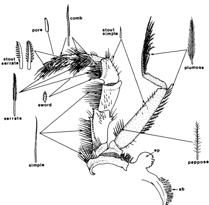
SETAL MORPHOLOGY
Before we describe the gnathothorax and associated appendages, we must first mention the types of setae found thereon. Setal morphology in aeglids is mostly unremarkable and similar to that in other anomuran and brachyuran decapods.
Most setal types are found on the third maxilliped and this appendage is used (Figure 8) for illustrative purposes. Setal types include comb, serrate, stout serrate, sword, plumose, pappose, pore, simple, and stout simple. Many setae appear intermediate in form and may represent "immature" or undifferentiated stages of more complex types; there is a gradation in length in simple setae and in serrate setae, and a gradation from shorter pappose setae to true comb setae. Some setae are intermediate in form between serrate and stout serrate types.
Terminology of setal types in the above section and in the descriptive sections below follows that of Kunze and Anderson (1979) for pagurid hermit crabs. This is by design; not only the setal types but their location on the mouthparts are essentially identical in hermits and aeglids (see Martin and Felgenhauer, 1986). Possibly unique to aeglids are the composite setae of the fifth pereiopod, and the sword and pore setae of the third maxilliped. Kunze and Anderson (1979) also noted that the eight setal types recognized in pagurid mouthparts are not discrete types; transitional forms exist there as in aeglids.
stout serrate
serrate
simple
plumose
pappose
FIGURE 8.—Third maxilliped of A. platensis, inner surface, with setal types mentioned in descriptions of aeglid mouthparts. Arrows indicate areas where occurrence of a given setal type is common, and are not meant to be exclusive of presence of a setal type.
GNATHOTHORAX
Snodgrass (1952b) considered under this heading "the carapace, the branchial chambers and the pleura, the mouth, the ventral skeleton, and the pleurosternal skeleton," or "the part of the animal covered by the carapace." The aeglid carapace was described previously. The pleura (= epimere,
epimeron, pleurepimere, pleurite, pleuron, pleural lobe, tergal fold; see McLaughlin, 1980) are described by McLaughlin (1980) as "each lateral part of integument of somite." In other words, these various terms have been advanced to designate the integumemt of the body where it corresponds to pre-existing somites. Although we recognize a functional gnathothoracic tagma in the fusion of thoracic and gnathal
NUMBER 453 15 components under the single dorsal carapace, we treat the
gnathal and thoracic regions separately. Although Snodgrass (1952b) chose to treat separately the appendages, we have included description of the appendage with its corresponding somite for the gnathal region.
Gnathal Region
MANDIBLES (Figures la,b,d, 9g,h).—The mandibles stem from the third cephalic somite and are similar to those of many other anomuran and brachyuran decapods. The molar process has been reduced to the point where it is barely visible as a small bump on the interior ventral border (Figure 9g); for practical purposes it is absent. The incisor process is strongly sclerotized and asymmetrical, the right side usually bearing a large blunt tooth that corresponds to an indentation on the left mandible (Figure Id). The mandible tapers gradually toward the marginal hinge of the epistome (mh) and the articulation point of the mandible (ar) on the maxillary bridge. The mandibular palp (pip, Figures Id, 9g) is two-segmented. The proximal segment bears several simple and plumose setae on the distal half of the dorsal border; the distal segment is flattened and ovate with many simple, pappose and plumose setae along the entire border. The two segments are approximately equal in length.
The mandibles extend ventrally through a gap between the epistome and the maxillary pleural bridge; their articulation with the gnathothoracic skeleton is described previously under the heading "Epistome".
PARAGNATHS (Figure ld,e).—The paragnaths (pg) are membranous extensions of the metastomal region. They arise from below (posterior to) the labrum and are situated on either side of the mouth. They are extremely flaccid and weak and their functional role is not readily apparent Snodgrass (1950) felt that the paragnaths were outgrowths of the metastomal plates (met), structures unique to brachyurans and anomurans.
In Aegla the distal margin of the paragnath bears few scattered plumose setae; the number may vary somewhat from side to side in a single individual. The metastomal plates (met) are triangular and weakly calcified; they do not extend into the paragnath region but undoubtedly lend support to the paragnaths. These plates arise just posterior to the mouth (mth) and extend anteriorly to just behind the paragnaths. The anteromesial borders bear small simple setae.
FIRST MAXILLA (maxillule) (Figures la, 9f).—The first maxilla arises from the fourth cephalic somite and extends ventrally; it is closely adhered to the mandible. The appendage is thin and membranous. The endopod (en) is indistinctly bilobed, with the proximal portion bearing few, long and short, simple setae. The distal portion of the endopod bears few, short, simple setae. The distal endite (de) is fringed with short simple setae and spines; the proximal endite (pe) is spatulate with numerous simple and pappose setae and few short spines.
The basal area bears several long, plumose, and simple setae.
SECOND MAXILLA (Figures la, 9e).—The second maxilla stems from the fifth cephalic somite and is modified for pumping water over the anterior branchial surfaces. The endopod, distal and proximal endites extend ventrally alongside the maxillule. The endopod (en) is elongate and bears few scattered, simple setae. The distal endite (de) is bilobed, with the distalmost lobe larger and more setose; both lobes of the distal endite bear simple, pappose, and plumose setae. The proximal endite (pe) is bilobed with the proximal lobe much larger, both lobes of the proximal endite bear simple, pappose, and plumose setae. The scaphognathite (scaph) is large and flattened and bordered with numerous plumose setae. The posterior lobe of the scaphognathite extends posteriorly into a respiratory chamber termed by Snodgrass (1952a,b) the pumping chamber (pch; see Figure 14a). This pumping chamber apparently functions in creating high water pressure to facilitate water flow over the posterior branchiae; it is considerably more narrow than the posterior branchial chamber covered by the branchiostegite.
The second maxilla arises far posterior to the first maxilla and appears to be situated posterior to or at the same somite level with the first maxilliped (Figure la); its foramen borders on the mesial margin of the maxillary pleural bridge. This is undoubtedly an adaptation for increased respiratory efficiency and is seen in a variety of reptant decapods. The only other illustration of an aeglid gnathal area is that of Snodgrass (1950, fig. 11), in which the foramen of the second maxilla of A.
prado is also shown posterior to the maxillule and nearly lateral to the first maxilliped.
Thorax
The first three of the eight thoracic segments bear appendages secondarily modified for feeding: the first, second, and third maxillipeds. These appendages and their correspond- ing somites are described first because of their affiliation with the true gnathal regions. The functional thorax, i.e., the five pairs of ambulatory appendages and their sterna, are described in the following sections.
FIRST MAXILLIPED (Figures la, 9d).—The sixth somite (first thoracic somite) gives rise to the first of three walking appendages that have been secondarily modified for feeding;
these are traditionally termed maxillipeds. The first maxilliped, which is thin and only slightly larger than the maxillae, arises posterior to the second maxilla but appears to arise mesial to it (see Figure la and second maxilla, above); the foramen is actually located in the narrow confluence of two mesial extensions of the maxillary pleural bridge. The exopod (ex) is 2-segmented with the distal segment a multiarticu- lated flagellum (fl); both segments bear pappose and plumose setae. The basal portion of the exopod is produced into a large lamellar lobe with plumose setae on its borders. The endopod (en) consists of a reduced, palp-like, terminal lobe and well-developed distal (de) and proximal (pe) endites. The
proximal endite is small and ovoid, with numerous pappose and spinose and few comb setae; the distal endite is larger and subrectangular with numerous pappose, simple, and stout sim- ple setae. The epipod is absent
SECOND MAXILLIPED (Figures la, 9a).—The seventh (second thoracic) somite gives rise to the second maxilliped. The second maxilliped is much larger than the first and assumes more of the grooming and feeding functions, as opposed to being primarily respiratory. This change in function is reflected in the form and setation of this appendage. The maxilliped is pediform, extends anteriorly from the gnathothoracic skeleton, and is not so ventrally oriented as the preceding appendages.
The exopod is 2-segmented, with the distal segment developed into a long multiarticulate flagellum bearing numerous, paired, plumose setae. The proximal segment of the exopod is flattened and bears a row of pappose and plumose setae along the lateral margin. The endopod is 5-segmented and more cylindrical in cross section, especially the terminal segments. The dactylus (d) is rounded terminally and bears simple, pappose, serrate, and stout serrate setae; these latter function in grooming as well as feeding. The propodus (p) is short, nearly cylindrical, and bears the same four setal types on the distal border. The carpus (c) is short and articulates with the propodus and merus so as to form nearly a right angle, directing the distal two segments inward toward the mouth; the carpus bears only simple and pappose setae. The merus (m) and fused basi-ischium (bi) are elongate, more flattened than the preceeding segments, and have long simple setae and few pappose setae. The coxal area (coxa) bears long, simple setae and pappose setae. The epipod is absent.
THIRD MAXILLIPED (Figures la, 8, 9b,c).—The third maxilliped is pediform and well developed; it functions in grooming and feeding (see Martin and Felgenhauer, 1986). In addition, it is often extended anteriorly when the aeglid appears to be searching for food or shelter and thus may have some sensory capabilities as well. This appendage arises from the eighth (third thoracic) somite and is firmly attached to the ventral thoracic sterna by the coxal segment (see Figure \0a,b).
The dactylus is subcylindrical and armed with simple, stout simple, pappose, pore, and serrate setae; most of the serrate setae are located on the distal half. The propodus is slightly longer than the dactylus and bears fewer serrate setae; a circular field of dense setae on the inner surface contains pappose, sword, serrate, and stout serrate setae. The carpus is short and thick with few setae on the outer surface but with a circular field of dense pappose, sword, serrate, and stout serrate setae on the inner surface. This circular field of setae, like that of the propodus, is slightly elevated relative to other areas of the scgmenL The two circular fields of dense setae on the propodus and carpus combine with the serrate setae of the dactylus to function in grooming and feeding. The merus is subcylindrical and bears numerous long, simple setae along its mesial margin, with scattered simple and pappose setae on the inner and outer surfaces; it is slightly dilated distally. The distal portion of the
merus is concave and allows the carpus to fold tightly against i t The fused basi-ischium is nearly triangular in cross section, and has scattered simple and pappose setae. The mesial margin bears a sharp, corneous, subterminal tooth and many long simple setae. The inner surface is similar to that of many other anomurans in having a row of well-developed corneous tubercles, the crista dentata (cd, Figure 9c). These tubercles become progressively larger toward the distal end of the row and terminate in a large sharp spine. The lateral margin of the basi-ischium bears a blunt subterminal protuberance armed with few simple setae. The coxal area bears simple, pappose, and plumose setae. The epipod is present as a small membranous bud extending posteriorly from the arthrodial membrane proximal to the coxa. There is a small arthrobranch present that is firmly attached to the epimeral plate and is usually lost when the maxilliped is removed. The only other illustrations of aeglid mouthparts are those of Ortmann (1892), which do not allow detailed comparison.
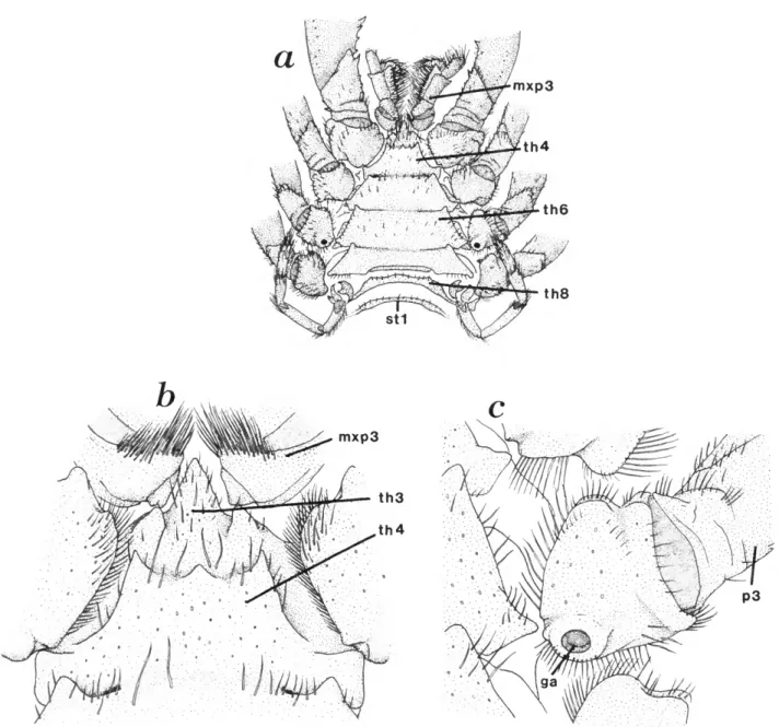
Thoracic Sterna
The sternal regions of the first and second maxillipeds are reduced and represented by a thin median extension of the maxillary pleural bridge (Figure la,b\ see also "Gnathal Region" above). The third maxillipeds are closely approxi- mated basally and the corresponding sternum is reduced to a small but well-calcified, conical sclerite, which in some species may be minutely bifurcate or heavily setose. This sclerite forms the apex of a large, sternal, triangular plate (Figure \0a,b) that extends posteriorly to the level of the 7th thoracic somite (pereiopod 4). The sternal plate becomes progressively wider posteriorly. The fourth thoracic sternum, between the coxae of the chelipeds, is of some systematic value in that some species have an anteromedial tubercle or spine on this sternum. This tubercle may be low and rounded (Figure 10b) or it may be sharp and distally cornified (e.g., A. bahamondei Jara and A. perobae Hebling and Rodrigues). In many species no such tubercle is seen, and the anterior margin of the fourth thoracic sternum is nearly smooth. All thoracic sterna bear rather blunt anterolateral projections; those of the third somite articulate with the coxa of the third maxillipeds (Figure \0b).
The posterolateral borders of the sterna articulate with a shallow groove or depression in the coxa of the corresponding pereiopod (Figure 10b,c). In A. platensis, A. uruguayana, and A. jujuyana, and probably in all other species, the sternal surfaces are marked by minute pits and scattered simple setae;
setation is most dense at lateral and anterolateral sternal borders. All thoracic sterna are fused except for that of the eighth thoracic somite (th8), which is connected to the sternal
FIGURE 9.—Mouthparts of A. platensis: a, second maxilliped; b, third maxilliped; c, ischial portion of fused basi-ischium with crista dcniata; d, first maxilliped; e, maxilla; / maxillule (first maxilla); g, inner view of right mandible; h, external view of right mandible.
scaph
a
mxp3
th6
t h 8
mxp3
FIGURE 10.—Ventral view of thorax of A. platensis: a, ventral view of thorax showing last thoracic segment unfused and first abdominal sternum; b, high magnification of sternum of thoracic segment four showing median tubercle; c, high magnification of coxa of female third pereiopod showing genital aperture.
plate of the anterior somites by a narrow membranous band (Figures 10a, 16b).
Pereiopods
The first pereiopods (chelipeds) are large chelate appen- dages. The second through the fourth are achelate and similar;
pereiopods will be treated separately.
FIRST PEREIOPOD (Figures 2, 3, 11).—The first pereiopod is a large chelate appendage that varies in form among species.
The cheliped is larger in males than in females, and almost always larger on the left side. Features of the cheliped have been used extensively as taxonomic characters, although it is the fifth are chelate and reduced. These three types of known that many characters vary within a species and from
NUMBER 453 19
coxa
FIGURE 11.—Cheliped of aeglid crabs: a, lateral view of left (major) cheliped of A. uruguayana; b, mesial view of same; c, dorsomesial view of right (minor) cheliped of A. uruguayana; d, coxa and fused basi-ischium of A.
platensis; e. coxa and fused basi-ischium of A. uruguayana (note distomesial spine); / chela and carpus of male A. platensis.
side to side in one individual.
The dactylus (d) is typically short and heavy with the distal end curved inward. The outer surface is smooth or lightly granulate with scattered, stout, simple setae arising from circular depressions. The cutting margin is bordered with smooth, low, corneous scales (cs) or tubercles and may or may not have a lobular basal tooth; in A. singularis the cutting border of the dactylus is actually indented rather than produced basally. The dorsal border is usually smooth or has a slightly elevated ridge of tubercles; in many species (e.g., A. rostrata, A. bahamondei, A. plana, A. camargoi, A. leptodactyla, etc.) there may be a small, spiniform, dorsal tooth on the proximal
!A of this segment.
The propodus (p) is large and inflated. The fixed finger has a row of corneous scales along the cutting edge and except for a few species (A. humahuaca, A. jujuyana, A. plana, A.
sanlorenzo, A. camargoi, and A. leptodactyla) has a large basal tooth also covered with tubercles; this is often indistinct or absent on the minor (right) cheliped. Scattered simple setae arise from circular depressions on either side of the corneous border of the cutting edge. The dorsal border of the propodus may be nearly smooth (e.g., Figure 11) or it may be compressed and elevated into a palmar crest or lobe (pc; see also Figure 2). This lobe most often takes the form of a serrate or tuberculate ridge, as in A. araucaniensis, A. manni, A.
bahamondei, A. denticulata, and many others, but it may be expanded into an excavated spoon-shaped structure. This condition, which is seen in A. castro, A. odebrechtii (both subspecies), and to a lesser degree in many other species, is best developed in the Brazilian A. senmitti (see Hobbs, 1979, and Figure 19). The lobe may be extremely variable within a species (e.g., see Hobbs, 1979). The approximation of the posterior edge of this palmar crest with the distal edge of the carpus can form a sinus termed by Ringuelet (1949a) the precrestal sinus (not illustrated). There is almost always a distinct dorsal groove just proximal to the articulation with the dactylus. Another groove extends proximally and ventrally from just below the distodorsal propodus border along the outer surface; this shallow groove appears to curve toward the upper margin of the fixed finger of the propodus. The ventral border of the propodus is usually smooth or tuberculate although in some species (e.g., A. parana and A. lenitica) the tubercles may give rise to small spines. The inner surface of the propodus (Figure 1 \b) may be smooth and slightly inflated or it may have a series of irregular longitudinal depressions (this condition illustrated). The posterior border of the inner surface of the propodus has a small, rectangular, detached sclerite that rests in the membranous area of the joint.
The carpus (c) is a short stout segment of variable form and ornamentation. The dorsal border typically bears a row of 4-5 heavy spines with corneous tips. These spines are typically of equal or subequal size, although there is considerable variation within a species (e.g., sec Hobbs, 1979). Most often they increase in size distally to the subterminal (largest) spine. The
distalmost spine (termed the antero-internal lobe by Ringuelet, 1949a; carpal lobe of Schmitt, 1942b) is usually reduced, does not taper as sharply, and may be terminally rounded or broadly spinose (e.g., A. jujuyana, A. n. neuquensis, A. affinis, and A.
alacalufi); often this terminal spine or tooth is separated from the remainder of the carpus by distinct grooves. The outer surface of the carpus is marked by a curved ridge of tubercles or small spines termed the carpal ridge (cr, Figure 1 If). This ridge may be low and broad (illustrated) or it may be more sharply defined and bear corneous spines. Occasionally (e.g., A. parana) there may be a second row of spines ventral to the carpal ridge. Often the anterior edge of the carpal ridge merges with a shallow groove'along the distal border of the outer surface. The ventral border of the carpus may be smooth or may bear 1-2 corneous spines (Figure 1 \a,b). The inner surface of the carpus almost always bears a single, sharp, heavy spine directed antero-mesially (Figure \\b,c). The posterior border of the inner surface of the carpus bears a subrectangular sinus in which rests a detached sclerite, similar to that seen in the propodus-carpus articulation (Figure Mb).
The merus (m) is heavy and triangular in cross section, with the dorsolateral and ventromesial edges armed with a row of heavy spines. The cheliped is slightly rotated inward so that the outer (dorsolateral) row of spines assumes a dorsal position and the smooth dorsomesial surface (see Figure l i e ) is appressed to the pterygostomial region of the carapace (Figures 2 and 3). The ventrolateral border of the merus bears only one or two distal heavy spines rather than a row of spines (Figure Mb). The dorsal margin almost always bears a distinct transverse groove just proximal to the articulation with the carpus; this groove is connected to the distal border of the merus by a short longitudinal groove and usually is continued around the entire distal perimeter of the segment (Figure l l a - c ) . The ventromesial and lateral rows of spines terminate proximal to this groove.
The ischium and basis are fused into a basi-ischium (bi);
this short segment articulates diagonally with the merus. The ventromesial border is often used as a taxonomic character; it may be nearly smooth, as in A. platensis (Figure Md), or it may bear one to two distal basi-ischial spines (e.g., A.
uruguayana, bis, Figure Me). In some species (e.g., A.
jujuyana, A. parana, A. sanlorenzo, and A. prado) the two basi-ischial spines may be widely separated, while other species may have a series of small spinules. Ringuelet (1948b, 1949a) has shown that at least in Argentine aeglids this character varies widely. The proximal end of the basi-ischium bears three indistinct lines, with the center line being a fracture plane (fp, Figure Me) for autotomy of the cheliped.
The coxa is a heavy globose segment that articulates with the thoracic sternum by a produced, posteroventral, indented lobe (Figures 10, Md,e).
The epipod is present as a reduced setose tubercle (ep, Figure 14b) loosely articulating with the coxa.
SECOND THROUGH FOURTH PEREIOPODS (Figure \2a-d).—