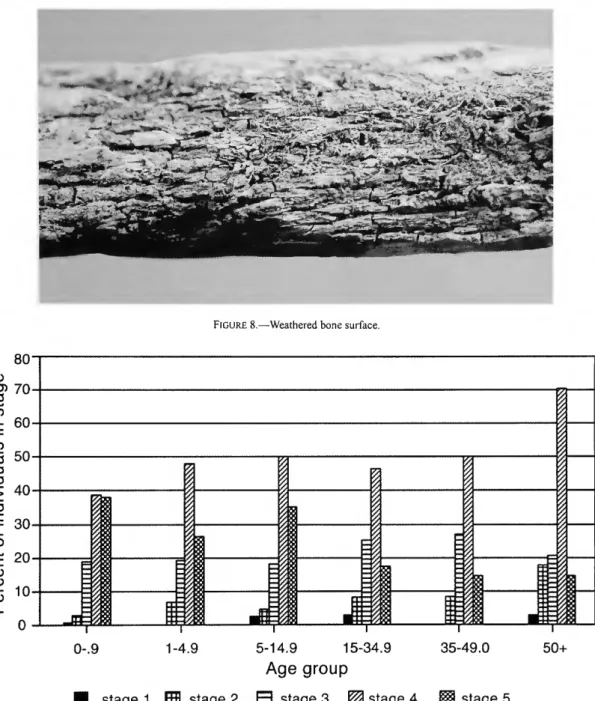
Analysis of the human remains revealed highly variable preservation with extensive weathering, particularly among adults. Most of the Voegtly Cemetery casket hardware was likely manufactured during the mid-1800s, earlier. This section focuses specifically on the analysis of human remains submitted to the Smithsonian Institution.
The report includes data on skeletal biology and an excellent literature review. Snow and Fitzpatrick (1989) summarized their analysis of the remains of 34 soldiers killed in the 1876 battle.

SMITHSONIAN CONTRIBUTIONS TO ANTHROPOLOGY
Green, copper staining was most often found on the bones of the skull (32 cases), followed by vertebrae and ribs (21 cases); hands, shoulders and collarbones (20 cases); The skull was the most frequently stained surface in each of the age and sex categories. Mortuary records from the cemetery of the German Evangelical United Church were translated from the original German text by Dzodin and Luff (1989) (Appendix 1).
ULHL
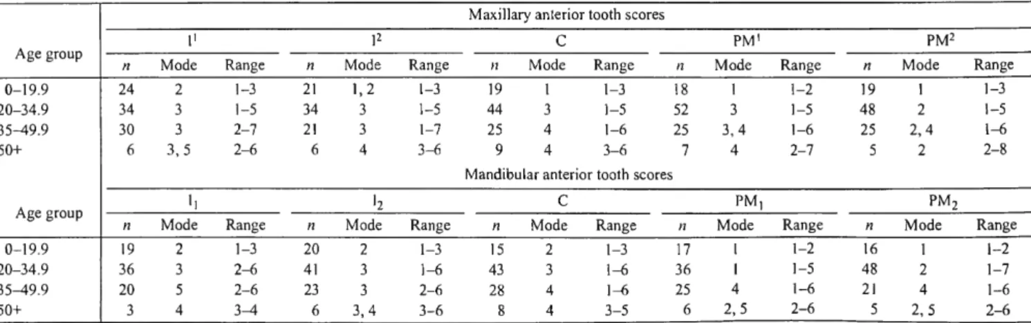
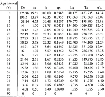
The values in these age ranges are then weighted by proportionally assigning them to individuals of unknown age, so that the total number of individuals in the sample corresponds to the number of individuals listed in the life table. Dx=total number of individuals per age interval, dx=percentage of individuals dying in each age interval, lx= . number of survivors from a theoretical cohort of 100 individuals entering each age interval, qx=probability of death in each age interval, Lx=total number of years lived in each age interval, Tx=total number of years lived after a lifetime for all individuals entering each age interval, e°x = life expectancy of all individuals entering each age interval.). Dx=total number of individuals per age interval, dx=percentage of individuals dying in each age interval, lx=number of survivors from a theoretical cohort of 100 individuals entering each age interval, qx=probability of death in each age interval, Lx=total number of years lived in each age interval, Tx=total number of years lived after a lifespan for all individuals entering each age interval, e°x=expected life expectancy for all individuals entering each age interval.).
Because this information was not always provided, the number of deceased siblings was definitely higher, as many surnames are quite common. Dx=total number of individuals per age interval, dx=proportion of individuals dying in each age interval, lx=number of survivors of a theoretical cohort of 100 individuals entering each age interval, qx=probability of death in each age interval, Lx=total number of years lived during each age interval, Tx=total years lived after a lifetime of all individuals entering each age interval, e°x=life expectancy of all individuals entering each age interval.).
Another method of determining family mortality is to use information provided by the pastor when a parent dies. Based only on information about the number of children born to each couple, it is possible to construct a graph showing that most individuals had 3 children at the time of death (Figure 12).
IIIIIIIII inn imr
Thirty-five were under the age of 15 and had lived in the United States an average of 2.8 years, with an average age at death of 5.7 years. For all immigrants, the average time spent in the United States was 10.2 years, with an average age at death of 39.3 years. The number of individuals in other age groups is roughly comparable between the two samples until the groups over 50 are examined.
Dx=total number of individuals per age interval, dx=percentage of individuals dying in each age interval, lx=number of survivors of a theoretical cohort of 100 individuals entering each age interval, qx=probability of death in each age interval, Lx=total number of years lived during each age interval, Tx=total number of years lived after a lifetime of all individuals entering each age interval, e°x=life expectancy of all individuals entering each age interval.). Dx=total number of individuals per age interval, dx=percentage of individuals dying in each age interval, lx=number of survivors of a theoretical cohort of 100 individuals entering each age interval, qx=probability of death in each age interval, Lx=total number of years lived during each age interval, Tx=total number of years lived after a lifetime of all individuals entering each age interval, e°x=life expectancy of all individuals entering each age interval.).

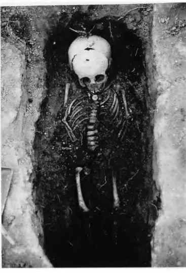
BURIAL 25.—FN: Nails and wood were the only artifacts found in association with this burial
BURIAL 26.—FN: A coffin outline was the only artifact asso- ciated with the human remains
BURIAL 27.—FN: Nails were the only associated artifacts re- covered
BURIAL 28.—FN: A coffin outline was associated with this burial, which had been disturbed in the northern portion by a
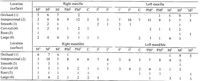
Green staining, perhaps from contact with copper, is on the lateral midshaft surface of the left ulna. Carious lesions in milk teeth are on the interproximal surfaces of the maxillary second molars. Carious lesions in baby teeth are on the interproximal surfaces of the maxillary left first molar and mandibular right canine.
The brown color is on the mesial surface of the upper left third molar and the distal surface of the right second premolar of the mandible. One carious lesion is on the interproximal surface of the decidual right maxillary canine.
BURIAL 60.—FN: Nails were the only associated artifacts re- covered
Bones present are the skull (3); right clavicle (3); right scapula (3); right ilium (3); both patellae (1); three cervical vertebrae; two thoracic vertebrae; four lumbar vertebrae; one right rib; middle (3) and distal (2) thirds and distal epiphysis (1) of the left humerus; proximal epiphysis (3) and proximal (3), middle (2), and distal (1) thirds and distal epiphysis (1) of the right humerus; left radius (1); distal epiphysis (3) and rest (1) of the right radius;. The size of the bones indicates a probable age at death from newborns to 1 year.

EpKyfl
Carious lesions are located on the occlusal surfaces of the maxillary second molars (two lesions each) and the left first premolar of the mandible; interproximal surfaces of the mandibular right second premolar and mandibular right first premolar (two lesions); Bones present are the frontal (2); parietals (2); occipital (2); both temporary (1); mandible (1); both collarbones (1); left scapula (1); left ilium (2); two cervical vertebrae; three thoracic vertebrae; both first ribs; four left and four right other ribs; proximal third (2) of the left humerus; The bones present are the frontal (2), both parietal (3), occipital (3), left (3) and right (2) temporal parts, mandible (3), right clavicle (2), right scapula (2), three cervical bones vertebrae, five thoracic vertebrae, four lumbar vertebrae, first right rib, three left and five right ribs, proximal epiphysis (1) of the left humerus, proximal epiphysis (2) and proximal (2) and distal (2) third of the right humerus, proximal epiphysis (2) and diaphysis (2) of the left femur, proximal (2) and middle third (2) of the right femur, and proximal epiphysis (1) of the right tibia.
Bones present are frontal (2), both parietals (3), occipital (3), left (3) and right (2) temporal bone, mandible (2), second cervical vertebra, three other cervical vertebrae, middle thirds (3 ) of both femurs, the proximal (3) and middle (3) thirds of the right tibia and the middle third (3) of the left tibia. The bones present are occipital (3), left temporal (3), both zygomatics (1), lower jaw (2), right clavicle (2), right scapula (2), four cervical vertebrae, five thoracic vertebrae, both first ribs, three other left and right ribs, the proximal (3) and middle third (2) of the right humerus, and the middle third (3) of the left femur. Bones present are the temporal bones (3), mandible (3), both shoulder blades (3), both ischia (3), middle thirds (3) of humerus, proximal epiphysis (3) of left radius, proximal epiphysis (3) and proximal third (3) left ulna, proximal epiphyses (3) and middle third (3) of femur and middle third (3) of tibia.
Bones present are parietal (2), occipital (2), left temporal (2), right clavicle (2), right scapula (2), three left ribs and additional rib fragments, right humerus (1), proximal epiphysis (2) . ) and the proximal third (1) of the right radius and the proximal epiphysis (1) and the proximal third (1) of the right ulna. Carious lesions are on the occlusal surfaces of the upper left second molar and the mandibular second molar. The bones of the cranial vault (2), mandible (2), left ilium (3), middle (2) and distal (2) thirds of the left humerus, diaphysis (2) of the right humerus, proximal third (3) of the left radius, middle third are present (3) of right radius, proximal (2) and middle (2) thirds of left ulna, proximal third (2) of right ulna, proximal epiphysis (3) and diaphysis (2) of femur, and one talus (side undetermined).
Bones present are temporal bones (2), mandible (1), left clavicle (2), both shoulder blades (3), fragments of vertebrae, left first rib, five left and two right second ribs, proximal and middle third (2) left humerus and the proximal third (2) of the right humerus. The bones present are the occipital (2), both temporals (2), the right scapula (2), eight left and six right ribs, and the diaphysis (2) of the right humerus. Carious lesions are on the occlusal surfaces of the upper right first molar and the mandibular third molar.
Bones present are frontal (3); sacrum (3); left (2) and right (3) ilia; four lumbar vertebrae; middle thirds (3) of humeri; middle third of left radius; distal third of right radius; middle third of left ulna; both femora (2) minus the distal epiphyses; distal epiphysis (2) and diaphysis (2) of left tibia; diaphysis (2) of right tibia; and middle thirds (2) of fibulae. Bones present are frontal (3), occipital (3), both temples (3), right zygomatic (2), both maxillae (3), both ilia (3), rib fragments, all cervical vertebrae, proximal epiphyses of femora (3), proximal (3) and distal (3) thirds of left tibia, diaphysis (3) of right tibia and middle third (3) of right fibula. Bones present are frontal (2), both parietal (3), occipital (2), left (3) and right (2) temple, mandible (2), sacrum (3), left acetabulum (3), first two cervical vertebrae, another cervical vertebra, two lumbar vertebrae, proximal thirds (3) of humeri, proximal thirds (3) of left ulna and proximal epiphyses (3) and proximal thirds (3) of femora.
The bones present are the frontal (3), both parietal (2), occipital (1), both temporal (2), both ilia (3), first cervical vertebra, proximal (3) and middle third (3) of the right humerus, proximal third (3) of the left radius, proximal (3) and middle (3) third of the left femur, distal epiphysis (2) of the right femur, proximal (3) and middle (3) third of the left tibia and the proximal third (3) of the right tibia.