To the former members of the Dougherty lab with whom I have had the pleasure of overlapping (Dr. Clint Reagan, Dr. Oliver Shafaat, Dr. Mike Post, Dr. Matt Rienzo, Dr. Matt Davis, Dr. Betty Wong, Dr (Catie Blunt, David Paul Walton, Dr. John Bryce Jarman, Dr. Annet Blum, and Dr. Richard Mosesso, Gabby Tender, and Kate Kutzer), thank you for making this lab a home.
Introduction
The Nicotinic Acetylcholine Receptors and Allosteric Modulators
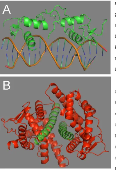
The α4β2 nAChR is particularly important as it is the most prominent heteromeric subtype in the brain.11 The α4β2 nAChR can be found in two stoichiometries: high-affinity (α4)2(β2)3 or low-affinity (α4)3. (β2)2. Unfortunately, recent work has shown that tobacco and flavoring ENDS are NAMs of the α4β2 nAChR.12,16 The most prominent of these Figure 1.2 How allosteric modulators act.
The Opioid Receptors and Protein Trafficking
In this sense, if desensitization occurs without endocytosis and subsequent recycling, endocytosis is not desirable. In Chapter Five, we perform new experiments investigating the role of C-terminal phosphorylation in morphine- and fentanyl-induced endocytosis.
![Figure 1.4. Overview of [cAMP] changes during (A) normal function, (B) upon initial opioid exposure, (C) upon development of tolerance, (D) during withdrawal](https://thumb-ap.123doks.com/thumbv2/123dok/10412797.0/23.918.173.751.123.975/figure-overview-changes-function-exposure-development-tolerance-withdrawal.webp)
Epithelial Sodium Channels’ Role in COVID-19
Chapter six documents our efforts to investigate how SARS-CoV-2 E and S proteins influence ENaC activity. We believe that the SARS-CoV-1 S protein appears to reduce ENaC currents in the Ji et al.
Major Experiments
- Two-electrode voltage-clamp electrophysiology on Xenopus oocytes
- NCAA mutagenesis
- ERES monitoring in SH-SY5Y cells
Finally, the NCAA could not be recognized by any of the endogenous synthetases or tRNAs. One could not do the same with the orthogonal tRNA/synthetase method, because phenylalanine is Figure 1.6 Overview of the TECV and in vitro aminoacylation technique in Xenopus.
Summary of Dissertation Work
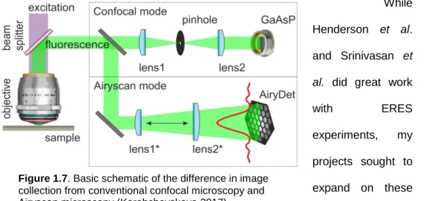
Points two and three are connected, as the z-stacks were only possible because we switched from a decades-old Nikon microscope to a state-of-the-art Zeiss LSM 880 with a Fast Airyscan module (Fig. The Airyscan principle is that instead of simply rejecting any light that is not captured by the confocal pinhole, additional detector elements will collect this light and reconstruct the image with this included light to improve resolution. Combined with the LSM 880 with the fast Airyscan, we have improved image quality and speed to obtain the entire z-band in live cells before significant movement of ERES was observed.
Chronic exposure to morphine and naltrexone induces changes in catecholaminergic neurotransmission in rat brain without altering μ-opioid receptor sensitivity. Inverse agonist upregulates the constitutively active D3.49(164)Q mutant of the rat μ-opioid receptor by stabilizing the structure and blocking constitutive internalization and downregulation.
Synthesis and incorporation of a solvatochromic amino acid into
- Abstract
- Introduction
- Results and Discussion
- Synthesis of 4-DMN and ligation onto THG73
- Confirming 4-DMN incorporation in mm nAChR
- Efforts to observe 4-DMN incorporated into mm nAChRs in Xenopus
- Conclusions
- Materials and Methods
- Synthesis
- Mouse muscle nAChR molecular biology
- Microinjection
- References
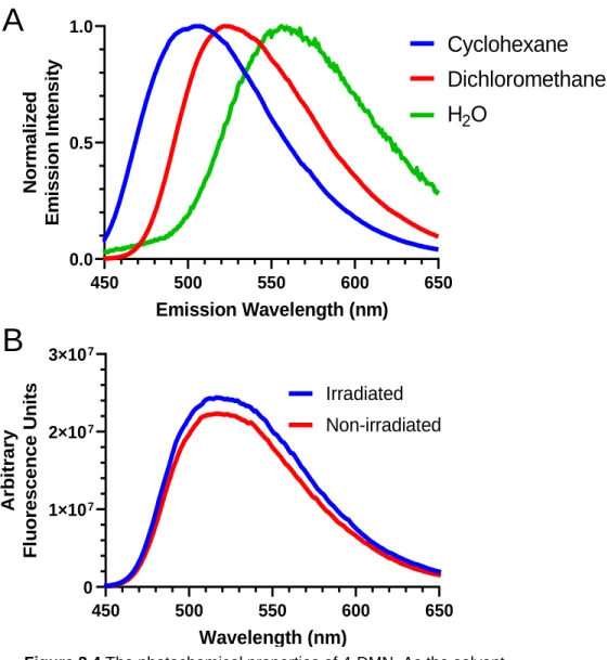
Excitation of the fluorophore causes a change in the dipole moment of the molecule (lower left to upper left). This usually only allows visualization of the plasma membrane and everything very close to it. Normalized emission intensity Cyclohexane Dichloromethane H2O. Figure 2.4 Photochemical properties of 4-DMN. environment) becomes more polar, the emission wavelength increases.
Identification and biophysical analysis of the menthol binding site
- Abstract
- Introduction
- Results and Discussion
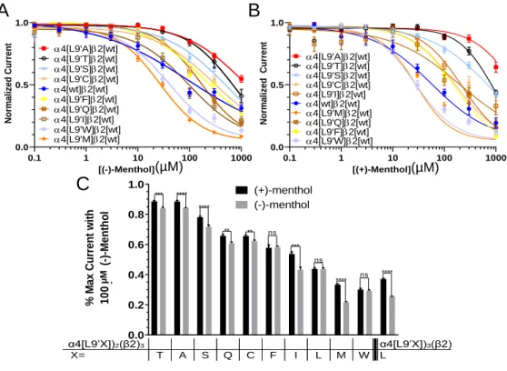
- Changing the residue at the 9’ site in the M2 helix alter’s menthol’s
- Menthol inhibition is directly related to the length of the 9’
- Only one menthol molecule is required for receptor inhibition
- Conclusions
- Materials and Methods
- Reagents
- Oocyte preparation and injection
- Oocyte electrophysiology
- References
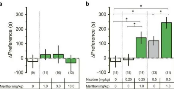
Combined with the importance of α4β2 nAChRs in the brain, we decided to focus specifically on α4β2 nAChRs. This inhibition is directly related to the concentration of menthol in the solution, where a higher concentration of menthol will inhibit the channel more. Role of lipid sensor M4 in folding, trafficking, and allosteric modulation of nicotinic acetylcholine receptors.
Opioid receptor antagonists pharmacologically chaperone a
Abstract
Introduction
This study examines the pharmacological chaperoning hypothesis.18 Pharmacological chaperoning occurs when a pharmacophore binds to a nascent protein and promotes proper folding, thereby aiding its exit from the ER.19 Pharmacological chaperoning participates in therapeutic approaches when suboptimal protein levels disrupt the plasma membrane reach .19-22 Agonists and antagonists are able to chaperone in various systems, and several studies show that ligands promote trafficking in δ-opioid receptors.23,24. Research examining pharmacological chaperoning has not yet examined important early events in MOR surface expression, in part because MORs traffic to the plasma membrane quite efficiently. In previous experiments with nicotinic receptors, pharmacological chaperoning causes increases in ERES levels.26,27 We determined that Sec24D, one of four Sec24 isoforms that can participate in ERES formation, does interact with MORs, suggesting that Sec24D MORs commute.
Results
- MOR[N190K] reaches wild-type plasma membrane densities after
- MOR interacts with Sec24D
- Ntx and naloxone, but not agonists, induce increases in ERES in SH-
- N-methyl-naltrexone does not cause a significant shift in ERES levels
- The lack of significant chaperoning by agonists is not due to
- Ntx does not induce a rise in [cAMP]
- Ntx pharmacological chaperoning depends on COPI
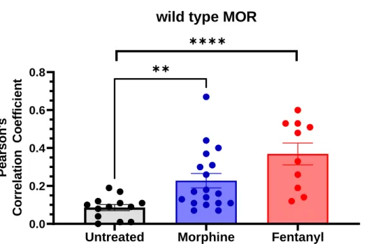
Indeed, unlike Ntx, Me-Ntx did not induce a significant shift in ERES levels in SH-SY5Y cells overexpressing MOR[N190K] (Figure 4.4). Ntx, but not agonists, increase ERES levels in SH-SY5Y cells despite abolishing the S375 phosphorylation site. However, morphine and fentanyl do not significantly increase ERES levels in SH-SY5Y cells overexpressing MOR[N190K][S375A] (Figure 4.5).
![Figure 4.1 MOR[N190K] appears on the plasma membrane after 24 h incubation in 10 μM Ntx or naloxone](https://thumb-ap.123doks.com/thumbv2/123dok/10412797.0/82.918.162.761.166.351/figure-mor-n190k-appears-plasma-membrane-incubation-naloxone.webp)
Discussion
Previous work in our laboratory and others has shown that COPI vesicles may play a role in pharmacological chaperoning.18,43 Here we find a similar dependence, as Ntx and naloxone no longer increase ERES levels if the cells are co-treated with brefeldin A ( BFA), a COPI inhibitor (Figure 4.4, purple).18,32,44 These results suggest that some of the ERES observed in the non-BFA-treated cells have already traveled to the Golgi. This retrograde trafficking dependence is not uncommon for some cargoes, and it appears that the MORs also depend on some ER-Golgi cycling before they are fully ready to go to the plasma membrane. To the extent that pharmacological chaperoning controls upregulation/supersensitivity to naltrexone, we note that Me-Ntx is FDA-approved to suppress opioid-induced constipation (OIC) when opioids are used for chronic non-cancer pain.
Materials and Methods
- Reagents
- SH-SY5Y cell culture and transfection
- Z-stack confocal microscopy
- Sensitized Emission Förster resonance energy transfer (FRET)
- Image analysis
- Competitive ELISA for [cAMP] measurement
- Statistical analysis
All images and z-stacks were processed using Airyscan processing in the Zen Blue software package (Zeiss). Using the AER and DER values calculated in samples expressing a single fluorescent protein, the following equation was used to calculate the corrected FRET intensity, cFRET. This same intensity threshold was used to label ERES in 3D images that were analyzed in Imaris (Bitplane).
Ligand-induced internalization of opioid receptors in enteric neurons after chronic treatment with the opiate fentanyl. Opioid receptor pharmacological chaperones act by binding and stabilizing newly synthesized receptors in the endoplasmic reticulum. Export from the endoplasmic reticulum represents a rate-limiting step in the maturation and cell surface expression of the human delta opioid receptor.
Agonist-induced µ-opioid receptor endocytosis is dependent on
Abstract
Introduction
Unlike binding assays, this measurement will look specifically at endocytosis and will not rely on potential complications of different states of the MOR that may inhibit DAMGO binding. For example, prolonged exposure to agonists can cause desensitization, which can alter the binding properties of the receptor.26 Because we rely only on fluorescence, the conformation of the MOR is not important in our assay. Furthermore, our assay is specific to endocytosis, while binding assays examine general decreases in MOR plasma membrane density.
Results and Discussion
- Morphine and fentanyl induce endocytosis of MORs in SH-SY5Y cells
- Fentanyl will, but morphine will not increase endocytosis of
- Inhibiting fentanyl induced endocytosis requires the abolition of
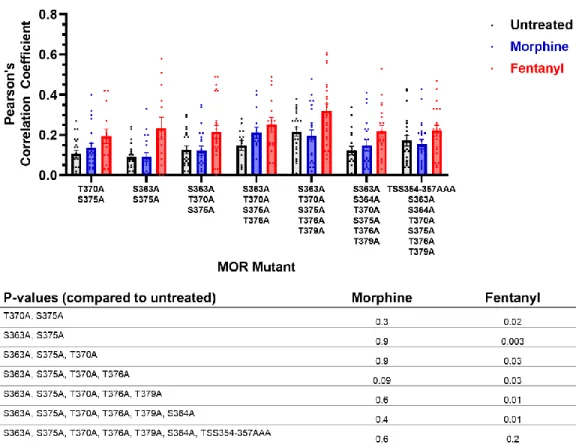
These results are consistent with observations that morphine only phosphorylates S375 and that phosphorylation is important for MOR endocytosis.11,22,24 Meanwhile, we know that fentanyl will phosphorylate sites other than S375, which appears to be sufficient to recruit β-arrestin and endocytosis cause. 21. We then progressively mutated phosphorylation sites to alanine and proceeded to test both fentanyl- and morphine-induced endocytosis. These results suggest that fentanyl-induced endocytosis will not occur if most of the C-terminal serine and threonine residues cannot be phosphorylated.
Conclusions
Further efforts may turn to binding assays or other biochemical experiments to evaluate MOR density on the plasma membranes.
Materials and Methods
- Reagents, materials, and plasmids
- SH-SY5Y cell culture and transfection
- Imaging and analysis
Imaging was performed on a Zeiss LSM 880 with Fast Airyscan. Fast Airyscan improves the signal-to-noise ratio by including photons traditionally rejected by the confocal pinhole to aid in image reconstruction. The 5% most intense pixels in each channel were used to calculate Pearson's correlation coefficient.
Disruption of the Na+ ion binding site as a mechanism for positive allosteric modulation of the µ-opioid receptor. Phosphorylation of Ser363, Thr370, and Ser375 residues within the carboxyl tail differentially regulates μ-opioid receptor internalization. Multisite phosphorylation is required for sustained interaction with GRKs and arrestins during rapid µ-opioid receptor desensitization.
Abstract
Regulation of Epithelial Sodium Channel Activity by SARS-CoV-1 and SARS-CoV-2 Proteins Biophysical Journal, 2021. Extends previous proposal that SARS proteins affect ENaC currents via protein kinase C (PKC) activation, PKC activation via phorbol 12-myristate 13 -acetate (PMA) decreases ENaC and α3β4 activity. We conclude that SARS-CoV-1 and SARS-CoV-2 proteins alter the function of human plasma membrane channels via incompletely understood mechanisms.
Introduction
It is important to understand how the various proteins encoded by SARS-CoV-2 interact with endogenous human proteins.2,6,7 SARS-CoV-2 must interact with host proteins to replicate. 8 These interactions begin when the SARS-CoV-2 S protein binds to the human angiotensin-converting enzyme 2 (ACE2) protein; this interaction leads to viral entry.9-11 Many subsequent interactions have been studied between SARS-CoV-2 and human proteins. Studies using mass spectrometry and in silico methods have identified many protein-protein interactions between SARS-CoV-2 and humans.7,12 Furthermore, researchers looked at work done on SARS-CoV-1, the related beta -coronavirus responsible for the SARS epidemic in 2002 and 2003.13. We are also interested in how the E and S proteins of SARS-CoV-2 affect ENaC function.
Results
- SARS-CoV proteins do not form ion channels in the plasma
- SARS proteins are expressed following mRNA injection
- SARS-CoV-1 E, SARS-CoV-2 E, and SARS-CoV-2 S proteins
- SARS-CoV-1 and SARS-CoV-2 E proteins inhibit α3β4 currents 104
- Mutating the furin cleavage site in SARS-CoV-2 S protein improves
- PKC activation decreases ENaC and α3β4 currents
- PKC inhibition does not abolish SARS-CoV-1 E, SARS-CoV-2 E, or
However, in contrast to our ENaC experiments, SARS-CoV-2S protein does not significantly reduce α3β4 currents. We find that if the SARS-CoV mRNA is injected 24 hours after the ENaC mRNA, there is no ENaC inhibition (Figure 6.5). However, currents were not fully restored, suggesting that other factors influence ENaC inhibition by SARS-CoV-2 S protein.

Discussion
We show that inhibition does not occur when SARS-CoV protein mRNAs are injected 24 hours after ENaC mRNA, indicating that SARS-CoV proteins affect early steps in the functional expression of channel proteins (Figure 6.5). Furthermore, when we blocked PKC activation by treatment with Gö-6976, we did not block the inhibitory effects of SARS-CoV-1 E, SARS-CoV-2 E, or SARS-CoV-2 S proteins (Figure 6.9). Among the currently known lineages of SARS-CoV-2 variants, we did not observe mutations at positions 55-56 and 69-70.

Conclusion
Several members of the B.1.351 lineage have a P71L mutation; this position is near the 69-70 sequence and immediately upstream of a candidate PDZ domain binding motif at the C terminus. Measuring the interactions made by the S and E proteins with human proteins could be crucial to understanding the COVID-19 and SARS-CoV-2 variants. Although our findings may be meaningful and important, they represent only part of the necessary knowledge about SARS-CoV-2 and COVID-19.
Materials and Methods
- cDNA and mRNA
- Protein expression in oocytes
- Immunoblotting
- Electrophysiology
For SARS-CoV-1 or SARS-CoV-2 proteins, 20 ng mRNA was injected into oocytes together with ENaC or α3β4 nAChR mRNA. In oocytes injected with a single SARS-CoV-2 protein mRNA, each oocyte received 20 ng of mRNA. In oocytes injected with nine SARS-CoV-2 and two SARS-CoV-1 protein mRNAs, 3 ng of each were injected into each oocyte.
SARS-CoV proteins reduce the levels and activity of human ENaC via activation of various PKC isoforms. Identification of a Golgi complex-targeted signal in the cytoplasmic tail of the severe acute respiratory syndrome coronavirus envelope protein. Activation of protein kinase C by phorbol ester induces downregulation of the Na+/K(+)-ATPase in Xenopus laevis oocytes.
Investigating protein-protein interactions in the estrogen
In cells transfected with ERα[His516TAG], we found that cells treated with media Figure A1.3 Verification of the presence of functional ERα. Finally, we found significantly greater chemiluminescence from cells transfected with ERα[His516TAG] and N3Phe tRNA/synthetase relative to those not transfected with tRNA/synthetase (Figure A1.3C). We decided to go through with the 275 site as it was closer to the center of Figure A1.6 Both tamoxifen and fulvestrant induce dimerization.