PAYING ATTENTION TO THE DETAILS: RARE GENETIC VARIATION IN THE DOPAMINE TRANSPORTER AND ADHD
By
Dhananjay Sakrikar
Dissertation
Submitted to the Faculty of the Graduate School of Vanderbilt University
in partial fulfillment of the requirements for the degree of
DOCTOR OF PHILOSOPHY in
Neuroscience May, 2012 Nashville, Tennessee
Approved:
Roger Colbran Aurelio Galli Gregg Stanwood
Ann Richmond Randy Blakely
Acknowledgements
To start, I would like to acknowledge the funding that supported my work. This includes training grant slot and fellowship from Vanderbilt Chemical and Physical Biology Program and Neuroscience Graduate program respectively. Further support was contributed by National Institute of Health grants to my mentor, Dr.
Randy Blakely (DA027739, HL56693).
I would like to express my gratitude to my mentor, Dr. Randy Blakely. Randy was one of the reasons I decided to join the Vanderbilt University for graduate studies. He has been a great mentor, always putting a positive spin on the experimental outcomes and encouraged me when the research was slow going.
He has always pushed me to pursue my own interest and encouraged me to attend and present my work in scientific meetings. I have never seen a person getting so excited by science and his enthusiasm will always be in inspiration for me.
I would also like to thank my thesis committee: Dr. Roger Colbran, Dr. Aurelio Galli, Dr. Gregg Stanwood, and Dr. Ann Richmond. Their input and guidance in my progress was invaluable, and their patience and constant availability was much appreciated. Their efforts to help me focus my thoughts and experiments were extraordinarily helpful.
I would also like to recognize our collaborator Dr. Michael Gill (Trinity College, Dublin). Dr. Gillʼs team was instrumental in examining and collecting samples
variants studied here. I want to acknowledge the efforts of Dr. Michelle Mazei- Robison and Marc Mergy for carefully screening and identifying the coding variants that lead to these studies. I would also like to thank Drs. Aurelio Galli and Erica Bowton as well as Peter Hamilton for their contributions in generating electrophysiological data associated with my studies.
The Blakely lab members have been amazing. I would like to thank members of the Blakely lab, both past and present for creating a serious, yet fun work environment. I owe many thanks to my friends and colleagues in the lab, in no particular order: Brett English, Raajaram Gowrishankar, Jen Steiner, Andrew Hardaway, Marc Mergy, Leah Miller, Ana Carneiro. The technical support has also been excellent and has helped me greatly during my tenure in the Blakely lab, so many thanks to Chris, Jane, Qiao, Angela, Tracy, and Kathryn for keeping lab running smoothly.
I have been blessed with a wonderful and supportive family. I would like to thank them for their faith in my decisions and abilities. I know my father who is present with us in spirit would have been very proud on my achievements. I cannot say enough thanks to my mother for constant support and love. I am my motherʼs boy and I hope I have made her proud. Last, but not least, I would like to thank my best friend, and my wife, Nidhi. I am incredibly grateful to her for helping me keep my life in balance and keeping everything in perspective. I would be forever in debt to her for giving me our son, Sameer, who inspired both of us to focus on finishing the tasks at hand. Nidhiʼs steadiest love and
confidence is my abilities have helped me push forward and finish this process with confidence.
TABLE OF CONTENTS
ACKNOWLEDGEMENTS ... II LIST OF FIGURES ... VIII LIST OF ABBREVIATIONS ... XI
I. INTRODUCTION ... 1
Overview of Dopamine and the Dopaminergic System ... 1
Discovery of Presynaptic DA Transport and Demonstration of Specific DA Transporter (DAT) Binding Sites ... 6
Cloning of DAT cDNAs ... 8
Dopamine Transporter Structure and Function ... 11
Regulation of the Dopamine Transporter ... 16
Presynaptic receptors ... 17
DAT-interacting proteins ... 20
DAT-membrane microdomain associations ... 28
DAT regulation by intracellular signaling pathways ... 31
Impact of Psychostimulants on DAT ... 36
Dopamine Transporter and Human Disorders ... 39
Specific Aims ... 44
II. MATERIALS AND METHODS ... 45
Materials ... 45
ADHD subject collection and ascertainment ... 45
PCR amplification of DAT exons and polymorphisms screening via temperature gradient capillary electrophoresis (TGCE) ... 46
Cell culture, transfections, and stable cell line generation ... 46
DA transport assays ... 47
IDT 307 Uptake Assays ... 48
Cell surface biotinylation, biotinylation internalization, and biotinylation recycling assays ... 49
Co-immunoprecipitation (co-IP) assays ... 51
DAT-CaMKII co-IP ... 51
DAT-flotillin-1 co-IP ... 52
Metabolic labeling to assess DAT phosphorylation ... 52
Cholera toxin B (CTxB) labeling and confocal microscopy ... 53
Detection of palmitoylated DAT using click chemistry ... 54
Detection of AMPH using High-Pressure Liquid Chromatography (HPLC) ... 55
Amperometry ... 55
Quantification and statistics ... 56
III. ALTERED REGULATION AND TRAFFICKING ASSOCIATED WITH THE ADHD-ASSOCIATED HUMAN DAT VARIANT R615C ... 58
Introduction ... 58
Results ... 60
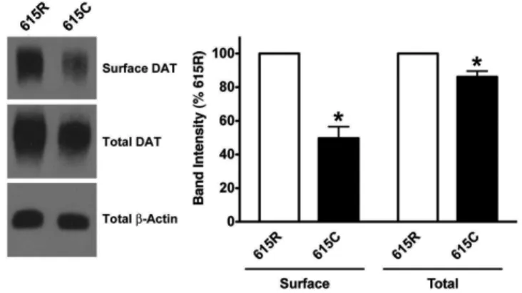
Identification of a functional DAT coding variant in an ADHD subject ... 60
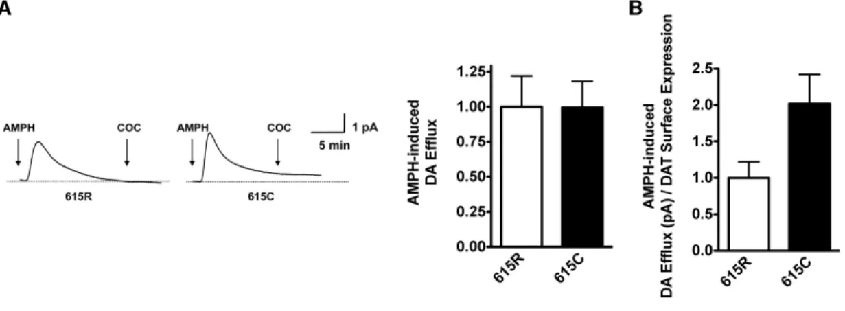
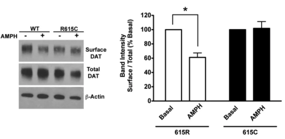
Anomalous modulation of DAT 615C by AMPH ... 64
DAT R615C exhibits accelerated rates of constitutive endocytosis and recycling. ... 69
Discussion ... 72
IV. ROLE OF SIGNALING PATHWAYS, MICRODOMAINS, AND THE C-TERMINUS IN THE ALTERED BEHAVIOR OF DAT 615C ... 76
Introduction ... 76
Results ... 77
DAT 615C exhibits a CaMKII-dependent state of functional inactivation. . 77
DAT 615C demonstrates altered localization to membrane microdomains. ... 83
DAT 615C acts dominantly via generation of local negative charge to disrupt AMPH actions ... 85
Discussion ... 89
V. HYPERPHOSPHORYLATION AND LACK OF AMPH ACTION IN THE HUMAN DAT A559V CODING VARIANT ... 96
Introduction ... 96
Results ... 98
Discussion ... 103
VI. SUMMARY AND FUTURE DIRECTIONS ... 107
INTRODUCTION TO APPENDICES ... 121
Further Characterization of DAT 615C-Associated Altered regulation ... 121
APPENDIX A: Effect of PKCβ inhibition on AMPH-mediated reduction in DA uptake ... 122 APPENDIX B: Determination of Ectopic Palmitoylation Due to the
APPENDIX C: Effect of Nitrous Oxide Synthase (Nos) Activators and Nitric Oxide (No) Scavengers on AMPH-mediated Reduction
in DA Uptake ... 127 APPENDIX D: Effect of Dynasore on DA Uptake ... 131 APPENDIX E: Development And Validation of a Fluorescent-based
Assay to Monitor DAT Function and AMPH-mediated Substrate Efflux .... 133 APPENDIX F: Impact of DAT Mutation on Intracellular Ca2+ ... 137
REFERENCES ... 139
LIST OF FIGURES
Figure Page
1 Biosynthesis and Degradation of DA …..………..………….….2
2 Schematic Illustration of Dopaminergic Projections …….……….…….3
3 Schematic Illustration of DAT Topology……….……..………10
4 Schematic Topology of DAT Based on LeuTaA crystal structure.………14
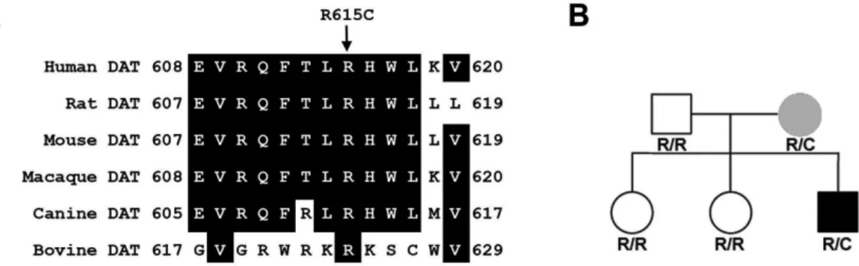
5 Sequence Alignment and Pedigree Analysis of DAT 615C ………….…...61
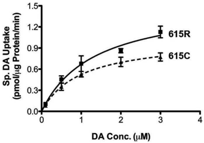
6 Transport Saturation Kinetic Analysis ……….……..……….62
7 Analysis of Total and Surface DAT Protein ……...………...63
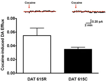
8 Amperometric Detection of Basal DA Efflux ………...…..………...….64
9 AMPH-induced DA Efflux ………..………...65
10 Effect of AMPH DAT Surface Expression ……..………66
11 Effect of AMPH DA Uptake Activity …………..………..……67
12 Effect of AMPH on DA uptake and DAT Surface Expression in Transiently Transfected CAD cells …...………..68
13 Effect of β-PMA on DA Uptake and DAT Surface Expression in Flp-In HEK Cells………...………..69
14 Accelerated constitutive Endocytosis Associated with DAT 615C …....…70
15 DAT 615C Exhibits Accelerated Recycling ……….………...…...71
16 Increased Basal CaMKII Association of the R615C Variant .…...……….78
17 Increased Basal Phosphorylation of the R615C Variant ……...…………..79
18 Effect of KN-93 and KN-92 on AMPH-mediated DA Transport Reduction……….81
19 Effect of KN-93 on AMPH-induced DAT Internalization ……….….82
20 Effect of KN-93+AMPH Treatment of DA Transport Kinetics of DAT 615C………...….…….83
21 Flotillin-1 Association is Reduced for DAT 615C ………..84
22 Decreased Localization of DAT 615C to GM1-containing Membrane Microdomains ……….……85
23 Effect of TAT-C24615R and TAT-C24615C Peptides on AMPH-mediated DA Transport Reduction.……….…….………….87
24 Effect of Amino Acid Substitutions at R615 Residue on AMPH-mediated DA Transport Reduction ….………….……….………88
25 Importance of T613 Residue in AMPH-mediated DA Transport Reduction….………..…….….89
26 Model Describing Differential Trafficking of DAT 615R and DAT 615C to the Regulated and Constitutive Endocytic Pathways and Biased Localization Toward GM1/Flotillin-1 Rich or Depleted Membrane Microdomains.………..………...94
27 Pedigree of the A559V Variant …..……….…...…...……..97
28 Increased Basal Phosphorylation of the A559V Variant ………...………..99
29 Effect of AMPH on DA Transport Reduction ………..….101
30 Effect of AMPH on DAT Surface Expression ….………....102
31 Effect of β-PMA on DA Transport Reduction………..….103
32 Effect of β-PMA on DAT Surface Expression....………..……...104
33 Colocalization of DAT with Ganglioside GM1………...…...105
34 Localization of ADHD-associated DAT Coding Variants………...109
35 Effect of PKCβ Inhibitor on AMPH-induced Reduction in DA Uptake.….123 36 Determination of Ectopic Palmitoylation Due to the Presence of Cysteine……….126
37 Effect of NOS Inhibition on DA Uptake ………..……..…128 38 Effect of NOS Inhibition on AMPH-mediated Reduction in the DA
Transport………...129 39 Effect of a NO Donor on AMPH-mediated Reduction in the DA
Transport………130 40 Effect of Dynasore on AMPH-mediated Reduction in the DA
Transport………...…132 41 Saturation Kinetic Analysis Using IDT 307 Uptake ………...…….………136 42 AMPH-mediated IDT 307 Efflux.………...……….…136 43 Measurement of Intracellular Ca2+……….138
LIST OF ABBREVIATIONS
5-HT-5-Hydroxytryptamine
AADC-Aromatic acid decarboxylase AChE- Acetylcholineesterase
ADE-Anomalous dopamine efflux
ADHD-Attention-deficit hyperactivity disorder Akt-Protein kinase B
AMPH-Amphetamine BPD-Bipolar disorder
CaMKII-Calcium/calmodulin-dependent protein kinase II CK2-Casein kinase 2
COMT Catechol-O-methyl transferase CTxB- Cholera toxin B subunit
DA- Dopamine
DAergic- Dopaminergic
DAT- Dopamine transporter/human dopamine transporter DIP- Dopamine transporter interacting protein
EL- Extracellular loop
ERK- Extracellular signal-regulated kinase fMRI- Functional magnetic resonance imaging GABA- gamma-aminobutyric acid
GAT1- GABA transporter 1
GLUT- Glucose transporter GLYT1- Glycine transporter 1 GPCR- G protein-coupled receptor
HPLC- High-pressure liquid chromatography ICQ- Intensity correlation quotient
IPD- Infantile Parkinsonian Dystonia KO- knock-out
LeuTAa- Aquifex aeolicus Leucine Transporter MAO- Monoamine oxidase
MAPK- Mitogen-activated protein kinase METH -Methamphetamine
mGluR- metabotropic glutamate receptor mPFC- medial prefrontal cortex
nAChR- Nicotinic acetylcholine receptor NE- Norepinephrine
NET- Norepinephrine transporter NO- Nitric oxide
NOS- Nitric oxide synthase
PET- Positron emission tomography PI3K- Phosphatidylinositol 3-kinase PICK1- Protein interacting with C kinase I PKA- Protein Kinase A
PKC- Protein Kinase C
PMA- Phorbol 12-myristate 13-acetate RACK1- Receptor for activated C kinase SERT- Serotonin transporter
SN- Substantia Nigra
SNAP-25- Synaptosomal-associated protein 25 SNP- Single nucleotide polymorphism
TAAR- Trace amine-associated receptor
TGCE- Temperature gradient capillary electrophoresis TH- Tyrosine hydroxylase
TMD- Transmembrane domain
VMAT- Vesicular monoamine transporter VNTR- Variable number tandem repteats VTA- Ventral tagmental area
WT- Wild type Y2H- Yeast 2-hybrid
CHAPTER I
INTRODUCTION
Overview of Dopamine and the Dopaminergic System
The neurotransmitter dopamine (3,4-dihydroxyphenethylamine, DA) makes a significant contribution to brain function through its modulatory role in pathways controlling reward, locomotor activity, and attention (Carlsson, 1987, Robbins, 2003). DA is synthesized in a two-step process involving first, the hydroxylation of tyrosine by tyrosine hydroxylase (TH) to yield L- dihydroxyphenylalanine (L-DOPA) that is subsequently decarboxylated by L- aromatic amino acid decarboxylase (AADC) to yield DA. The action of TH is rate- limiting in this reaction due to the higher turnover rate and lack of substrate saturation of AADC. In pathways that utilize DA as a neurotransmitter, DA is packaged for release or processed further intracellularly to synaptically inactive metabolites. DA is packaged for release into synaptic vesicles using vesicular monoamine transporter-2 (VMAT-2). DA is transported into the synaptic vesicles using proton gradient present across the synaptic vesicle membrane. VMAT-2 function and expression is often altered by use of psychostimulants (Little et al., 2003). Another VMAT isoform, VMAT-1, is mostly associated with large secretary granule vesicles, whereas VMAT-2 is predominantly present on small synaptic
intracellular DA is enzymatically degraded by isoforms of monoamine oxidase (MAO) into 3,4-dihydroxyphenylacetic acid (Jonason, 1969). In the prefrontal cortex, however, enzymatic breakdown of DA occurs by catechol-O-methyl transferase (COMT) into 3-methoxytyramine (Yavich et al., 2007). Other brain and peripheral pathways use DA as a precursor to produce norepinephrine (NE) and epinephrine. Synthesis and degradation of DA is summarized in Figure 1.
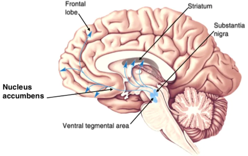
Four major brain pathways utilize DA as a neurotransmitter: the tuberoinfundibular pathway, the nigrostriatal pathway, the mesocortical pathway, and the mesolimbic pathway (Figure 2). The tuberoinfundibular pathway originates from of neurons in the hypothalamic arcuate nucleus and projects to
Figure 1 Biosynthesis and Degradation of Dopamine: (A) Biosynthesis of DA from the actions of enzymes, tyrosine hydroxylase, and dihydroxyphenylanine (DOPA) decarboxylase (also known as AADC due to its lack of specificity for DA). (B) Degradation of DA by monoamine oxidase and catechol-O-methyl transferase.
the median eminence, where DA secretion produces prolactin secretion from the anterior pituitary gland. The nigrostriatal pathway, which modulates movement, and that is lost in Parkinson's disease, originates in neurons of the substantia nigra (SN), so named for their pigmented appearance in unstained sections.
Dopaminergic (DAergic) axons from the SN terminate in nuclei of the basal ganglia, termed the striatum in rodents, but divided into the caudate nuclei and putamen in primates. Both the mesocortical and the mesolimbic pathways originate in the ventral tagmental area (VTA), projecting to the prefrontal cortex and nucleus accumbens, respectively. The mesocortical pathway is important in the modulation of motivation and emotion, whereas the mesolimbic pathway is implicated in reward and pleasure.
Figure 2 Schematic Illustration of Dopaminergic Projections: DA neurons in the VTA project to the nucleus accumbens and prefrontal cortex through the mesolimbic and mesocortical pathways, respectively. DA neurons in the SN project to the striatum via the nigrostriatal pathway.
The tuberoinfundibular pathway is not depicted. Adapted from Neuroanatomy, An Atlas of Structures, Sections, and Systems
Nucleus accumbens
Due to the actions of DA in the regulation of motor and cognitive function, as well as the recognition of incentive salience and reward, alterations in DAergic tone can lead to multiple neurological and psychiatric disorders. These include Parkinsonʼs disease (Chase et al., 1998), dystonia (Kurian et al., 2009), attention-deficit hyperactivity disorder (ADHD) (Mazei-Robison et al., 2005), addiction (Ritz et al., 1987), and schizophrenia (Horn and Snyder, 1971).
The first evidence for altered DA neurotransmission in a brain disorder involved the demonstration that rats given reserpine displayed features of Parkinsonʼs disease (Carlsson et al., 1966), findings that would ultimately bring Arvid Carlsson the Nobel Prize in 2000. Subsequently, Hornykiewicz established a loss of DA in the brains of Parkinson disease subjects (Ehringer and Hornykiewicz, 1960). The reversal of Parkinsonian symptoms by the DA precursor, L-DOPA, provided clear evidence of the role of DA in the disorder (Birkmayer and Hornykiewicz, 1962). Hornykiewicz would go on to show that Parkinsonʼs disease arises from degeneration of nigrostriatal DA neurons, resulting in denervation of basal ganglia that form the extrapyramidal motor system (Hornykiewicz, 1972).
Although Parkinsonʼs disease patients, particularly in the latter stages, often exhibit cognitive and emotional problems, the idea that DAergic dysfunction is involved in psychiatric disorders derives in large measure from research demonstrating altered DA metabolism and signaling in patients with schizophrenia (Hokfelt et al., 1974, Lindvall et al., 1974). Indeed, classical
antipsychotic drugs, including chlorpromazine and haloperidol, block D2 subtype DA receptors, thus suggesting a role for hyperdopaminergic signaling (Horn and Snyder, 1971). Another strong evidence of altered DAergic neurotransmission contributing to a neuropsychiatric disorder came from ADHD patients. ADHD is the most commonly diagnosed childhood disorder affecting 3-5% of school age children. Multiple studies point to a contribution of variation in genes expressed in DAergic neurons as influencing risk for ADHD (Gill et al., 1997, Qian et al., 2003, Bobb et al., 2005, Mazei-Robison et al., 2005). I will discuss relationship between ADHD and DAergic transmission later in this chapter. Whereas L-DOPA and chlorpromazine have therapeutic efficacy, other drugs targeting DA signaling can be highly addictive, including the psychostimulants cocaine and amphetamine (AMPH), Long-term use of these agents produces molecular, cellular and circuit- level plasticities that drive a desire and search for drug, despite the recognition of negative consequences (Kauer and Malenka, 2007, Dietz et al., 2009). Taken together, alterations in DA signaling can be seen to underlie multiple, devastating disorders that affect tens of millions of individuals worldwide. My thesis research derives from the belief that a better understanding of DA signaling mechanisms, and how these mechanisms are perturbed by disease states, can lead to advances in the prevention, diagnosis and therapy.
Discovery of Presynaptic DA Transport and Demonstration of Specific DA Transporter (DAT) Binding Sites
Acetylcholine, the first identified neurotransmitter, is inactivated by degradation via the extracellular enzyme acetylcholinesterase (AChE). Although metabolism of the catecholamine NE by MAO and COMT to inactive products has been established, acute inhibition of these enzymes does not impact synaptic DA inactivation. Axelrod and colleagues discovered that radiolabeled NE could be sequestered into the terminals of sympathetic neurons (Hertting and Axelrod, 1961), and subsequently demonstrated that NE accumulation could be blocked by both antidepressants and psychostimulants (Axelrod et al., 1961, Hertting et al., 1961). These studies lead to the hypothesis that synaptic NE inactivation was mediated by rapid clearance through a presynaptic uptake process (Axelrod, 1971). Later, investigators would demonstrate that brain slices could accumulate both NE and DA and that psychostimulants could inhibit this process (Carlsson et al., 1966, Glowinski and Axelrod, 1966, Ross and Renyi, 1967). Subsequently, Ross and Renyi demonstrated that the pharmacological profiles of DA uptake inhibition in striatum and cortex were distinct from that of NE uptake inhibition (Ross and Renyi, 1967). They demonstrated that whereas AMPH and cocaine were potent inhibitors of DA and NE uptake in both brain regions, desipramine was a weak inhibitor of DA uptake in striatum, but a potent inhibitor of NE in cortex (Ross and Renyi, 1967). These findings suggested that the DA uptake mechanism was distinct from that supporting NE clearance,
contributing further to the idea that DA was not simply a precursor for NE but also a neurotransmitter in its own right. Snyder and Coyle would go on to demonstrate that the specificities of the DA and NE uptake processes for isomers of AMPH were distinct (DA uptake equivalently inhibited by D- and L-AMPH, NE uptake preferentially inhibited by D-AMPH) (Snyder and Coyle, 1969). Ritz and coworkers provided further dissociation of the actions of cocaine on DA and NE uptake sites by demonstrating cocaineʼs potency to inhibit DA uptake in rat brain extracts was significantly higher than cocaineʼs potency for inhibiting NE uptake (Ritz et al., 1987). Ritz and colleagues also reported important in vitro findings that the antagonist potency in blocking brain DA uptake correlated well with the potency of cocaine determined from self-administration studies with non-human primates (Ritz et al., 1987), further strengthening the hypothesis that the abuse liability of the psychostimulant derived from an inhibition of DA transport and inactivation.
The studies described above prompted efforts to identify the DA transporter, first by demonstrating the presence of specific cocaine binding sites in brain and their relationship to the DA reuptake process. Autoradiographic studies using the cocaine analog [3H] 2 beta-carbomethoxy-3 beta-(4- fluorophenyl)tropane ([3H]CFT) demonstrated specific binding presence in monkey brain, specifically in the caudate and putamen, a dopamine-rich area, and one that had been previously implicated in the behavioral effects and abuse potential of cocaine (Canfield et al., 1990). Kennedy and Hanbauer, using rat
striatal synaptosomes, demonstrated the presence of presynaptic, Na+- dependent cocaine binding sites (Kennedy and Hanbauer, 1983). Today, these cocaine binding sites are known to reflect the presence of DAT protein. Such binding approaches remain valuable as they are used with in vitro studies of DAT regulation as well as for in vivo analyses, as with positron emission tomography (PET) imaging techniques to monitor DAergic nerve terminals in Parkinsonʼs disease (Antonini et al., 2001, Song et al., 2012).
Cloning of DAT cDNAs
In 1988, Blakely and colleagues demonstrated the transport of radiolabeled L-glutamate, gamma-aminobutyric acid (GABA), glycine, DA, serotonin (5-HT), and choline in Xenopus laevis oocytes microinjected with brain mRNAs. mRNAs competent for expression of neurotransmitter transporter demonstrated a regional enrichment consistent with the anatomical distribution of neurotransmitter synthesizing soma and thereby demonstrated the opportunity for expression cloning of neurotransmitter transporter proteins (Blakely et al., 1988). Following the cloning of the norepinephrine (NET) (Pacholczyk et al., 1991) and serotonin transporters (SERT) (Blakely et al., 1991), four groups reported the cloning of cDNAs encoding rat and bovine DATs (Giros et al., 1991, Kilty et al., 1991, Shimada et al., 1991, Usdin et al., 1991). Using in situ hybridization, several of these groups demonstrated that DAT mRNA was expressed in the SN and VTA, consistent with the localization of DA neuron cell
bodies (Giros et al., 1991, Kilty et al., 1991, Shimada et al., 1991). Heterologous expression of rat DAT cDNA revealed the induction of a Na+- and Cl- dependent, DA uptake process that was absent in non-transfected cells. The transport of DA displayed a high-affinity for DA, with a KM estimated at between 300 and 900 nM.
Finally, the in vivo DA uptake blockers, GBR12909 (IC50=12 nM), mazindol (IC50=27 nM), cocaine (IC50=336 nM), and D-AMPH (IC50=881 nM) inhibited DA uptake (Giros et al., 1991) with the same potencies previously reported with rat brain synaptosomes (Ritz et al., 1987). Together, these studies validated the contention that the DAT transcript produced a protein with the properties expected for the DA uptake process, including its antagonist-binding site, in the CNS. They also revealed that no additional, brain-specific subunits were required to produce functional DAT protein.
Oligonucleotides based on rat DAT cDNA sequence were used to design probes to clone human dopamine transporter (DAT) cDNA from SN libraries (Giros et al., 1992, Vandenbergh et al., 1992). DAT is now understood to be a 620 amino acid protein that displays ~92% amino acid identity to rat DAT. DAT displays reduced, but significant identity to other members of the Na+/Cl-- dependent neurotransmitter transporter family (SLC6) members, with highest amino acid sequence identify observed with hNET (66%) and the human serotonin transporter (hSERT, 50%). Hydropathy analysis of DAT, like that of other SLC6 transporters, predicts 12 transmembrane domains (TMDs) with N- and C- termini located intracellularly (Figure 3). Three sites for N-linked
glycosylation are located in extracellular loop 2 (EL2) that connects TMDs 3 and 4. Mutating all three canonical glycosylation sites as well as enzymatic degradation and blockade of glycosylation resulted in reduced DAT surface expression; significantly diminished catalytic activity and inhibitor sensitivity compared to wildtype (WT) DAT (Li et al., 2004). Recently, a novel isoform of DAT produced by alternative splicing was discovered from human blood cells (Sogawa et al., 2010). The spliced isoform lacked exon 6, resulting in the deletion of TMD5 and an extracellular C-terminus. Further characterization showed expression differences between brain and peripheral tissues, indicating tissue- specific, alternative splicing. Expression of the spliced isoform resulted in loss of DAT activity and when expressed with the WT DAT, the spliced isoform acted
Figure 3. Schematic Illustration of the hDAT Topology: The hDAT protein contains 12 TMDs with N-and C-termini located intracellularly. Large EL2 contains 3 N-linked glycosylation sites. There are numerous consensus phosphorylation sites depicted by P in black circle on the N-terminus.
Figure 3 Schematic Illustration of DAT Topology: The DAT protein contains 12 TMDs with N- and C-termini located intracellularly. EL2 contains 3 N-linked glycosylation sites. Numerous consensus phosphorylation sites, depicted by P in a black circle, are found on the N- and C- termini as well as intracellular loops.
dominantly, reducing overall DA uptake (Sogawa et al., 2010). The functional significance of these observations in vivo, however, is unknown. Additionally, in schizophrenia patients, Talkowaski and colleagues identified a novel cassette exon within intron 3 of DAT gene that is conserved among primates (Talkowski et al., 2010). The presence of this cassette exon introduces multiple in-frame stop codons resulting in a truncated product, and that may undergo nonsense- mediated decay. The presence of this spliced variant was confirmed using RNA from postmortem human SN. Factors that increase use of this cassette exon splice variant could reduce the amount of unspliced RNA encoding WT DAT, with attendant functional consequences (Talkowski et al., 2010).
Dopamine Transporter Structure and Function
The characterization of DAT structure/function relationships has largely relied on heterologous expression studies performed in vitro. Data from studies expressing DAT and NET chimeras provided initial evidence that discrete domains within the transporter were involved in distinct functions such as substrate recognition, translocation, and affinity (Buck and Amara, 1994, Giros et al., 1994, Syringas et al., 2000). In these studies, TMDs 1-3 and 10-11 were identified as important for determining substrate affinity, whereas TMDs 5-8 were identified as critical for substrate translocation and the selectivity of inhibitors (Buck and Amara, 1994, Giros et al., 1994). Using similar approaches, TMDs 1-3
and 9-12 were identified as important for the Na+ and Cl- dependence exhibited by DAT, as well as other SLC6 transporters (Syringas et al., 2000).
Whereas chimera studies implicated broad regions of the transporter in key DAT properties, site-directed mutagenesis studies provided insights into more defined structure-function relationships, identifying in some cases single residues important for transport function and regulation. These studies indicated that, aside from support for N-glycosylation, EL2 appears to play a role, or be influenced by, inhibitor interactions, as inhibitor binding could be shown to protect DAT from trypsin digestion in this region (Gaffaney and Vaughan, 2004). In contrast, DAT substrates do not protect against proteolytic digestion (Gaffaney and Vaughan, 2004). Additionally, zinc immobilization of EL2 was shown to block DA transport through coordination of residues of EL2 and EL4, suggesting that movement of EL2 might be crucial for substrate translocation, possibly by structural movement of TMDs 3 and 4 (Norregaard et al., 1998). Interestingly, zinc immobilization of EL2 and EL4 increases ion conductance, which negatively impacts transport and stimulates efflux (Meinild et al., 2004). The later studies highlight the importance of electrophysiological approaches in providing mechanistic insights of mutation-induced changes in DAT function.
To further our understanding as to how both substrates like DA and antagonists like cocaine interact with DAT, mutagenesis studies have been conducted to identify residues critical for ligand coordination. In these studies, alanine substitution was used to modify residues in or near TMDs, including all
tryptophan (Lin et al., 2000b), proline (Lin et al., 2000a), and phenylalanine (Lin et al., 1999) residues. The importance of Asp79 (Kitayama et al., 1992) and Phe105 (Wu and Gu, 2003) residues in cocaine recognition has been studied in detail. DAT immunogold labeling has shown that in the nigrostriatal neurons, these transporters are strategically localized to extrasynaptic plasma membrane to re-uptake diffused DA back into the presynaptic terminal (Nirenberg et al., 1997). Using chemical cross-linkers, Fluorescent Resonance Energy Transfer (FRET) analysis, and expression studies with differentially tagged DAT constructs, DAT have been shown to exist in oligomeric form at the plasma membrane (Hastrup et al., 2001, Sorkina et al., 2003b).
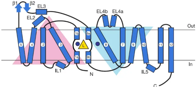
Although the three dimensional structure of DAT is unknown, the generation of a high-resolution crystal structure for a bacterial SLC6 family member from Aquifex aeolicus, the leucine transporter (LeuTAa) supports the prediction of a 12 TM topology for DAT and homologs (Yamashita et al., 2005).
The orientation and arrangement of TMDs of LeuTAa is depicted in Figure 4.
Unfortunately the leucine transporter has short cytoplasmic N- and C-termini that are unstructured in the crystal structure. Molecular modeling efforts based on the information obtained from the crystal structure of LeuTAa helped Beuming and colleagues investigate the relationship between DA and cocaine binding sites on DAT (Beuming et al., 2008). Modeling and mutagenesis approaches provided evidence that the binding sites for cocaine and cocaine analogs are buried deeply between TMDs 1, 3, 6, and 8 and overlap with the binding site for DA and
AMPH (Beuming et al., 2008). Recently, using steered molecular dynamics simulation and homology modeling, a second DA binding site has been proposed to lie in the DAT extracellular vestibule and that this second substrate-binding site allosterically modulates transport mechanism (Shan et al., 2011). This proposal remains a significant point of debate among investigators studying the structural determinants of the function of SLC6 family members (Piscitelli et al., 2010).
The cytosolic tails and intracellular loops of DAT contain numerous serine,
and threonine residues, some of which are located in consensus sites for protein kinase C (PKC), protein kinase A (PKA), and calcium calmodulin-dependent kinase II (CaMKII). However, the exact residues phosphorylated by these kinases remains an active area of investigation (see below).
Figure 4 Schematic Topology of DAT Based on LeuTaA crystal structure: TMDs1-5 and 6- 10 form a pseudo two-fold axis in the membrane plane (inverted triangle configuration) and fold over forming substrate translocation pathway. Yellow triangle with L and blue circles depict leucine and sodium ions respectively. TMDs 11 and 12 flank the outer surface of TMDs 9 and 10, and possibly contributing to the oligomerization. Adapted from Yamashita et al., 2005.
The transport of DA is Na+ and Cl- dependent, with a predicted stoichiometry of 2Na+, 1Cl- and one DA+ (DA is positively charged at physiological pH) (Amara and Kuhar, 1993). In keeping with the proposed net charge transfer per transport cycle, DA transport through DAT is electrogenic and a voltage dependent process, where hyperpolarization increases and depolarization decreases DA transport (Sonders et al., 1997).
Electrophysiological studies reveal that DA transport through DAT produces a larger current than is expected for stoichiometric charge transfer determined by the ion-dependence of DA transport. This observation appears to reflect the presence of channel-like states that operate to permit ion permeation during the DA transport cycle (Carvelli et al., 2004, Kahlig et al., 2005).
As a DAT substrate, AMPH competitively inhibits DA reuptake, causing an increase in synaptic DA that otherwise would have been cleared by re-uptake.
Additionally, AMPH stimulates the release of DA through DAT in the brain (Fischer and Cho, 1979, Jones et al., 1998) and in transfected cells expressing DAT (Khoshbouei et al., 2003).AMPH-induced DA efflux has been considered to be mediated by a facilitated-exchange diffusion process, in which the inward transport of AMPH increases the availability of inward-facing binding sites of the transporter (Fischer and Cho, 1979), thereby leading to increased efflux of cytosolic DA. As noted below, this perspective is altogether too simple as multiple kinase-mediated signaling mechanisms can shift DAT into an “efflux willing”
state. Finally, AMPH is a weak base and as a VMAT substrate, can collapse the
synaptic vesicle proton gradient needed for VMAT to package DA (Sulzer et al., 1995). Via this process, AMPH can elevate cytoplasmic DA levels and enhance the probability of DAT-mediated DA efflux.
Regulation of the Dopamine Transporter
Although DAT was recognized early in its documentation as a target for psychoactive drugs, the transporter was discussed more as a static component of presynaptic homeostasis, inexorably clearing DA upon synaptic release.
Mechanisms that could influence DAT activity and clearance were unknown. An early hint that DAT might be locally regulated arose from studies of Kuharʼs group, where irreversible inhibitor manipulation of striatal DAT revealed that the time for newly synthesized DAT to replace inactivated transporters in terminals is very long, on a synaptic transmission scale, with only ~50% of DAT replaced in 6 days (Fleckenstein et al., 1996). Although later studies reduced this figure to 2-3 days (Kimmel et al., 2000), the point remains that somatic biosynthetic mechanisms are too slow to effect significant changes in DAT-mediated DA clearance to match the more rapid changes in the firing rates of DA neurons (Goto et al., 2007). Today, evidence exits that rapid (sec-minutes) DAT regulation is imposed by presynaptic receptors, associated proteins and intracellular signaling networks, with evidence that both functional changes and/or membrane environment dictate the capacity for DA clearance (Zahniser and Doolen, 2001, Torres, 2006, Chen et al., 2010).
Presynaptic receptors
DA acts on both post- and pre-synaptic receptors, with presynaptic D2 receptors belonging to the Gi/Go family of G-protein coupled receptors (GPCR) that decrease adenylate cyclase activity (among other actions). D2 receptors have two isoforms as a result of alternative splicing with different spliced forms expressed pre or post-synaptically. D2 long (D2L) receptors, which have additional 29 amino acids in third intracellular loop and are exclusively expressed on postsynaptic neurons (Giros et al., 1989). On the other hand, D2 short (D2S) receptors that lack 29 amino acid insert are expressed presynaptically and act as DA autoreceptors. In response to elevated extracellular DA, D2S receptor provide inhibitory feedback in altering DA synthesis, release, and re-uptake (Meiergerd et al., 1993), with DAT regulation mediated by initiation of a second messenger cascade (Meiergerd et al., 1993). D2S receptors have been reported to increase DAT activity through activation of extracellular signal-regulated kinases (ERK1 and 2) (Bolan et al., 2007). The same group also provided evidence that D2S receptor modulation of DAT involves a phosphoinositide-3 kinase (PI3K)- independent mechanism (Bolan et al., 2007).
Type 1 metabotropic glutamate receptors (mGluRI) have been reported to regulate DAT expressed by dendrites of substantia nigra DA neurons (Falkenburger et al., 2001). These investigators demonstrated that stimulation of subthalamic nucleus afferents resulted in DAT-mediated DA efflux.
Pharmacological inhibition of DAT, as well as antagonism of mGluR1 receptors,
attenuated DA release from DA neurons. In another study, mGluR5 was reported to regulate DAT in vitro. Activation of mGluR5 by its selective agonist (S)-3,5- dihydrophenylglycine (DHPG) decreased DA transport by DAT in striatal synaptosomes (Page et al., 2001). In these studies, mGluR5-dependent inhibition of DAT activity was dependent on signaling by CaMKII and PKC pathways.
Nicotinic acetylcholine receptors (nAChRs) have been reported to regulate DAT function. In vivo voltammetry studies provide evidence that systemic nicotine administration increases DA clearance in striatum, medial prefrontal cortex (mPFC) (Middleton et al., 2004), and nucleus accumbens (Hart and Ksir, 1996). The dose-response profile of nicotine in modulating DA clearance in striatum and mPFC is different, possibly owing to differential expression of nAChR subunits. AMPH-stimulated DA release from prefrontal cortex brain slices was reported to increased following 5 µM nicotine application (Drew et al., 2000).
AMPH-mediated DAT downregulation following nicotine administration were found to be PKC-dependent but CaMKII-independent (Drew and Werling, 2001).
Thus, activation of PKC via mGluRI and nAChR may drive opposite effects on DAT activity. One possible explanation for this could be that processes supporting DA uptake and DAT-mediated DA efflux may be governed by different mechanisms.
Regulation of DAT by σ2 receptors (formally classified as opioid receptors) has also been reported. Activation of σ2 receptor by (+)-pentazocine enhances AMPH-stimulated DAT-dependent DA release in striatal slices (Izenwasser et al.,
1998), as well as in PC12 cells (Weatherspoon and Werling, 1999). σ2 receptors have been suggested to regulate intracellular Ca2+ levels through their action on endoplasmic reticulum Ca2+ stores (Vilner and Bowen, 2000). In rat striatal slices, activation of σ2 receptors has been reporter to act through Ca2+ and PKC- dependent mechanisms to stimulate DAT activity (Derbez et al., 2002).
Conversely, in PC12 cells, σ2 receptors activation was found to increase AMPH- stimulated DA efflux, stimulation dependent on CaMKII activation (Weatherspoon and Werling, 1999). Choice of model system and experimental designs likely dictate these regulatory differences. These studies with σ2 receptors reinforce the complexity in DAT regulation and demonstrate a need for more in-depth investigation using more precise pharmacological and genetic tools.
A recent addition to the list of receptors reported to regulate DAT is the trace amine-associated receptor 1 (TAAR1). TAAR1 is a Gs-coupled GPCR that signals through increasing cAMP production (Wainscott et al., 2007). TAAR1 is activated by trace amines like beta-phenylethylamine, as well as classic biogenic amines, and amphetamine-related psychostimulants. TAAR1 is expressed by substantia nigra DA neurons in primates and rodents (Xie and Miller, 2007, Xie et al., 2007) and its activation by DA or methamphetamine (METH) appears to lead to a PKA- and PKC-mediated reduction in DAT-mediated DA uptake. TAAR1 activation also induced an increase in DA efflux via a PKC-dependent mechanism (Xie and Miller, 2007). Further studies of DAT regulation by TAAR1 may be of clinical relevance in the treatment of METH addiction.
DAT-interacting proteins
Multiple DAT-interacting proteins (DIPs) have been identified using yeast two-hybrid (Y2H) screens with specific DAT sequences as bait, with confirmation of interactions pursued in cells or brain preparations by co-immunoprecipitation (Torres, 2006). DIPs are theorized to regulate DAT somatic export, synaptic localization, and surface trafficking, as well as DAT functional properties. None of these interactions has as yet been shown to influence DAT in vivo.
Torres and colleagues, using the DAT C-terminus as bait in a Y2H screen, described the first DIP, Protein Interacting with C kinase-1 (PICK1) (Torres et al., 2001). PICK1 is a PDZ (postsynaptic density 95/Discs large/zona occludens 1) domain containing protein that has been argued to be important for synaptic localization of DAT. The PDZ domain of PICK1 binds to a large number of membrane proteins especially proteins containing C-terminus PDZ type II binding motif. PICK1 also has a BAR (Bin/amphiphysin/Rvs) domain. The BAR domain of PICK1 binds to lipids, mainly phosphoinositides (Xu and Xia, 2006). PICK1 was originally identified as a PKC binding protein (Staudinger et al., 1995) and therefore, the PICK1-DAT interaction may be important in PKC-mediated regulation and trafficking of DAT. The Torres studies, the interactions between PICK1 and DAT were found to be mediated by the PDZ domain of PICK1 and the class II PDZ binding motif (LKV) that ends the C-terminus of DAT. PICK1/DAT interactions were reported to increase the number of surface DAT molecules, leading to an increase in DA uptake (Torres et al., 2001). However, more recent
studies from Getherʼs lab argue that surface targeting of transporter does not depend on PICK1 binding to the transporterʼs PDZ binding motif, but rather derives from residues (RHW) upstream of these sequences (Bjerggaard et al., 2004). Moreover, these authors suggest that the ability of PICK1 to modulate DAT surface expression may derive from an effect on the export of DAT from ER/Golgi biosynthetic stores. Recent studies by Madsen and colleagues show that deletion of PDZ domain from PICK1 results in clustering of PICK1 and BAR domain-dependent redistribution of PICK1 to endosomal compartments (Madsen et al., 2008). These authors further show that under basal conditions, the PDZ domain and the linker region connecting PDZ domain and BAR domain fold over leading to inhibit interactions of the BAR domain with its targets. Disinhibition of the BAR domain leads to membrane interactions, membrane curvature, and altered protein trafficking (Madsen et al., 2008). These findings raise the possibility that the action of PICK1 with respect to DAT may be constrained by BAR domain interactions with the PICK1 PDZ domain, and thus be one target of the regulatory cascades impacting DAT trafficking out of the ER/Golgi or to and from the cell surface.
In the Y2H screen where PICK1 was identified, Caronʼs group identified a second DIP, the multiple Lin-11, Isl-1, and Mec-3 (LIM) domain-containing adaptor protein, Hic-5 (Carneiro et al., 2002). Hic-5 is a member of a focal adhesion-associated adaptor protein family and is closely related to paxillin. In addition to its membrane localization, Hic-5 has also been found localized to
nuclear compartments (Shibanuma et al., 2004), with suggestion that the protein may cycle between these compartments, though a functional significance of this idea has not been determined with respect to DAT. Hic-5/DAT interactions arise through Hic-5 LIM domains and membrane-proximal sequences of the DAT C- terminus. Carneiro and colleagues reported that expression of DAT and Hic-5 in HEK 293 cells results in reduced DA uptake activity that arose from decreased DAT surface expression (Carneiro et al., 2002). Even though the physiological importance of DAT/Hic-5 interaction in vivo is unknown, Hic-5 is targeted by numerous signaling molecules (Shibanuma et al., 2012), and thus DAT interactions could connect the transporter to multiple intracellular signaling pathways. Recently, Carneiro and Blakely extended the generality of interactions of Hic-5 to other SLC6 family transporters, as well as their dynamic nature and occurrence in native preparations, with demonstration of PKC-dependent, SERT/Hic-5 interactions in human and mouse platelets (Carneiro and Blakely, 2006). Co-immunoprecipitation (co-IP) and 5-HT transport studies revealed that Hic-5 produces SERT inactivation and targets SERT to a membrane compartment that is permissive for PKC-dependent relocation of transporters from the cell surface. Interestingly, SERT/Hic-5 associations were reduced by PKC activation over the same time course as SERT internalization. Since association of intracellular SERT with Hic-5 was not detected, these authors concluded that Hic-5 participates in chaperoning SERT to membrane domains competent for PKC-dependent endocytosis (Carneiro and Blakely, 2006, Steiner
et al., 2008) and then dissociates thereafter or shortly following SERT endocytosis. Studies examining DAT/Hic-5 interactions in DA terminals are needed to determine whether the SERT/Hic-5 model extends to DAT.
The synaptic vesicle associated protein α-synuclein is yet another protein found to interact with DAT, once again mediated by sequences in the transporterʼs C-terminus (Lee et al., 2001). Mutations in α-synuclein have been implicated in familial forms of Parkinsonʼs disease (Higuchi et al., 1998, Kruger et al., 1998), a disorder characterized by the degeneration of DA neurons. The physiological role of α-synuclein in vivo remains an active area of investigation (Chandra et al., 2005, Jin et al., 2011). DAT/α-synuclein interactions were first identified via a Y2H screen, and thereafter confirmed in neurons and co- transfected cells (Lee et al., 2001, Lee et al., 2007). DAT/α-synuclein interactions have been found to cluster the transporter at the plasma membrane, to increase uptake activity and to enhance DA-induced cellular apoptosis (Lee et al., 2001), possibly via enhanced oxidative stress produced by oxidation of the neurotransmitter. The functional outcome of this interaction is somewhat controversial as one group has reported that DAT/α-synuclein interactions following heterologous expression of these molecules leads to decreased DAT activity (Wersinger and Sidhu, 2003). The same group also reported that disruption of microtubule networks results in increased DAT activity as a consequence of DAT-α-synuclein interactions (Wersinger and Sidhu, 2005). As these studies have largely been conducted via heterologous expression, the
importance of DAT/α-synuclein interactions for presynaptic DA homeostasis and signaling remains unclear. Indeed, α-synuclein knockout mice do not show significant changes in DAT activity or protein expression (Abeliovich et al., 2000), though regulatory control of DAT has yet to be examined in this model.
In another Y2H screen using the DAT C-terminus as bait, Fog and colleagues identified CaMKIIα as a DIP (Fog et al., 2006). DAT/CaMKIIα interactions were confirmed using GST pulldown experiments and co-IP studies of both transfected cells and brain lysates, as well as through immunocytochemical colocalization studies. These researchers demonstrated that CaMKIIα binds to the DAT C-terminus and leads to phosphorylation of the transporter, likely through sites in the N-terminus of DAT. Additionally, CaMKII binding and DAT phosphorylation were shown to be necessary for AMPH- mediated DA efflux through DAT (Fog et al., 2006), providing evidence that conformational changes in the transporter that mediate DA efflux do not simply arise from substrate-mediated conformational changes, but involve a stabilized transition of DAT to an “efflux-willing” state. Whether DAT/CaMKIIα associations are constitutive, regulated or sensitive to disease-associated DAT mutations is unknown.
Recently, Melikianʼs group used a Y2H screen with a bait that encoded DAT C-terminal sequences 587-596 to identify a Ras-like GTPase, Rin, as a DIP.
This interaction was confirmed through GST pull-downs, co-IP studies, and co- localization studies using heterologous expression system. DAT/Rin interactions
are suggested to be essential for PKC-induced DAT downregulation and internalization (Navaroli et al., 2011). The actual mechanism by which DAT-Rin interaction leads to PKC-mediated DAT downregulation is still under investigation. Rin binds to, and activates calmodulin (Lee et al., 1996). Since CaMKIIα also interacts with the DAT C-terminus, it is possible that Rin may serve to activate DAT-bound CaMKII via its activation of calmodulin. Rin is also required for nerve growth factor (NGF)-stimulated neurite extension (Spencer et al., 2002) and p38 mitogen-activated protein kinase (MAPK) activation in response to NGF signaling (Shi et al., 2005). Since DAT trafficking has also been linked to MAPK signaling (Moron et al., 2003, Bolan et al., 2007), studies are needed to determine if Rin participates in this process.
In summary, the DAT C-terminus is a site of interaction for multiple cytosolic proteins that have been argued to regulate DAT activity and expression.
Understanding whether all of these associations occur together, are constitutive, or arise at different phases in the life cycle of the transporter needs further study.
Since artificial systems provide the bulk of the data supporting the physiological significance of these findings, additional studies with DA neurons in vitro and in vivo are needed.
Using the N-terminus of DAT as Y2H bait, two additional DIPs have been identified; Receptor for Activated C Kinase (RACK1), and syntaxin 1A. RACK1 was originally identified as protein that binds activated PKC and recruits it to the plasma membrane, in proximity of its substrates (Ron et al., 1994). Co-
immunoprecipitation from rat brain extracts has shown the presence of a ternary complex involving DAT-RACK1-syntaxin 1A (Lee et al., 2004). It is tempting to speculate that this interaction between DAT and RACK1 may modulate function of the transporter via recruiting PKC to the membrane. Syntaxin 1A is a major component of the SNARE-mediated mechanism of synaptic vesicle fusion and neurotransmitter release (Sudhof and Rothman, 2009). GABA transporters (GAT1) (Beckman et al., 1998), glycine transporters (GLYT1) (Geerlings et al., 2000), NET (Sung et al., 2003b) and SERT (Quick, 2002), have also been shown to interact with syntaxin 1A. Cervinski and colleagues have shown that syntaxin 1A/DAT complexes are present in rat striatal synaptosomes, that syntaxin 1A cleavage enhances DAT activity, supporting a role for these complexes in DA neurons, though other mechanisms downstream of syntaxin 1A cleavage are possible (Cervinski et al., 2010).
Syntaxin 1A transfection into NET expressing cells leads to increased transporter surface expression, likely due to the SNARE-mediated fusion role of syntaxin 1A. In contrast, this expression also leads to a decrease in NE uptake capacity, accompanied by a reduction in NE-activated NET currents (Sung et al., 2003a). These authors showed that activation of PKC leads to a dissociation of NET and syntaxin 1A paralleled by NET internalization. Cervinski and colleagues have found that in rat striatal synaptosomes, cleavage of syntaxin 1A does not preclude PKC-dependent downregulation of DAT (Cervinski et al., 2010).
Together, these findings suggest that NET and DAT interactions with syntaxin 1A
are likely to be part of a dynamic cycle by which transporters are brought to the presynaptic membrane, possibly to specialized microdomains (see below), and then regulated to provide for neural-activity dependent chances in transport function.
The ability of syntaxin 1A to modulate currents has also been reported with SERT, where syntaxin 1A can dictate whether the transporter exhibits an electroneutral or electrogenic 5-HT transport cycle (Quick, 2003). Recent studies have shown that DAT/syntaxin 1A associations promote AMPH-induced DA efflux (Binda et al., 2008) and can regulate DAT channel activity (Carvelli et al., 2008). In the C. elegans model, DAT/syntaxin 1A interactions have been reported to impact DA neurotransmission and behavior (Carvelli et al., 2008), to date the only evidence that these complexes are important in vivo As to the dynamic nature of transporter/syntaxin 1A interactions, Quick demonstrated that PKC activation with phorbol esters destabilized GAT1/syntaxin 1A interactions, possibly through phosphorylation of the syntaxin 1A-associated protein munc-18 (Beckman et al., 1998). Recent studies by the Galli lab suggest that phosphorylation of syntaxin 1A by casein kinase 2 (CK2) destabilize DAT/syntaxin 1A interactions, leading to enhanced AMPH-mediated DA efflux (Cartier 2011). How these findings reflect changes in second messenger-based signaling pathways, such as those mediated by PKC and CaMKII requires further analysis.
With regard to coordinating DA release and reuptake, Lee and colleagues have reported that a direct interaction between DAT and D2S receptors, mediated by the DAT N-terminus, can modulate the DAT function (Lee et al., 2007). This protein-protein interaction has great significance as both proteins are present at DA terminals and the associations may be part of a mechanism to synchronize DA release and clearance.
DAT-membrane microdomain associations
The term “Lipid raft” refers to the dynamic clustering of sphingolipids and cholesterol to form membrane microdomains that within the lipid bilayer. Rafts are proposed to function as platforms for the attachment of proteins when membranes are moved around inside the cell and during signal transduction (Simons and Ikonen, 1997, Lingwood et al., 2009). For a long time, membrane microdomain associations have been speculated to impact DAT expression, function, trafficking, and lateral mobility on the cell surface. Recently, Adkins and colleagues demonstrated the localization of DAT in specialized membrane microdomains, limits the lateral mobility of DAT and regulates DA transport capacity (Adkins et al., 2007). Since then a number of groups have confirmed this finding (Foster et al., 2008, Hong and Amara, 2010, Cremona et al., 2011).
Foster and colleagues have suggested that PKC activation by β-PMA leads to DAT internalization through a non-raft fraction and that raft-associated DAT might undergo cholesterol-dependent, trafficking-independent regulation (Foster et al., 2008).
With respect to DAT trafficking, the redistribution of DAT from the plasma membrane to intracellular compartments in response to PKC activation has been reported to be a clathrin-dependent and dynamin-dependent endocytic mechanism (Daniels and Amara, 1999, Sorkina et al., 2005, Foster et al., 2008).
AMPH-induced DAT trafficking has also been reported to be clathrin-dependent.
These studies suggest that membrane domains supporting PKC and psychostimulant-induced endocytosis may originate from a common, or similar, membrane compartment, discussed further below.
Hong and Amara recently demonstrated the effects of cholesterol depletion on DAT conformation (Hong and Amara, 2010). After a mild-detergent extraction, DAT was found in cholesterol-rich lipid rafts. Increasing membrane cholesterol content using water-soluble cholesterol, these investigators observed an increase in DAT binding to cocaine analogs without a parallel increase in DAT surface expression. Using accessibility of DAT cysteine residues to a membrane- impermeable maleimide-biotin conjugate, DAT was found to favor an outward- facing conformation in lipid rafts, which may indicate a modulatory role of surrounding lipid content on DAT ligand interactions (Hong and Amara, 2010).
The authors suggest that harsher treatments to remove cholesterol in earlier studies may have led to changes in DAT activity through non-specific mechanisms. The methods described in this study should be very useful in future studies that probe the functional significance and dynamic status of DAT in membrane microdomains.
Consistent with the findings of Hong and Amara, Cremona and colleagues demonstrated an association of DAT with the lipid-raft associated protein flotillin- 1, (Cremona et al., 2011). DAT/flotillin-1 interactions were shown to be required for both the localization of DAT to lipid rafts and AMPH-induced DA efflux, as well as for PKC-mediated DAT internalization. Treatment of cells with phorbol esters was found to lead to phosphorylation of flotillin-1 at Ser 315, a modification that was found to be essential for DAT endocytosis. These studies may explain the finding that PKC activation leads to DAT internalization in a DAT phosphorylation-independent manner (Granas et al., 2003). Although DAT is phosphorylated in cells and synaptosomes by activators of PKC (Huff et al., 1997, Vaughan et al., 1997), this phosphorylation may play a role in DAT function versus DAT trafficking, with phosphorylation on raft-associated proteins of greater significance in endocytosis. Since AMPH-induced DA efflux, as noted above, is dependent on DAT N-terminal phosphorylation and CaMKII activation(Khoshbouei et al., 2004, Fog et al., 2006), a critical role for flottilin-1 reinforces the idea that DAT membrane compartmentation, trafficking, and catalytic function are coordinated via membrane microdomains. Cremona and colleagues also provide evidence that flotillin-1 participates in PKC-mediated DAT endocytosis. Both SERT and DAT demonstrate endocytic and functional regulation, though the signaling pathways supporting their regulation appear somewhat distinct (Steiner et al., 2008), and SERT-mediated 5-HT efflux has not received the same degree of attention as DAT-mediated DA efflux.
DAT regulation by intracellular signaling pathways
In addition to (but likely in concordance with) regulation of DAT by DIPs, DAT is believed to be regulated through covalent modifications, including phosphorylation, ubiquitination and lipid modification. The importance of these modifications for DAT trafficking, function and turnover are, to date, based on in vitro studies, most conducted following heterologous expression though several studies have been conducted ex vivo with brain synaptosomes (Vaughan et al., 1997). The development of transporter knock-in mouse models (Herrstedt Hansen, 2011, Mergy, 2011), as well as studies of DAT in powerful invertebrate models, including C. elegans (Jayanthi et al., 1998, Nass et al., 2001, Carvelli et al., 2004, McDonald et al., 2006) and Drosophila melanogaster (Porzgen et al., 2001), portend an increased opportunity to tackle questions related to in vivo relevance of DAT post-translational modifications in the coming years.
Canonical Ser/Thr phosphorylation sites, and many potential noncanonical Ser/Thr sites, are found on the DAT N- and C-termini, as well as on intracellular loops. Numerous studies demonstrate that DAT activity is associated with activation or inhibition of Ser/Thr protein kinases, including PKC, PKA, MAPKs, such as ERK1 and 2, CaMKII, and PI3K (Zapata and Shippenberg, 2002, Bolan et al., 2007, Blakely and Edwards, 2012). No studies to date have reported evidence of basal or regulated DAT phosphorylation following activation of tyrosine kinases. Although evidence supporting insulin receptor modulation of DA clearance and DAT trafficking has been advanced (Carvelli et al., 2002, Daws et
al., 2011), it is unclear whether this modulation is constitutive or dynamic since the paradigms examined so far either exploit treatments with PI-3K inhibitors or monitor DAT following insulin deprivation, followed by rescue of normal DAT trafficking/function with insulin restoration (see below).
Activation of PKC, either by exogenous application of phorbol esters such as β-PMA (Huff et al., 1997, Vaughan et al., 1997, Zhang et al., 1997, Zhu et al., 1997, Daniels and Amara, 1999, Loder and Melikian, 2003, Foster et al., 2008), or indirect stimulation through the G protein-coupled substance P receptor (Granas et al., 2003), has been reported to produce significant DAT downregulation along with decreased cell surface expression. PKC-dependent downregulation of DAT activity is thought to be primarily due to the intracellular accumulation of the transporter (Zhang et al., 1997, Zhu et al., 1997, Melikian and Buckley, 1999, Granas et al., 2003, Loder and Melikian, 2003), and was initially believed to involve PKC-dependent DAT phosphorylation. Although PKC activation using β-PMA treatment on striatal synaptosomes does increase DAT phosphorylation (Huff et al., 1997, Vaughan et al., 1997, Foster et al., 2002), more recent evidence suggests that DAT phosphorylation is not necessary for DAT downregulation (Foster et al., 2002, Granas et al., 2003). Possibly, N- terminal phosphorylation part of a redundant mechanism that insures appropriate control of transporter trafficking. We must also admit that our tools are likely quite limited with respect to following more subtle properties of “DAT activity”, as well as in detecting nuances in DAT trafficking pathways that may be altered by loss
of phosphorylation sites. We also have to recognize that no studies investigating a role of DAT phosphorylation involve an in vivo manipulation of DAT phosphorylation sites. Thus, current models, even ones reported in the present thesis, should be cautiously interpreted and will need extending through more physiologically relevant preparations.
The lack of a requirement for DAT phosphorylation in PKC-mediated DAT downregulation led to a search for other post-translational modifications that might be critical for regulated DAT trafficking. In this regard, PKC activation has been found to increase DAT N-terminal ubiquitination. In MDCK cells, DAT ubiqutination has been associated with lysosomal degradation (Daniels and Amara, 1999), though in other cells, the modification has been associated with endocytosis of the transporter, without a discernible reduction in protein levels (Miranda et al., 2005, Miranda et al., 2007).
Despite a lack of a direct role in DAT trafficking, PKC-induced phosphorylation of DAT may be critical for other aspects of DAT function, such as DA efflux (Kantor and Gnegy, 1998, Johnson et al., 2005b). Indeed, Cowell and colleagues showed in striatal slices that DAT phosphorylation following PKC activation is paralleled by DAT-dependent DA efflux (Cowell et al., 2000). These data are consistent with both trafficking-dependent and independent roles for PKC in regulating DAT function. Ramamoorthyʼs (Jayanthi et al., 2004) and Blakelyʼs groups (Zhu et al., 2005, Steiner et al., 2008) have provided evidence that NET and SERT undergoes both trafficking-dependent and trafficking-
independent modes of regulation, suggesting that the control of DAT described here may be further evidence of a more general phenomenon.
The finding by Carvelli and co-workers that a 5 hour treatment of serum- starved, DAT-transfected HEK-293 cells with 1 µM insulin stimulates DAT activity, lead to an association of PI-3 kinase-linked pathways to DAT regulation (Carvelli et al., 2002). Garcia and colleagues demonstrated that a dominant negative form of Akt, an effector downstream of PI-3K, was able to block the effects of insulin of DAT expressed by these cells (Garcia et al., 2005).
Consistent with these findings, studies by Galliʼs lab have demonstrated that treatment of rat brain synaptosomes with a PI-3K inhibitor reduces DAT function (Carvelli et al., 2002). The findings that insulin application to cells expressing DAT displayed increased DA uptake (Carvelli et al., 2002), and that rats made hypoinsulinemic by fasting exhibited decreased rate of DA uptake (Patterson et al., 1998) is consistent with a supportive role of insulin signaling and PI-3K linked pathways in sustaining DAT function.
Interestingly, AMPH self-administration is reduced in rats that are made hypoinsulinemic by injection of streptozotocin (Owens et al., 2005). Collectively, In vitro studies suggest that hypoinsulinemia may regulate the actions of AMPH by inhibiting the insulin downstream effectors PI3K and Akt, which we have previously shown are able to fine-tune DAT cell-surface expression (Carvelli et al., 2002, Garcia et al., 2005, Wei et al., 2007b) Finally, in vivo, using functional magnetic resonance imaging (fMRI) the ability of AMPH to elicit positive blood