This thesis addresses the challenge of minimizing the toxicity of therapeutic protein-protein crosslinking of the cornea and sclera. The current study demonstrates that protein-protein cross-linking can be achieved with minimal toxicity by using eosin Y activated by visible light (eosin Y/visible). The diffusivity and partition coefficient values, together with the cross-linking kinetics, were used in a predictive model of the cross-linking profile as a function of treatment parameters.
Introduction
P ROTEIN -P ROTEIN C ROSS - LINKS
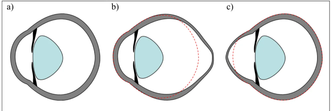
The shape of the eye is maintained by the ocular coat, which consists of the cornea and sclera. Similarities Between Degenerative Myopia and Keratoconus – Similar to the sclera in degenerative myopia, change in the structure of the cornea is a significant factor in. We differ from Wollensak et al. in choosing a sensitizer that minimizes cytotoxic effects. photosensitizer eosin Y is effective in strengthening the cornea and the sclera.
Reaction Pathways for Photodynamic Collagen Cross-linking
M ETHODS
The intensity profile as a function of position at the top of the quartz window was characterized using an optical fiber with a "cosine corrector" (Ocean Optics Jaz) and was found to differ by less than 5% from the value at the center of the 8-mm diameter sample surface. The upper tool was then lowered to bring the sample into contact with the lower plate. The gap was reduced to 90% to ensure good contact between the sample and the tool.

R ESULTS
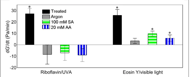
Collagen cross-linking activated by eosin Y with visible light exhibits very similar behavior to riboflavin/UVA. In particular, oxygen is required for crosslinking and the addition of singlet oxygen quenchers (sodium azide and ascorbic acid) inhibits crosslinking (Figure 2.2). Thus, the photosensitized cross-linking reactions for both riboflavin/UVA and eosin Y/visible proceed via the singlet oxygen pathway.
Motivated by the importance of protein-protein crosslinking in aging, photodynamic therapy and tissue engineering, extensive studies have investigated crosslinking induced by singlet oxygen in crystalline ribonuclease A[10], spectrin[46], fibrin[47], fibrinogen. [47], and collagen[48]. While methionine is reactive towards singlet oxygen and is present in collagen type I, it does not participate in the cross-linking reactions. Furthermore, dityrosine formation was not observed in the crosslinking process mediated by singlet oxygen in proteins, peptides or model tyrosine copolymers.
The specific rate of cross-linking reactions depends on the proximity of one of these amino acids to a photo-oxidized histidine and the degree of "exposure" of the side chains for reaction[13]. Peroxide species are intermediates in photodynamic crosslinking – Photooxidation of tryptophan and tyrosine residues has been investigated, establishing the structures of the peroxide intermediates formed and of some of their decomposition products[59-63]. The present experimental results, together with previous literature, suggest that collagen cross-linking induced by riboflavin/UVA and eosin Y/visible light are both mediated by singlet oxygen.
In addition, histidine is the most likely amino acid to play an important role in collagen cross-linking reactions in the cornea and sclera.
Collagen Cross-linking Kinetics
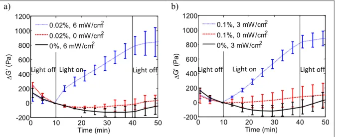
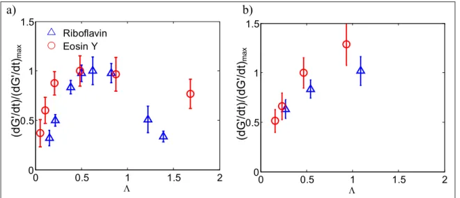
Regarding the application of collagen cross-linking to treat degenerative myopia [14, 15], in vivo studies of eosin Y activated by visible light showed no retinal toxicity in a guinea pig model [11], contrary to early in vivo results with riboflavin/ UVA[4]. At a given photosensitizer concentration and sample thickness, the cross-linking rate (manifested by dG'/dt) increases monotonically with irradiation intensity for both eosin Y and riboflavin, approaching a plateau rate (Figure 3.3a). Collagen gel photorheology can be used to efficiently characterize the effects of irradiation intensity, photosensitizer concentration and sample thickness on the rate of collagen cross-linking.
Collagen cross-linking can also be achieved by chemical or physical techniques without photoactivation. Light activation of the drug also allows control over the depth of crosslinking within the tissue. Thus, the clinical radiation intensity (3 mW/cm2) corresponds to the lowest value that causes the highest degree of cross-linking.
In contrast to riboflavin/UVA treatment, collagen cross-linking activated by eosin Y using visible light has relatively low toxicity (Chapter 6). Photorheology can be used to efficiently characterize the effects of treatment parameters (including photosensitizer concentration and irradiation intensity) on the cross-linking rate of therapeutic collagen cross-linking. In the specific case of eosin Y activated by green light, photorheology indicates that the rate and extent of collagen cross-linking can do this.
Mazzotta, C., Balestrazzi, A., Baiocchi, S., Traversi, C., Caporossi, A., Foschia stromale dopo reticolazione combinata del collagene corneale riboflavina/UVA.
Transcorneal and Transcleral Transport of Eosin Y and Riboflavin
R ESULTS
Therefore, we approximate the total number of drug molecules delivered to the cornea during tc as the sum of the number of drug molecules in the three extracts (Figure 4.6a). Therefore, we also approximate the total number of drug molecules delivered to the sclera during tc as the sum of the number of drug molecules in all extracts (Figure 4.6b). a). A quantitative description of the number of drug molecules delivered to the target tissue is essential in terms of drug delivery for the development of safe and effective therapies.
The values of the partition coefficient and diffusion coefficient enable prediction of the amount of drug transferred to the tissue and the distribution of drug in the tissue, which is very important for safety and efficacy in therapeutics. The permeability, P, is equal to the product of the partition coefficient and diffusion coefficient divided by the tissue thickness[36]. The observed values of the partition coefficients for the two drugs in the cornea and sclera provide interesting information about the interaction between the drugs and the constituents of the tissue.
In the case of the stroma of the cornea and sclera, the main components are collagen fibrils embedded in a matrix of Accurate partition coefficient values are important for understanding drug transport in tissues. The greater the affinity of the drug for proteins or proteoglycans in tissues, the lower the effective diffusion coefficient.
The choice of delivery agents for corneal treatment in vivo can be guided by quantitative comparison of the amount of drug transferred to the tissue.

C ONCLUSION
The selected viscosity enhancers (hyaluronic acid[53, 54], carboxymethylcellulose[55, 56], sodium alginate[57] and methylcellulose[58] have been widely used in various ocular drug delivery systems. Wollensak, G., Iomdina, E ., Long-term biomechanical properties of rabbit sclera after collagen cross-linking using riboflavin and ultraviolet A (UVA).Raiskup-Wolf, F., Hoyer, A., Spoerl, E., Pillunat, L.E., Collagen cross-linking with riboflavin and ultraviolet- A light in Keratoconus: Long-term results.
Klein, S.R., Epstein, R.J., Randleman, J.B., Stulting, R.D., Corneal Ectasia After Laser In Situ Keratomileusis hos patienter uden tilsyneladende præoperative risikofaktorer. Maren, T.H., Jankowska, L., Sanyal, G., Edelhauser, H.F., The Transcorneal Permeability of Sulfonamide Carbonic Anhydrase Inhibitors and their effect on Aqueous Humor Sekretion. Edwards, A., Prausnitz, M.R., Fiber Matrix Model of Sclera and Corneal Stroma for Drug Delivery to the Eye.
Boubriak, O.A., Urban, J.P., Akhtar, S., Meek, K.M., Bron, A.J., The effect of hydration and matrix composition on solute distribution in rabbit sclera. Geroski, D.H., Measurement and Prediction of Transient Transport Across Sclera for Drug Delivery to the Eye. Korb, D.R., Scaffidi, R.C., Greiner, J.V., Kenyon, K.R., Herman, J.P., Blackie, C.A., et al., The effect of two new lubricant eye drops on tear film lipid layer thickness in subjects with dry eye symptoms.
Lin, H.R., Sung, K.C., Vong, W.J., In Situ Gelling of Alginate/Pluronic Solutions for Ophthalmic Administration of Pilocarpine.
A Model for the Photodynamic Collagen Cross-linking Treatment
D ISCUSSION
Several studies have investigated the extent of cross-linking in the anterior and posterior corneal stroma due to riboflavin/UVA treatment by measuring changes in resistance to enzymatic degradation[2], thermomechanical[13], collagen fiber diameter[14] and hydration. to compare. [15], and biomechanical behavior[7]. This result is consistent with the model predicting a monotonically decreasing cross-linking profile across a 500 µm thick cornea for riboflavin/UVA treatment (Figure 5.4c). This is consistent with the observed behavior where there is a large increase in the shrinkage temperature in the front part due to a higher degree of cross-linking compared to the back part.
The clinical concentration yields a degree of crosslinking that is only 81% of the optimal degree (collagen gel photorheology estimated 78% of the optimal degree, Chapter 3). In addition to providing a higher ∆G'avg, the optimal conditions also produce a more uniform crosslinking profile. Therefore, it is desirable to deliver an amount of drug to the tissue that allows for a rapid reaction and a more uniform light intensity profile, resulting in a more uniform crosslinking profile.
The average optimal drug concentration within tissues is 0.016%, and concentrations within them have correlation rates that are within 90% of optimal. Selection of Irradiation Protocol – Given the drug concentration profile, the intensity and duration of irradiation determine the amount of cross-linking, but not the distribution of cross-linking. Wollensak, G., Spoerl, E., Seiler, T., Stress measurements of human and porcine porcines after riboflavin-ultraviolet-A-induced cross-linking.
Wollensak, G., Spoerl, E., Wilsch, M., Seiler, T., Keratocyte apoptosis after cross-linking of corneal collagen using riboflavin/UVA treatment.

Cross-linking the Cornea with Minimal Toxicity
D ISCUSSION
Crosslinks induced by EY/vis are expected to be equivalent to those formed by R/UVA (Chapter 2). It is therefore worth investigating the expectation that EY/vis will also provide long-term performance. Treatment with EY/vis resulted in little or no corneal opacification, consistent with the histological results showing a normal distribution of keratocytes throughout most of the stroma (Table 6.4 and Figure 6.3a).
The EY/vis treatment was very well tolerated by the endothelial cell layer in rabbit corneas. If clinical trials prove this, EY/vis treatment could also be safe for post-LASIK ectasia patients, who often have thin corneas due to the removal of corneal tissue during LASIK[29]. Major differences are observed between the two treatments in terms of corneal toxicity, with little phototoxicity observed in the EY/vis treatment.
Of particular interest, no endothelial toxicity was observed with EY/fish in a rabbit model, even though the cornea is less than 400 µm thick. Therefore, future clinical studies are recommended to determine whether EY/vis treatment is safe for patients with corneas thinner than 400 μm. Compared to the usual R/UVA clinical protocol, the EY/vis protocol requires 1/4 of the treatment time (15 minutes).
If clinical studies confirm the results observed in the rabbit model, treatment with EY/vis may reduce patient discomfort and treatment costs relative to R/UVA.