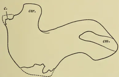
Another dugongi, represented by a significant part of the skeleton, was excavated in a Fuller earth mine. In conducting this study, no reference to the Cenozoic development history of the Sirenia on the Pacific coast of North America was considered. Compared to that of the recent dugong, the brain cavity is more elongated, somewhat com-.
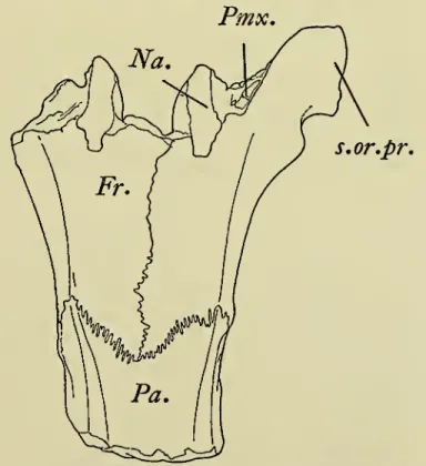
NEW iSPECIBS OF EXTTNCT MIOCENE SIRENIA 73 At the posterior end of the mesorostral fossa of Cari-. On each side, the premaxillary bones form most of the downward-facing rostrum. The posttympanic process of the squamosal is extended ventrally beyond the level of the basi.
The jugal of this fossil skull is relatively much longer and less semicircularly curved than that of the recent dugong Dugong (USNM 284443). This anterior portion of the jaw and its external palatal projection and jugal enclose a very large infraorbital foramen. The vomer apparently did not extend unusually to the height of the anterior end of the row of cheek teeth.
On the skull of the Recent dugong, the pair of rather small palatine bones are visible in the palate between the posterior molars.

NEW SPECIES OF EXTTNOT MIOCENE SIRENIA 77 The facet for contact with the facet on the cms longum of
The absence of incisors is described by Simpson as a remarkable feature of the middle Miocene Hawthorn. According to Reinhart (1959, p. 31) the Californian Halianassa [=^ Metaxytherium]. vanderhoof lacks incisor teeth on the anterior margins of the premaxillaries. Annular ridges on the circumference of the dentin core are exposed from the outer cementum slip zone.
All of the characteristics described above, with the exception of the dimensions, can be matched by a Calvert tusk (USNM 8457) as well as a Florida Tertiary tusk (AMNH 9852). On the palate of the Lower Miocene (Burdigalian), Halianassa studeri fourcheek teeth are in place in addition to four root cavities on one or two missing incisors. A depression on the lingual and buccal sides of the worn crown of M' marks the location of the transverse valley.
This Calvert M' does not closely resemble the corresponding molar of the Helvetian Metaxytherium cuvierii. The image of the three molars published by Flot appears to have been reversed by the engraver. A consistent cross pattern is not easily observed when comparing the upper third molars of three individuals (USNM.
On the posterior side of the crown of the left M2 tooth on the buccal side there is a worn cone considered the hypoconulid. The hind claw is large and minutely pointed; this part of the crown is less sanded. The tips of the protoconid and metaconid do not converge; This anterior portion of the crown is also sharply separated from the posterior portion by a deep transverse valley.
The posteromedian cuspule is located partially anterior to the hypoconid, which in turn is separated by a nasal fissure from the entoconid on the lingual side of the crown. For these deciduous teeth (USNM 16630) the terminology of the permanent molars is applied topographically in light of the uncertain homologies of the cusps on the crowns of the deciduous cheek teeth. Incomplete right and left scapulae have been associated with this skeleton, although the right one lacks only a portion of the anterior border.
USNM USNM USNM
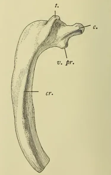
The facet for articulation with the tubercle of the corresponding rib at the end of the diapophysis becomes. A much less prominently developed crest can be recognized on the first rib of the dugong. No noticeable narrowing of the vertebral portion of the thick shaft is discernible, the maximum.
The curvature of the shaft is accentuated at the vertebral one-third and the distal tip is bent backwards.
THE SOUTH CAROLINA MIOCENE DUGONG
92 UNITED STATES NATIONAL MUSEUM BULLETIN 247 donated by the Nerac Museum in 1866, measured 260.4
THE COLOMBIA TERTIARY DUGONG
34;The geological description 'grey to green black mottled sandstone' suggests the Honda Formation whose outcrops are present in the general area of Ortega. As will be noted in the descriptive part of the text, the reduction of the upper molar to three molars would be an unusual achievement as early as the Lower Miocene. On the basis of currently recorded geological occurrences of fossil dugongs, this reduction to three upper molars would logically have been expected in the Upper Miocene. atal part of left upper jaw containing M', M^ and M^. Horizon and locality: Ortega, north of the mouth of the Rio Saldana, DepartamentoTolima, Colombia.
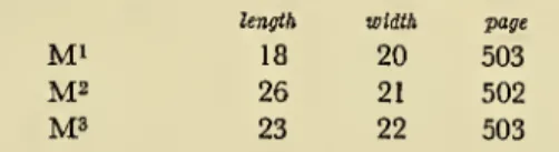
The molars of the Ortega dugong are larger than those of Felsinotherium serresii (Deperet and Roman, 1920, p. 12, pi. The upper first and second molars of F. serresii are more subquadrate than the same molars of the Ortega dugong; the placement of the cusps, however, are quite similar, but the anterior lacuna between the cingulum and the paracone in the Montpellier teeth is very narrow. The serresii does not appear to have fully erupted. The length of the molar teeth in the right and left maxillae of specimens in the Gennevaux collection, Universite de Lyon, published by Deperetand Roman (1920, p. 12) are as follows: M\ 18mm.; M^ 21mm.; M^, 22 mm.
Each of these molars has an anterior half of the crown separated from the posterior half by a deep transverse valley, only slightly obstructed by the metaconule. An obvious indentation on the buccal side of the M' crown marks the outer end of a deep, narrow transverse valley. This first molar has an enlarged paracone, which is connected to an anterior slightly cuspate cingulum with a short cusp, and is medially continuous with a small protocone, which, however, is not severed from the crescentic margin of the lingual protocone.
A deep transverse lake intervenes between the anterior crest of the cingulum and the continuous enamel connecting isthmus or anterior wall of the paracone, protoconule and protocone occlusal surface. The deep anterior lake between these three cusps and the anterior cingulum is relatively small. The dimensions of the anterobuccal parastyle are comparable to those of this cusp on M^ and this style is likewise separated from the rather broad anterior cingulum by a thin fissure.
The deep transverse valley is slightly deflected forward, the cupolae are, however, raised above the smaller middle part in the center by the more forward placement of the cupolae. Unclad by the metacone and hypocone respectively by a thin tip of the metacone and hypocone are not raised slits.
BIBLIOGRAPHY
The quadrangular anterior more is larger than In contrast to M^ there are three distinct cuspules on the. The more infront of the posterior cingulum narrower above the metaconule; however, the hypocones were printed as those of M-. Le Felsinotheriumserresides sable Pliocenes de Montpellier et les rameaux phyletiques des sireniens fossiles de I'ancien monde.
1. [Sur vin Cetace fossile voisin des dugongs etdes lamantins, gefunden in Raedersdorf, im Departement Haut-Rhin, S. 1848. betreffend die fossilen Überreste von Säugetieren aus den Tertiärlagerstätten in der Gegend um die Landeshauptstadt Linz in Oberösterreich. Bericht über die in den Sandablagerungen von Linz gefundenen fossilen Überreste eines prähistorischen Madenwurms (Halitherium crisiolii).
96 UNITED STATES NATIONAL MUSEUM BULLETIN 247 Gervais, Franqois Louis Paul
A New Odontocete From the Calvert Miocene of Maryland
These mandibles belonged to a cetacean with teeth somewhat icier than any previously recorded calvertodontocete characterized by an elongated rostrum. Diagnosis: Mandibles thick, robust, ankylosed anteriorly by symphyseal joint; large mandibular alveoli, with antero-posterior diameter of 18-23 mm., separated by 5-8 mm. The shortest part (length, 233 mm.) of the left mandible has two teeth with worn crowns in place and 4 empty alveoli.
The largest alveolus in the symphysis part of the left mandible measures anteroposteriorly 23. at the edge, and the smallest posterior alveolus in the right mandible is 18.5 mm. Judging by the dimensions of the alveoli, the anterior teeth were slightly larger than those at or near the posterior end of the tooth. The inner basal shelf became progressively smaller towards the anterior end of the tooth row and is barely visible on the posteriormost of the two teeth retained on the left.
The basal margin of the enamel crown appears to extend ventrally further on the inner surface than on the outer surface, although the distance from the conical apex to the ventral margin is approximately the same on both surfaces. The root of the extracted mandibular tooth (pi.. 45, . fig. 3) is widest near the middle of its length, bent back distally, and compressed from side to side at the distal third. The end of the root is wrinkled, with at least sLx tubers; the pulp cavity is finally closed.
The symphysial part of the ankylotic rami also tapers toward the anterior end, with its transverse diameter becoming less than 45 mm. Dorsoventral diameter of symphysis ankylosis at fractured anterior end, 39 mm; dorsoventral diameter of symphysis at the level of the fourth alveolus, counting back from the anterior end, 46 mm. The outer surface of the right mandible is nearly vertical near the aborted posterior end and becomes more convex anteriorly and gradually shifts to a more oblique slope from the alveolar ridge to the ventral midline.
Although the rostrum has not yet been recorded, the width of the mandibular symphysis of the middle Miocene Florida genus, Megalodelphis magnidens (Kellogg, 1944, p. 445), indicates a skull of somewhat larger dimensions, particularly the width of the rostrum, than the skull. Calvert Miocene odontocete. The antero-posterior diameter (18-20 mm) of the alveoli posterior to the symphysis (USNM 23408) is slightly smaller than the corresponding alveoli (23-25 mm) of Megalodelphis magnidens (MCZ 17883) and the interspaces or septa (5-8 mm. ) between the alveoli are much narrower than those (10-12 mm.) magnidensa. The rostral fragment representing the type of Champsodelphis valenciennesii (see Kellogg, 1944, p. from the Helvetic shelly marl at Sort, 8 kilometers from Dax, DepartementLandes, France, should correspond in general configuration and dimensions to the missing rostrum of this Calvertian odontocete.
A NEW CALVERT MIOCENE ODONTOCETE 101
S. National Museum Bulletin 247, Plate 33
S. National Museum Bulletin 247, Plate 35
S. National Museum Bulletin 247, Plate 37
S. National Museum Bulletin 247, Plate 39
S. National Museum Bulletin 247, Plate 40
S. National Museum Bulletin
S. National Museum Bulletin 247, Plate 43
9 Mandibles, USNM 23408, Hadrodelphis calvertense
S. National Museum Bulletin 247, Plate 45