Yersinia pseudotuberculosis is a gram-negative pathogenic bacterium believed to be an evolutionary ancestor of Yersinia pestis, a deadly pathogen that causes bubonic plague. Further studies on NuLi1 human airway epithelial cells revealed morphological changes induced by purified MVs and were shown to be cytotoxic after they were internalized into the cells by successfully delivering the 60 kDa Chaperone and cytotoxic necrosis factor (CNFy). Flow cytometric analysis confirmed that apoptosis is not a major event in cytotoxicity with only 2% of cells found to be apoptotic.
Introduction
Host-microbe interaction and pathogenesis
The delivery of virulence factors via secretion systems
Membrane vesicles (MVs)
Similar studies have revealed the chemical composition of MVs present in outer membrane components and other cellular compartments. Although several mechanisms for vesiculation have been proposed, vesiculation is thought to occur by a well-conserved mechanism21; however, different types of MVs may exist and originate from different mechanisms. An important feature of MVs is that the proteins associated with them exhibit biological activities.
Biogenesis of membrane vesicles (MVs)
Localized concentration of curvature-inducing molecules leads to rupture of the underlying OM peptidoglycan bonds after those membranes bleed. Furthermore, molecules that induce curvature will interact with specific periplasmic proteins and cause enrichment of certain MV cargo. In each situation, vesicle production is regulated by the accumulation and diffusion of envelope proteins.
Factors influencing vesicle production
Alternatively or in addition to periplasmic turgor pressure, molecules that promote curvature can cause membrane bulging. This event leads to the enrichment of these molecules in MVs, as observed with P molecules. It is often assumed that MVs are released from the cell when the bud grows to the point at which membrane curvature forces separation33.
Virulence factors associated with membrane vesicles
Specific accumulation of particular proteins at these sites would enrich these proteins in MVs21. MVs are found in a range of sizes, indicating that no critical curvature threshold precipitates their release.
Advantage of membrane vesicles over bacterial secretion systems
The genus Yersinia
Yersinia pseudotuberculosis and its virulence
Recently published studies have described how bacterial pathogens use MVs as vehicles to transport and deliver virulence factors to host cells.71 As such, MVs represent a novel and powerful bacterial secretion mechanism that circumvents the need for direct contact between the bacterium and the host cell,21, 72 does not require typical secretion machinery such as the T3SS and is displayed by diverse pathogens.53, 73 Moreover, one can easily imagine a scenario in which a pathogen incorporating invasive factors into its outer membrane, such as the AilA protein of Y.
Abstract
- Bacterial strain and growth condition
- Culture of human airway epithelial cell line (NuLi-1 Cells)
- Culture of human breast epithelial cell line (MCF-10A Cells)
- Cloning of 60kDa chaperonin and CNFy
- Membrane vesicle isolation
- Observation of MVs using TEM and super-resolution microscopy (SR-SIM)
- Visualization of internalized MVs by confocal microscopy
- Proteomic Analysis of Y. pseudotuberculosis Vesicles
- Criteria for protein identification
- Cell viability and cytotoxicity assays
- Immunostaining of F-actin
- Semi-quantitative analysis of the chaperonin in MVs
- Quantification of CNFy by ELISA
Cells were then washed with sterile DPBS and trypsinized for 5 min to detach from the flask. After incubation, the cells were washed twice with PBS and added to 0.5 ml BGEM medium. Cells were washed with PBS and nuclei were stained with DAPI (1 µg/ml in PBS) for 5 min at room temperature.
Quantification of multinucleation
- Confirmation of packaging of the 60 kDa Chaperonin
- Yersinia pseudotuberculosis Secretes membrane vesicles (MVs)
- Y. pseudotuberculosis MVs are Double MVs
- Y. pseudotuberculosis MVs Invade Human Cells and Cause Multinucleation
- Y. pseudotuberculosis MVs Transport CNFy into Human Cells
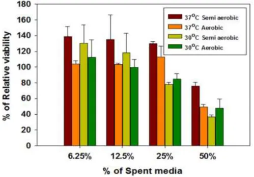
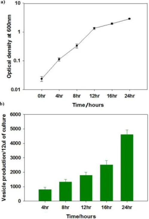
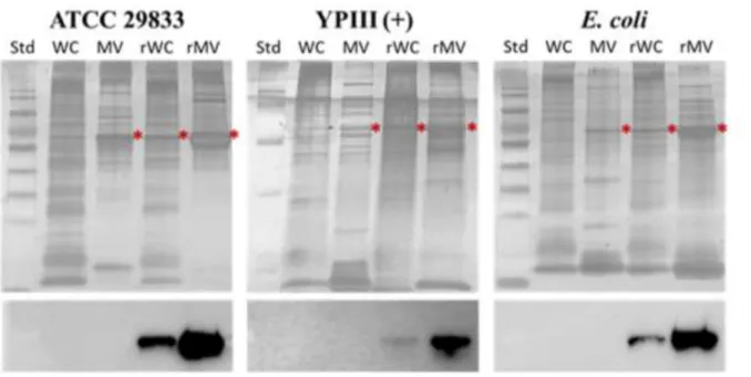
Filtered (0.22 µm) cell-free spent media from cultures of Yersinia pseudotuberculosis ATCC 29833 grown overnight under several different conditions (Figure 2.1) were found to be harmful to NuLi-1 airway epithelial cells and reduce their viability ( figure 2.2). Although the sizes of MVs in (Figure 2.3) and other TEM images varied greatly between about 20 nm to 400 nm in diameter, the sizes were more clearly visible after cleaning (Figure 2.6). MVs are clearly associated with and bud from bacterial cells (scale bar 500 nm).
Semi-quantification of the chaperonin present in the MVs was performed using purified recombinant protein as a standard (Fig 2.6) and suggests that the MVs can up to ten times more chaperonin per microgram of protein than the whole cells contains. This was confirmed in Fig 2B as the recombinant chaperonin could be detected within the infected human cells by means of western blotting (Fig 2.13). Not only are virulence factors transported by MVs, but they also appear to be active as clear dose-dependent phenotypic responses were elicited from the exposed NuLi-1 epithelial cells (Figs 2.14 and 2.15).
Furthermore, pretreatment of the purified MVs with heat or proteinase K before their addition significantly or completely alleviated their toxicity (Fig. 2.14). As shown in (Fig. 2.18), expression of the full-length CNFy gene in this host led to significantly more multinucleation and at a level analogous to that observed in Y. To confirm the presence of CNFy in the MVs , initially Western blot analysis was unsuccessful due to CNFy. Furthermore, the MV sample was subjected to ELISA (Fig. 2.15) and it was confirmed that CNFy was present in the MVs.
Semi-quantification of the chaperonin present in the MVs was performed using purified recombinant protein as a standard.
2hours 12 hours 24 hours
Summary
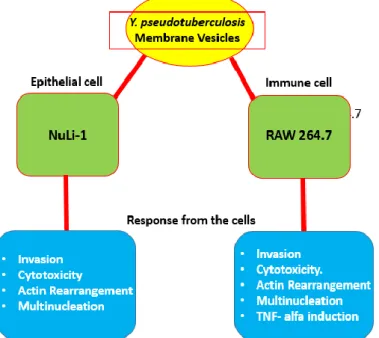
The adverse effect of Yersinia pseudotuberculosis membrane vesicles (MVs) on human airway epithelial cells (NuLi-1) was confirmed in the previous study. The current study focused on the response of mouse macrophage RAW264.7 immune cells to MVs. The result suggested that MVs are also associated with mouse macrophages after 2 hours of treatment.
Furthermore, treatment with MVs leads to actin rearrangement after 3 h of incubation, and there was a significant loss in viability after 48 h of exposure to the MVs. Further investigation showed that macrophages induced a high level of TNFα response to purified vesicles (5209 pg/ml), and 5214 pg/ml TNFα was detected in both ATCC and YPIII(+) vesicle treated cells, and untreated was 190.6 pg/ml. ml after 24 hours of exposure.
Introduction
- Bacteria growth and culture condition
- Mouse leukemic monocyte/macrophage RAW 264.7 cell culture
- MVs isolation and purification
- Invasion of Yersinia pseudotuberculosis and purified MVs
- Cytotoxicity assay
- Apoptosis assay
- Immunostaining of F-actin
- Observation of multinucleation
- Quantification of TNF-alfa
- Y. pseudotuberculosis MVs associated with macrophages
- Y. pseudotuberculosis MVs is cytotoxic to murine macrophage
- Apoptosis is not major event in cytotoxicity
- Y. pseudotuberculosis MVs treatment leads to morphological changes in murine macrophages RAW 264.7
Cells were grown in DMEM F12 medium (Gibco; Life Technologies, NY, USA) supplemented with 10% fetal bovine serum (FBS; ATCC, USA) and 100 mg/ml hormone (InvivoGen, USA) at 37 °C with 5 % CO2 incubator. After incubation, the cells were washed twice with PBS and added to 0.5 ml of DMEM F12 medium. After incubation, cells were washed twice with DPBS and then infected with 1 ml of purified MVs in DMEM F12 in 10 µg/ml medium without MVs as a control.
For F-actin staining, a monolayer of RAW cells was prepared in 8 well chamber coverslips by seeding 2.5X10⁴ /well and incubated in a CO2 incubator at 37ºC for 24 hours. After fixation, cells were washed with DPBS and permeabilized with 0.2% Triton-X (Sigma-Aldrich, USA) in PBS for 5 min. RAW cells were grown in a 12-well plate (SPL, Korea) at 5X10⁴ cells/well for 24 h, and then the vesicle media was replaced with 10 μg/ml MVs (5 μg/well) without vesicles used as a control Cells of treated were incubated in a CO2 incubator at 37ºC for 24 hours.
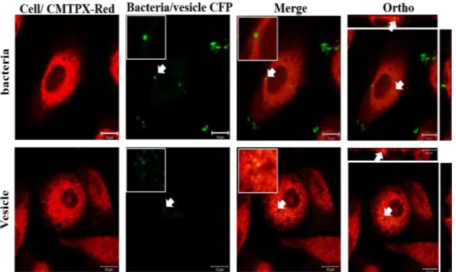
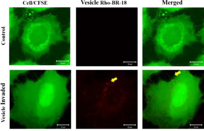
Further evaluation, the treated cells were incubated for 48 hours and stained with CMTPX red, and DAPI showed the observed giant cell phenotype and all cells were checked (Fig 3.6) the result suggested that Y. Further observation revealed that the cells crude were treated with MVs purified from YPIII(+) and ATCC 29833 after 48 h MVs showed multinucleation (Fig 3.6). Before treatment, macrophages were stained with CMPTX red, then treated with 10 μg/ml purified MV from ATCC29833 and YPIII (+) treated after treatment cells were incubated for 48 h.
Macrophage cells were grown on 12-well plate 5X10⁴ cells/well for 24 hours, and then replaced media with vesicle 10 µg/ml of MVs (5 µg/well) and incubated in a CO2 incubator at 37ºC for 24 hours.

Analysis of Post Translational Modifications (PTMs) Yersinia pseudotuberculosis Cytotoxic Necrotizing Factor (CNFy)
Summary
Introduction
- Culture of human airway epithelial cell line (NuLi-1)
- Cloning of CNFy ORF with promoter
- Expression and purification of His-tagged CNFy
- Quantification of CNFy by ELISA
- MTT assay to assess cytotoxicity of ECNFy and YCNFy
- Analysis of CNFy Proteins by LC-MS/MS
- CNFy is nontoxic to human airway epithelial cells
- PTM analysis revealed high level of post translational modification in YCNFy
- Lysine (K) and glutamic acid (E) methylation in CNFy
This domain was conserved in the crystal structure, where the third residue of the catalytic triad, Valine 833113, was identified. This is one of the post-translational modifications that affects several downstream signaling events, unlike other bacterial toxins113. Positively charged lysines are often found in protein subcellular localization signaling domains116 and their acetylation can regulate protein subcellular localization109.
Several studies have shown that CNFy is one of the toxins that leads to multinucleation in another type of cells108. The result is acceptable because recombinant protein expressed in different hosts has different activity due to the unfolding or the fixing of the protein will be compromised when it is shown in another host121. A previous study showed that I.C.V injection of the whole CNF toxin did not cause any lethal effects in animals109.
The viability of NuLi-1 cells was determined by MTT assay after 48-h post-treatment with different concentrations of ECNFy and YCNFy. The viability of NuLi-1 cells was determined after 48-hour treatment with different concentrations of E CNFy and YCNFy. Above all, YCNFy has various post-translational modifications, a total of 60 asparagine deamination is one of the dominant modifications present in both YCNFy and ECNFy has the highest modification.
It was hypothesized that methylation of glutamic acid plays a key role in protein folding and functions; it is expected that neutralization of the amino acids, negative charge, would have a significant impact on the proteins127.
Methylation Glutamic acid Methylation lysine
Methylation glutamine Acetylation lysine
Acetylation threionine Acetylation Asparagine
Deamidation of asparagine (N)
Deamidation of the asparagine was found to be the most abundant PTM in YCNFy, a total of 37 amino acids were deamidated, that is more than 50% PTMs in YCNFy. Deamidation of asparagine is also associated with ECNFy, but it is a relatively low number (Table 4.2). Deamidation is the critical protein post-translational modifications that contribute to various diseases celiac disease, urinary tract infection, cataract formation, cancer, Alzheimer's130. It is the one of the known non-enzymatic modifications, it occurs in both occurs both in vivo and in vitro131.
Based on previous reports, post-translational modification plays a key role in cell signaling during pathogenesis and disease progression, methylation of lysine (K), glutamic acid (E), and deamination of asparagine (N) and lysine (K) acetylation in the context of cell signaling or disease progression. In our study, CNFy is one of the major virulence factors that induces a very precise signaling event during the interaction with the host system. Here, therefore, we hypothesize that post-translational modification is a likely player in CNFy activity.
Further research should focus on identification of essential PTM for the activity and identification of enzyme required for the modification in Y.
Conclusion Conclusion
Chatterjee, D.; Chaudhuri, K., Association of cholera toxin with Vibrio cholerae outer membrane vesicles internalized by human intestinal epithelial cells. Brandenburg, K.; Whiteley, M., Interaction of quorum sensing signals with outer membrane lipids: insights into prokaryotic membrane vesicle formation. E.; Godinez, D.; de la Garza, M., Membrane vesicles released by Actinobacillus pleuropneumoniae contain proteases and Apx toxins.
P.; OHagan, S.; Dermott, E., Comparison of hemagglutination and enzymatic activity of Bacteroides fragilis whole cells and outer membrane vesicles. J.; Kadurugamuwa, J.; Walther-Rasmussen, J.; Hoiby, N., A chromosomal beta-lactamase packaged into membrane vesicles and secreted from Pseudomonas aeruginosa. Muraca, M.; Putignani, L.; Fierabracci, A.; Teti, A.; Perilongo, G., Gut microbiota-derived outer membrane vesicles: under-recognized major players in health and disease.
Winter, J.; Letley, D.; Rhead, J.; Atherton, J.; Robinson, K., Helicobacter pylori membrane vesicles stimulate innate pro- and anti-inflammatory responses and induce apoptosis in Jurkat T cells. Bjartell, A.; Morgelin, M.; Riesbeck, K., Multicomponent Moraxella catarrhalis outer membrane vesicles induce an inflammatory response and are internalized by human epithelial cells.