STUDY OF DIELECTRIC BARRIER DISCHARGE PLASMA JET (DBDJ) FOR BACTERICIDAL IN CONTAMINATED WOUNDS
PIPATH PORAMAPIJITWAT
MASTER OF SCIENCE IN NANOSCIENCE AND TECHNOLOGY MAEJO UNIVERSITY
2019
STUDY OF DIELECTRIC BARRIER DISCHARGE PLASMA JET (DBDJ) FOR BACTERICIDAL IN CONTAMINATED WOUNDS
PIPATH PORAMAPIJITWAT
A THESIS SUBMITTED IN PARTIAL FULFILLMENT
OF THE REQUIREMENTS FOR THE DEGREE OF MASTER OF SCIENCE IN NANOSCIENCE AND TECHNOLOGY
GRADUATE SCHOOL MAEJO UNIVERSITY 2019
Copyright of Maejo University
STUDY OF DIELECTRIC BARRIER DISCHARGE PLASMA JET (DBDJ) FOR BACTERICIDAL IN CONTAMINATED WOUNDS
PIPATH PORAMAPIJITWAT
THIS THESIS HAS BEEN APPROVED IN PARTIAL FULFLLMENT OF THE REQUIREMENTS FOR THE DEGREE OF MASTER OF SCIENCE
IN NANOSCIENCE AND TECHNOLOGY
APPROVED BY Advisory Committee
Chair
(Dr. Sureeporn Sarapirom) .../.../ ...
Committee
(Dr. Keratiya Janpong) .../.../ ...
Committee
(Associate Professor Dr. Wasin Charerntantanakul) .../.../ ...
Program Chair, Master of Science
in Nanoscience and Technology (Assistant Professor Dr. Viruntachar Kruefu) .../.../ ...
CERTIFIED BY GRADUATE SCHOOL
(Associate Professor Dr. Kriangsak Mengamphan) Dean of Graduate School
.../.../ ...
C
ABSTRACT (THAI)
ชื่อเรื่อง การศึกษาพลาสมาเจ็ทแบบไดอิเล็คทริกแบร์ริเออร์ดิสชาจ์นเพื่อการฆ่า เชื้อแบคทีเรียในแผลติดเชื้อ
ชื่อผู้เขียน นายพิพัฒน์ ปรมาพิจิตรวัฒน์
ชื่อปริญญา วิทยาศาสตรมหาบัณฑิต สาขาวิชาวิทยาศาสตร์และเทคโนโลยีนาโน อาจารย์ที่ปรึกษาหลัก อาจารย์ ดร.สุรีย์พร สราภิรมย์
บทคัดย่อ
พลาสมาความดันบรรยากาศ (CAPP) เป็นเทคนิคที่ได้รับการยอมรับการใช้งานทางด้าน การแพทย์ ซึ่งเป็นนวัตกรรมใหม่ที่มีการน าไปใช้งานทางด้านการแพทย์ เช่น การเพิ่มประสิทธิภาพ ของการรักษาแผลติดเชื้อพร้อมกับบรรเทาอาการเจ็บปวดของผู้ป่วยไม่มีผลข้างเคียง แผลติดเชื้อเป็น ปัญหาสุขภาพที่ส าคัญในหลายประเทศ ปัจจัยที่เป็นสาเหตุของปัญหาสุขภาพของผู้ป่วย ได้แก่
โรคเบาหวาน บาดแผลที่ปนเปื้อนแบคทีเรียและอื่น ๆ ผู้ป่วยต้องทนทุกข์ทรมานและต้องใช้เวลานาน ในการบ าบัดรักษาด้วยยาปฏิชีวนะหรือวิธีการรักษาอื่น ๆ นอกจากนี้การใช้ยาปฏิชีวนะเป็นระยะ เวลานานจะก่อให้เกิดการดื้อยาของแบคทีเรียเกิดขึ้น เช่น methicillin-resistant Staphylococcus aureus (MRSA) โดยเชื้อแบคทีเรียนั้นมีขีดความสามารถในการพัฒนาความต้านทานต่อยาปฏิชีวนะ ได้รวดเร็วกว่าการที่เราจะท าการพัฒนายาปฏิชีวนะตัวใหม่ออกมา ด้วยเหตุนี้จึงเป็นปัญหาหลักในการ รักษาแผลติดเชื้อ ในการศึกษานี้จะใช้พลาสมาเจ็ทแบบไดอิเล็คทริกแบร์ริเออร์ดิสชาจ์น (DBDJ) ใน การฆ่าเชื้อแบคทีเรียและทดลองกับ Primary Human Dermal Fibroblasts Adult (HDFa) cells เพื่อศึกษาผลข้างเคียงของ DBDJ ต่อเซลล์ โดย DBDJ ก าเนิดด้วยเงื่อนไขที่ไฟฟ้าแรงดันสูงความถี่ 20 กิโลเฮิร์ทซ์และอัตราการไหลของก๊าซฮีเลียม 1 ลิตรต่อนาที ก าลังของพลาสมาในช่วงระหว่าง 0.27 วัตต์ ถึง 0.50 วัตต์และระยะเวลาในการทดลองระหว่าง 15 วินาที ถึง 60 วินาที อนุมูลในพลาสมาจะ ใช้เครื่องมือวิเคราะห์สเปกตรัมทางแสง (OES) ในการตรวจวัด ผลสเปกตรัมจาก OES พบกลุ่มของ NO และ OH ซึ่งเป็นอนุมูลที่ส าคัญต่อการรักษาบาดแผลที่เกิดจากเชื้อแบคทีเรีย การเพิ่มขึ้นของ ปริมาณอนุมูลอิสระในพลาสมาขึ้นอยู่กับก าลังของพลาสมา โดยวิธี Colony Forming Unit (CFU) จะใช้เพื่อตรวจสอบประสิทธิภาพของการ ฆ่าแ บคที เรีย Staphylococcus aureus (S.
aureus) และ Pseudomonas aeruginosa (P. aeruginosa) ซึ่งใช้ในการศึกษาประสิทธิภาพการ ฆ่าเชื้อแบคทีเรีย ผลการทดลองแสดงให้เห็นว่าก าลังของพลาสมาและระยะเวลาที่ใช้เป็นปัจจัยส าคัญ ในการฆ่าเชื้อแบคทีเรีย เมื่อใช้ก าลังพลาสมาที่ 0.50 วัตต์และ ณ เวลาที่ 60 วินาที ผลของการฆ่าเชื้อ แบคทีเรียเพิ่มขึ้นถึง 100% ดังนั้นจึงเลือกที่สภาวะการทดลองดังกล่าวในการศึกษาผลกระทบของ
D พลาสมาต่อแบคทีเรียไบโอฟิล์มและเซลล์ HDFa ผลการทดลองจากกล้องจุลทรรศน์แบบฟลูออเรส เซนต์ โดยใช้เทคนิค live/dead assay แสดงให้เห็นว่าพลาสมามีประสิทธิภาพสูงในการก าจัด แบคทีเรียไบโอฟิล์ม การศึกษาผลกระทบของ DBDJ ต่อเซลล์ HDFa จะใช้กล้องจุลทรรศ์แบบเชิงแสง เทคนิค live/dead assay และเครื่อง Muse Cell Analyzer ร่วมกับชุด Muse Count & Viability Assay Kit และ Muse Annexin V and Dead Cell Assay Kit ผลที่ได้แสดงให้เห็นว่า DBDJ มี
ประสิทธิภาพสูงในการฆ่าเชื้อแบคทีเรียโดยไม่ก่อให้เกิดผลกระทบข้างเคียงต่อเซลล์ HDFa ภายใต้
สภาวะเงื่อนไขเดียวกัน ดังนั้น DBDJ ที่ก าลังพลาสมา 0.50 วัตต์และ ณ เวลา 60 วินาที มี
ประสิทธิภาพสูงในการฆ่าเชื้อแบคทีเรีย การก าจัดแบคทีเรียไบโอฟิล์มและไม่ก่อให้เกิดความเสียหาย หรือผลข้างเคียงต่อ การมีชีวิตของเซลล์ การเกิดอะพอพโทซิส และการตายของเซลล์ HDFa ในการ ทดลองซึ่งเป็นสิ่งที่ส าคัญ
ค าส าคัญ : พลาสมาเจ็ทแบบไดอิเล็คทริกแบร์ริเออร์ดิสชาจ์น, เซลล์ไฟโบรบลาสต์, การฆ่าเชื้อ แบคทีเรีย, การรักษาแผล, แบคทีเรียไบโอฟิล์ม, แผลติดเชื้อ
E
ABSTRACT (ENGLISH)
Title STUDY OF DIELECTRIC BARRIER DISCHARGE
PLASMA JET (DBDJ) FOR BACTERICIDAL IN CONTAMINATED WOUNDS
Author Mr. Pipath Poramapijitwat
Degree Master of Science in Nanoscience and
Technology
Advisory Committee Chairperson Dr. Sureeporn Sarapirom
ABSTRACT
The cold atmospheric pressure plasma (CAPP) technique has been recognized in medical fields to bring a new innovative approach in biomedical applications such a contaminated wound healing enhancement as well as relieving the patient’s pain without side effects. Contaminated wounds are a major health problem in many countries. Factors of patient health problems are such as diabetes, contaminated wounds, bacteria as well as others. The patients endures pain and requires long treatment periods using antibiotics or other therapies. Moreover, using antibiotics for a long period of time bacterial resistance occurs, such as methicillin- resistant Staphylococcus aureus (MRSA). Bacteria have the ability to develop resistance to antibiotics faster than one can develop new drugs. This being the main problem in contaminated wound healing. In this study, the dielectric barrier discharge plasma jet (DBDJ) was used for bactericidal and treated the Primary Human Dermal Fibroblasts Adult (HDFa) cells to study the side effect of DBDJ. This DBDJ is driven by high voltage dc pulse at 20 kHz and using 1 L/min of helium (He) as plasma gas. The DBDJ plasma varied the plasma dissipated power from 0.27 W to 0.50 W and the exposure time 15 s to 60 s. Plasma radical species were utilized by using an optical emission spectroscopy (OES). The results of the OES study found NO and OH radical groups, which play an important role in bactericidal and contaminated wound healing.
The increase of radical plasma density depends on the plasma dissipated power. The Colony Forming unit (CFU) method was used to monitor the efficiency of bacteria
F killing. Staphylococcus aureus (S. aureus) and Pseudomonas aeruginosa (P.
aeruginosa) were used in the vitro bacteria killing test. The results showed that the plasma dissipated power and exposure time were major factors in bactericidal. When increasing the plasma dissipated power to 0.50 W and exposure time to 60 s, the effect of bacteria killing increased to 100%. Therefore, this condition was choosing to study the effect of plasma on bacteria biofilm and HDFa cells. The result from fluorescent images by live/dead assay showed that DBDJ had high efficiency to removed biofilm.
Studying the effects of DBDJ on HDFa cells using optical microscopes, live/dead assay and Muse Cell Analyzer with the Muse Count & Viability Assay Kit and Muse Annexin V and Dead Cell Assay Kit, the result showed the DBDJ being highly efficient for bactericidal as well as not having any side effects on HDFa cells under the same conditions. Therefore, the DBDJ at plasma dissipated power 0.50 W and exposure time 60 s has a high efficiency to kill bacteria, destroy bacteria biofilm by without damage cell and also without side effect to cell viability, apoptosis and death of HDFa cells as in vitro is essential for clinical use.
Keywords : Dielectric Barrier Discharge Plasma Jet, Human Dermal Fibroblasts adult cells, Bactericidal, Wound Healing, Bacterial Biofilms, Contaminated Wounds
G
ACKNOWLEDGE MENTS
ACKNOWLEDGEMENTS
The authors are thankful to the following contributors: Plasma Medicine project in supporting the equipment and budget. The Dielectric Barrier Discharge Plasma Jet (DBDJ) from Dr.Chanchai Chutsirimongkol to investigate the bactericidal in contaminated wounds as in vitro. Thailand Institute of Scientific and Technological Research (TISTR) for support bacterial stain in my research. Thailand Center of Excellence in Physics (ThEP), STEM (Science, Technology, Engineering, Mathematics) Workforce and Graduate School Maejo University for my scholarships. Plasma and Beam Physics research facility (PBP) for equipment, laboratory, suggestion, guidelines and help in my research.
I would like to thank my thesis advisor Dr. Sureeporn Sarapirom for supporting, suggesting and giving me an opportunity to do this new subject research. The door of her office was always open whenever I ran into a trouble spot or had a question about my research or writing. She consistently forced me that this paper to be my own work and steered me in the right direction. Dr. Keratiya Janpong and Assoc. Prof. Dr. Wasin Charerntantanakul my thesis co-advisors in reading and publishing of my thesis, to them I am grateful and indebted for their valuable comments. A special mention to, Assoc.
Prof. Dr. Dheerawan Boonyawan for my scholarship and budget in my research.
Finally, I must express my gratitude to my parents for their supporting and continuous encouragement throughout my years of study. This accomplishment would not have been possible without them. Thank you.
Pipath Poramapijitwat
TABLE OF CONTENTS
Page ABSTRACT (THAI) ... C ABSTRACT (ENGLISH) ... E ACKNOWLEDGEMENTS ... G TABLE OF CONTENTS ... H LIST OF TABLES ... K LIST OF FIGURES ... L
CHAPTER 1 INTRODUCTION ... 1
1.1 Background ... 1
1.1.1 Atmospheric pressure plasma (APP) ... 2
1.1.2 Plasma medicine ... 2
1.2 Objectives ... 4
1.3 Expectation ... 4
CHAPTER 2 THEORY AND LITERATURE REVIEW ... 5
2.1 Plasma ... 5
2.1.1 Classical plasma ... 8
2.1.2 Degree of ionization in plasma ... 8
2.1.3 Collisions between particles... 9
2.1.4 Plasma parameters ... 10
2.1.5 Plasma thermal equilibrium ... 12
2.1.5.1 Thermal equilibrium plasma ... 12
2.1.5.2 Nonthermal equilibrium plasma ... 12
I
2.1.6 Plasma source ... 13
2.1.7 Atmospheric pressure plasma (APP) ... 14
2.2 Bacteria ... 17
2.2.1 Bacterial cells... 17
2.2.2 Staphylococcus aureus ... 18
2.2.3 Pseudomonas aeruginosa ... 18
2.3 Mechanisms of CAPP to bactericidal ... 19
CHAPTER 3 EXPERIMENT ... 23
3.1 The Dielectric Barrier Discharge Plasma Jet (DBDJ) ... 23
3.2 Plasma properties ... 24
3.2.1 Electrical properties ... 25
3.2.2 The reactive oxygen nitrogen species (RONS) ... 26
3.3 Microbiological test ... 28
3.3.1 Bactericidal ... 28
3.3.2 Bacteria biofilm ... 29
3.4 Human Dermal Fibroblasts adult (HDFa) cells ... 30
3.4.1 Cell culture ... 30
3.4.2 Cell morphology ... 30
3.5 Cell cytotoxicity assay ... 31
3.5.1 Hoechst 33342 and Propidium iodide (PI) double stain ... 31
3.5.2 Muse Cell Analyzer ... 32
3.5.3 Muse Count & Viability Assay ... 32
3.5.4 Muse Annexin V and Dead Cell Assay ... 33
3.5.5 Statistical analysis ... 34
CHAPTER 4 RESULTS AND DISCUSSION ... 35
4.1 The Dielectric Barrier Discharge Plasma Jet (DBDJ) ... 35
4.1.1 Electrical properties ... 35
4.1.2 The reactive oxygen nitrogen species (RONS) ... 38
4.2 Microbiological test ... 42
4.2.1 Bactericidal effect of DBDJ ... 42
4.2.2 Effect of DBDJ on bacterial biofilms ... 47
4.3 Human Dermal Fibroblasts adult (HDFa) cells ... 50
4.3.1 Cell morphology ... 50
4.3.2 Live/dead assay of HDFa cells ... 52
4.3.3 Muse Count & Viability Assay ... 54
4.3.4 Muse Annexin V and Dead Cell Assay ... 55
CHAPTER 5 CONCLUSION AND RECOMMENDATIONS ... 57
5.1 Conclusion ... 57
5.2 Recommendations ... 57
REFERENCES ... 59
APPENDIX ... 63
APPENDIX A PLASMA PROPERTIES ... 64
APPENDIX B BACTERICIDAL... 76
APPENDIX C BACTERIA BIOFILM... 82
APPENDIX D CELL CULTURE ... 89
APPENDIX E PUBLICATION ... 105
CURRICULUM VITAE ... 127
LIST OF TABLES
Page
Table 1 Types of plasma and plasma parameters (Nojiri, 2015). ... 11
Table 2 Surface reactions (Braithwaite, 2000)... 13
Table 3 Gas phase reactions involving electrons (Braithwaite, 2000). ... 13
Table 4 Gas phase reactions involving ions and neutrals (Braithwaite, 2000). ... 14
Table 5 Technical data of AvaSpec-2048. ... 27
Table 6 Duty%, area (Lissajous figure), plasma dissipated power and dose of DBDJ lv.1 to lv.7. ... 37
Table 7 Relative of dose to plasma dissipated power and exposure times... 38
LIST OF FIGURES
Page
Figure 1 The plasma applications on living tissue (von Woedtke et al., 2013). ... 3
Figure 2 The collision of electrons and neutral atoms generate ion (Nojiri, 2015). ... 6
Figure 3 Principle of gas discharge (Nojiri, 2015). ... 6
Figure 4 Model of glow discharge plasma (Nojiri, 2015). ... 7
Figure 5 Characteristic variation of the degree of ionization of an atomic gas at atmospheric pressure (Harry, 2010). ... 9
Figure 6 Atomic and molecular energy associated with different energy transitions (Harry, 2010). ... 10
Figure 7 Atmospheric-pressure plasma exposure in biomedical applications; (a) DBD, (b) plasma jet and (c) plasma effluent downstream (Setsuhara, 2016). ... 15
Figure 8 (A) DBDJ, (B) monitor display of power supply and (C) power supply of BIO Plasma. ... 23
Figure 9 (A) Internal components of DBDJ and (B) Plasma glow discharge of DBDJ. ... 24
Figure 10 Experiment diagram.... 25
Figure 11 Setup diagram of equipment. ... 26
Figure 12 The Lissajous figure of DBDJ at repetition rate 50 Hz or plasma dissipated power 0.27 W. (A) t = 10 ms, (B) t = 5 ms, (C) t = 1 ms, (D) t = 500 us, (E) t = 250 us and (F) t = 100 us. ... 36
Figure 13 A) The applied voltage signal (channel 1) the voltage across the 1 nF capacitor (channel 2) and B) the Lissajous figure of DBDJ the plasma dissipated power at 0.27 W. ... 36
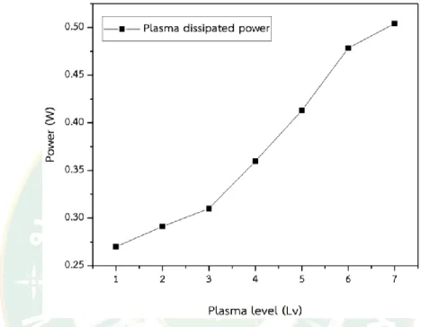
Figure 14 Increasing of plasma dissipated power influenced by plasma level 1 to level 7 (repetition rate at 50 Hz to 110 Hz). ... 37
M Figure 15 The emission spectra of DBDJ with plasma dissipated power 0.27 W to 0.50
W. ... 39
Figure 16 The emission spectra of the plasma dissipated power at 0.50 W. ... 39
Figure 17 The relative of the RONS intensity and the plasma dissipated power. ... 40
Figure 18 NO and OH intensity and the plasma dissipated power. ... 40
Figure 19 The relative between NO and O3 concentration and the plasma dissipated power. ... 41
Figure 20 The efficiency bactericidal S. aureus of DBDJ, the plasma dissipated power at 0.27 W and exposure time 15 s to 60 s (a, b, c and d), the plasma dissipated power at 0.50 W and exposure time 15 s to 60 s (e, f, g and h). ... 44
Figure 21 The percentage efficiency of bactericidal S. aureus by DBDJ. ... 44
Figure 22 The efficiency bactericidal P. aeruginosa of DBDJ, the plasma dissipated power at 0.27 W and exposure time 15 s to 60 s (a, b, c and d), the plasma dissipated power at 0.50 W and exposure time 15 s to 60 s (e, f, g and h). ... 45
Figure 23 The percentage efficiency of bactericidal P. aeruginosa by DBDJ. ... 45
Figure 24 The percentage efficiency bactericidal of DBDJ (A) S. aureus with plasma dissipated power at 0.27 W and 0.50 W, (B) P. aeruginosa with plasma dissipated power at 0.27 W and 0.50 W, (C) S. aureus and P. aeruginosa with plasma dissipated power at 0.27 W and (D) S. aureus and P. aeruginosa with plasma dissipated power at 0.50 W. ... 46
Figure 25 Bacterial biofilm S. aureus by Live/Dead assay with double stain Hoechst 33342 and Propidium iodide (PI). Control sample S. aureus (A, B and C) and plasma dissipated power at 0.5 W with exposure time 60 s for destroy bacterial biofilm S. aureus (D, E and F). ... 48 Figure 26 Bacterial biofilm P. aeruginosa by Live/Dead assay with double stain
Hoechst 33342 and Propidium iodide (PI). Control sample P. aeruginosa (A, B and C)
and plasma dissipated power at 0.5 W with exposure time 60 s for destroy bacterial biofilm P. aeruginosa (D, E and F). ... 48 Figure 27 Cell morphology of HDFa cells, (a,b) negative control or natural control as DMEM + 10% FBS, (c,d) positive control as DMEM + 50% DMSO 2 h, (e,f) treatment control as He gas blow 60 s and (g,h) DBDJ at plasma dissipated power 0.50 W with exposure time 60 s. ... 51 Figure 28 HDFa cells by Live/Dead assay with double stain Hoechst 33342 and Propidium iodide (PI) at magnification 200x. Negative control as DMEM + 10% FBS (a, b and c), positive control as DMEM + 50% DMSO 15 min (d, e and f), treatment control as He gas blow 60 s (g, h and k) and plasma dissipated power at 0.5 W with exposure time 60 s (j, k and l). ... 52 Figure 29 HDFa cells by Live/Dead assay with double stain Hoechst 33342 and Propidium iodide (PI) at magnification 400x. Negative control as DMEM + 10% FBS (a, b and c), positive control as DMEM + 50% DMSO 15 min (d, e and f), treatment control as He gas blow 60 s (g, h and k) and plasma dissipated power at 0.5 W with exposure time 60 s (j, k and l). ... 53 Figure 30 A) Representative figures from flow cytometry of HDFa cells with negative control as (DMEM + 10% FBS), treatment control (He gas blow 60 s), plasma
dissipated power at 0.5 W with exposure time 60 s and positive control (DMEM + 10% DMSO 2 h). B) Quantitative analysis of percentage cell viability from flow
cytometry. Data are means of 3 replicates and expressed as means ± SD. ** indicates P<0.01. ... 55 Figure 31 A) Representative figures from flow cytometry of HDFa cells with negative control as (DMEM + 10% FBS), treatment control (He gas blow 60 s), plasma
dissipated power at 0.5 W with exposure time 60 s and positive control (DMEM + 10% DMSO 2 h). B) Quantitative analysis of percentage total apoptosis from flow cytometry. Data are means of 3 replicates and expressed as means ± SD. ** indicates P<0.01. ... 56
CHAPTER 1 INTRODUCTION
1.1 Background
In recent years, the over-prescription and incorrect use of antibiotics has led to a rise of bacterial resistant to antibiotic. Antibiotic resistance in bacteria is a major problem and is on the rise worldwide, making some previously treatable infections incurable or even life-threatening. Bacteria are prokaryotic micro-organisms which have great adaptability to changing environments. Bacteria is the main cause of illnesses, wound infections, contaminated wound, meningitis, pneumonia etc. Bacteria can be divided into two groups according to the cell wall structure. Gram-positive bacteria have a relative thick peptidoglycan (20 to 80 nm) cell wall, while gram-negative bacteria have a thinner peptidoglycan (< 10 nm) cell wall (Lin et al., 2016; Mai- Prochnow et al., 2016). The difference of bacterial cell wall leads to different properties to the cell, especially the cell responses to external stressors such as heat, UV radiation and also antibiotics. One of the gram-positive bacterial strains is Staphylococcus aureus (S. aureus) and one gram-negative bacterial strains is Pseudomonas aeruginosa (P.
aeruginosa). These strains are causing public health concerns in postsurgical patients.
The S. aureus, P. aeruginosa and methicillin-resistant Staphylococcus aureus (MRSA) are the most commonly identified bacterium in wounds (Mohd Nasir et al., 2016).
Therefore, there is a growing need for the discovery and development of new antibacterial methods of sterilization. Preferably these methods should be easy to use, be cost effective, have low toxicity and no resistance from bacteria (Mai-Prochnow et al., 2015). One of those techniques is cold atmospheric pressure plasma treatment (CAPP). CAPP was widely studied and reported to have high efficiency to eradicate a wide range of pathogens such as fungi, viruses and bacteria. The CAPP sources have been operated at an excitation frequency, either in the several tens of kilohertz ac range or in the radio frequency (RF) range. Various factors including temperature, charged particle, ion and radical species can be manipulated to find suitable conditions. Therefore, it is important to understand the basics of physical and chemical
properties in plasma, such as power deposition and consumption, electromagnetic, electrical characteristics, optical emission spectrum, gas temperature and other parameter in plasma (Kim et al., 2009).
In this study, effects of plasma radicals on S. aureus, P. aeruginosa, bacteria biofilm and Primary Human Dermal Fibroblasts adult (HDFa) cells were investigated for contaminated wound healing model as in vitro.
1.1.1 Atmospheric pressure plasma (APP)
When more energy or heat is added to atoms or molecules, they turn to be ionized. An electron may gain enough energy to escape from an atom. After the escape of electron, atoms turn into ions. In adequately heated, energy to gas, ionization happens many times, creating clouds of ions and free electrons. This ionized gas mixture comprises of ions, electrons and neutral atoms, called “Plasma”. Plasma is the fourth state of matter.
Among the many distinctions between different kinds of plasmas, one is between thermal plasma and non-thermal plasma. Within thermal plasmas the electrons and the ions have the same energy. Non-thermal plasma gains its reactivity from the high energy electrons, while the ions and neutral species remain near room temperature (von Woedtke et al., 2013). Example of thermal plasma are sun and laser fusion plasma. Non-thermal plasma divides two kinds, vacuum pressure plasma or low- pressure plasma and atmospheric pressure plasma. APP is generated by applying an electrical field to a neutral gas or gas mixture. Low-pressure plasma is such as arc plasma, plasma torch, metal vapor vacuum arc and filtered cathodic vacuum arc. APP is such as dielectric barrier discharge (DBD), corona plasma, plasma effluent downstream and plasma jets.
1.1.2 Plasma medicine
Plasma medicine is a new way of plasma application. Physical plasmas are excited ionized gases, which contain different concentrations of low molecular reactive atoms, ions and molecules and emit several kinds of electromagnetic radiation including infrared, visible and ultraviolet (UV) light which are generated by energy
3 supplied to a neutral gas. Non-thermal plasma or low-temperature plasma (LTP) attributes are mostly technical ones whose composition and temperature are adjustable in a wide range by parameters like type of energy source, power of generator, type of gas, pressure of gas, rate of gas flow and composition. In most cases CAPP technical applications are generated by applying an electrical field to a neutral gas or gas mixture (von Woedtke et al., 2013).
In earlier stages, direct plasma applications on living cell in electro-surgery were based on very harsh interactions of plasma with cells and tissue leading finally to cellular eradication and local “sealing” of tissue. The new research field of plasma medicine considers the advantages and effects of LTP or CAP techniques, with much more differentiated interaction of specific plasma components and specific structural elements along with functionalities of living cells which can probably lead either to stimulation or inhibition of cellular function (Figure 1) (von Woedtke et al., 2013).
In CAPP sources, the main reactive components will consist of reactive neutral species such as reactive oxygen and nitrogen species and are called “reactive oxygen-nitrogen species (RONS)”, UV-radiation and electrical field. In some cases, charged species and electromagnetic fields may also play a role.
Figure 1 The plasma applications on living tissue (von Woedtke et al., 2013).
About 1% of the population in developed nations suffer from contaminated wounds (Etufugh and Phillips, 2007). Contaminated wounds may result from venous diseases, arterial diseases, diabetes mellitus, pyoderma gangrenosum, carcinoma and other factors. As the population ages, this occurrence is likely to increase. CAPP may help with the treatment though it is unlikely that plasmas will “heal” the underlying disease, The eliminating of bacterial and fungal infections by plasmas may well reduce the suffering, support the treatment and accelerate the recovery (Kong et al., 2009). In recent year, CAPP has shown potential to bactericidal and optimize wound healing process by RONS, free radicals, charge particles and ion in plasma.
It is interesting to note that physical plasma has been coined by Irving Langmuir. In repeating the characteristics of ionic liquids extensively in biology and medicine they are like the characteristics of ionized gas. Despite this historical connection, applications of plasma in medicine has not been explored until recently (Laroussi et al., 2003). This situation will surely change and in this research DBDJ were used to effectively treat bactericidal and bacteria biofilm without damaging HDFa cells in vitro.
1.2 Objectives
1.2.1 Optimize condition of dielectric barrier discharge plasma jet (DBDJ) for bactericidal as in vitro.
1.2.2 Dielectric barrier discharge plasma jet (DBDJ) for eradicated bacteria, bacterial biofilm and without side effect to Primary Human Dermal Fibroblasts adult (HDFa) cells as in vitro.
1.3 Expectation
The optimum condition of dielectric barrier discharge plasma jet (DBDJ) in killing bacteria, destroying bacterial biofilm by without damage cell and also without side effect to cell viability, apoptosis and death of HDFa cells as in vitro is essential for clinical use.
CHAPTER 2
THEORY AND LITERATURE REVIEW
2.1 Plasma
The first time the term “plasma” for an ionized gas was presented in 1927 by Irving Langmuir (1881-1957) (Mott-Smith, 1971). The American chemist, who won the Nobel Prize in 1932, studied electric discharges and their fluid characteristics at General Electric Research and Development Center. Natural plasma phenomena on earth such as lightning and the aurora borealis, a diffuse light displayed in the sky close to the polar circles, when the magnetosphere collide with atoms in the air by high energy charged particles initiated from solar wind.
Plasma refers to an “ionized gas,” in which approximately electrons and ions are of equal numbers. It has macroscopic viewpoint of an electrically neutral state.
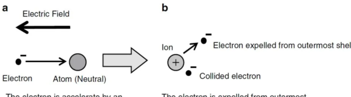
The electron density (𝑛𝑒) and ion density (𝑛𝑖) are extremely equal, and relative to the plasma density. Because within plasma electrons can move freely and have a conductive property. When applied radiofrequency (RF) power to a couple of electrodes in an etch chamber, electric fields are generated by the RF power and electrons are accelerated, acquire kinetic energy, and collide with atoms and molecules (Figure 2a). If the electron kinetic energy is greater than the ionization energy, the electron in the valence band are ejected from the atom or molecule.
Therefore, the neutral atom or the molecule change to ion (Figure 2b). However, the ejected electron from the molecule or the atom, adds to the first colliding electron and hence makes a total of two electrons. These electrons under the electric field are accelerated, collide with other atoms and molecules, and new ions and electrons are generated. The number of ions and electrons increased as in an avalanche and finally exceed a threshold level over which a resulting discharge begins and thus creates a plasma (Nojiri, 2015). This mechanism shows in Figure 3.
Figure 2 The collision of electrons and neutral atoms generate ion (Nojiri, 2015).
Figure 3 Principle of gas discharge (Nojiri, 2015).
The plasma classification is a completely ionized plasma, in which electrons and ions are ionized 100%, and a weakly ionized plasma with lower degree of ionization and hence a mixture of ions, electrons and where neutral atoms and molecules coexist. Glow discharge plasma is a weakly ionized plasma and consists of the same numbers of positive and negative charges, including electrically neutral atoms and molecules. Figure 3 shows a model of glow discharge plasma. In glow discharge plasma the degree of ionization is in the order of 10–6–10–4. In other words, the degree of ionization is around 1 in 10,000 at most. Most of the particles are neutral and contains only one ion and electron for every 10,000 neutral particles.
Consequently, it is called a weakly ionized plasma. At a pressure of 13.3 Pa (100 mTorr) the number of gas molecules is around 3.5 × 1015 cm–3 and the plasma density at an
7 ionization degree of 10–4 is 3.5 × 1011 cm–3. In the glow discharge plasma, the plasma density is within a range of 10–11 cm–3 (Nojiri, 2015).
Figure 4 Model of glow discharge plasma (Nojiri, 2015).
CAPP has been used for disinfection in many ways such as medical equipment, packaging in the food industry, implants, blood coagulation, etc. (Fridman et al., 2008).
CAPP has a high efficiency in eliminating bacteria which is partly due to their easy access into narrow and pent up spaces (Laroussi, 2002). In recent years, CAPP has a temperature lower than 40 oC and CAPP sources have been developed that provide the possibility to extend plasma treatment to soft tissue cells. CAPP can be generated in several way such as RF, microwave frequencies, high voltage AC or DC, etc. Excited species and reactive gases may be generated during the non-equilibrium processes and in plasma medical field these species have particular attention (Kong et al., 2009).
Plasma composes of electrons, positive ions, and neutral particles, and can be described based on the ionization degree, density, thermodynamic equilibrium, and so on; in consequence, plasma is classified in many ways.
2.1.1 Classical plasma
A plasma gas or called classical plasma, for plasma kinetic theory which has classical Boltzmann statistics, if the distance between gas particles is adequately large.
The distance between electrons in a plasma is adequately large if it is larger than the average electron, of mass 𝑚𝑒 and speed 𝑣𝑡ℎ from its thermal energy, de Broglie wavelength (K. Wiesemann, 2013):
𝜆𝐵 = ℎ
𝑚𝑒𝑣𝑡ℎ (1)
The de Broglie wavelength explains particles have wave-like properties, a particle of mass 𝑚 moving at speed 𝑣 will have the properties of a wave of the de Broglie wavelength, where ℎ is the Planck constant.
Furthermore, a plasma can be explained as an ideal gas, when particles only interact elastically, if the mutual potential energy of electrons and ions is smaller than the average kinetic energy:
3
2𝑘𝐵𝑇𝑒 >> 𝑒2
4𝜋𝜀0𝜆𝑒 (2)
If the distance between particles in the plasma large enough is the result to the electrostatic force between particles will be weak. Using the relations of 𝑘𝐵𝑇𝑒
and Debye-Hückel length, 𝜆𝑒 for plasma being in ideal gas state therefore we get the equivalent conditions (K. Wiesemann, 2013):
𝜆𝐷 >> 𝜆𝑒 = 1
𝑛𝑒1/3 (3)
2.1.2 Degree of ionization in plasma
The number of ionized atoms or molecules depend on the degree of ionization (normally the number of electrons 𝑛𝑒 or ions 𝑛𝑖 are equal) as a fraction 𝛼 of the total number 𝑛𝑡 of atoms or molecules:
𝛼 =𝑛𝑒
𝑛𝑖 (4)
The ionization degree is defined as 𝛼𝑖 = 𝑛𝑖/(𝑛𝑖 + 𝑛𝑛), where 𝑛𝑖 is the number density of ions and 𝑛𝑛 is the number density of neutrals. 𝛼𝑖 are determinant factor of the response of any plasma to a magnetic field as well as the electric
9 conductivity of plasma. Plasma with 10−6 < 𝛼𝑖 < 10−1 is weakly ionized. Because the electron temperature in the plasma is determinant to the degree of ionization, weakly ionized plasma is called low-temperature plasma. In common plasma-processing chambers, the degree of ionization is less than 10–4. The degree of ionization of inductively coupled plasma (ICP) and electron cyclotron resonance is a lot higher, about 10–2. Plasma with 𝛼𝑖 ≈ 1 is fully ionized, and is referred to as “hot” plasma or high-temperature plasma. Examples include fusion plasmas, solar wind (interplanetary medium), and stellar interiors (the Sun’s core).
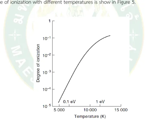
Only a small part of the atoms in an electric discharge turn to be ionized, typically one (1) in 105−106, which at a gas pressure of 133 Pa (1 Torr) corresponds to a particle density of 1022 m−3 and an electron density of 1016 m−3. The variation of the degree of ionization with different temperatures is show in Figure 5.
Figure 5 Characteristic variation of the degree of ionization of an atomic gas at atmospheric pressure (Harry, 2010).
2.1.3 Collisions between particles
Collisions of particles may be elastic, in which case momentum is preserved, or inelastic, where momentum is transferred to potential energy. In both cases, energy has exchanged. The energy to collision and the collision frequency
between atomic particles influence the rate at which energy can be coupled with or released from a plasma, the mass of an electron, 𝑚𝑒, is 9.11 × 10⁻³¹ kg, the nucleus atomic weight of a hydrogen atom (proton), 𝑚𝑝, is 1.67 × 10⁻²⁷ kg.
Figure 6 shows the energy levels of different atomic and molecular transitions. The energy gained by an electron (charge 1.6 × 10⁻¹⁹ C) accelerated through a potential of 1V is 1 eV or 1.6 × 10⁻¹⁹ J. The Maxwell–Boltzmann distribution indicates the small number of particles available with adequate energy for ionization.
Figure 6 Atomic and molecular energy associated with different energy transitions (Harry, 2010).
The electron small mass results to only an insignificant exchange of kinetic energy from electrons in an elastic collision, both particles conserve the magnitude of their momentum, but its direction will be changed.
2.1.4 Plasma parameters
Table 1 shows commonly values of plasma parameters correlated with an arc discharge plasma or strongly ionized plasma, and a glow discharge plasma or a weakly ionized plasma. A glow discharge plasma is characterized by being without
11 thermal equilibrium between the electron temperature (𝑇𝑒) and gas temperature (𝑇𝑔). An electron temperature correlates with the energy of the electrons, and its relationship to the kinetic energy 12𝑚𝑒𝑣𝑒2 is expressed as
1
2𝑚𝑒𝑣𝑒2 = 3
2𝑘𝑇𝑒 (5) Where 𝑚𝑒 is the electron mass, 𝑣𝑒 is the electron velocity, and 𝑘 is
Boltzmann’s constant.
Table 1 Types of plasma and plasma parameters (Nojiri, 2015).
Type of plasma
Plasma density (cm-3)
Electron temperature
𝑇𝑒 (K)
Ion temperature
𝑇𝑖 (K)
Gas temperature
𝑇𝑔 (K) Arc
discharge
Strongly ionized plasma (high-
temperature plasma)
>1014 6,000 6,000 6,000
Glow discharge
Weakly ionized plasma (low- temperature
plasma)
109-1012 ~104 300-1,000 300
Since electrons are very lightweight, electrons are accelerated by an electric field and receive large kinetic energy. The average electron energy in a glow discharge plasma is several electron-volts. Example the electron energy is 2 eV; then the electron temperature 𝑇𝑒 is 23,200 K, according to (5). On the contrary, the temperature of the neutral atoms and molecules has a gas temperature 𝑇𝑔, which is near room temperature (293 K). Otherwise, 𝑇𝑇𝑒
𝑔 is around 80, and the electron temperature 𝑇𝑒 and gas temperature 𝑇𝑔 are not in a thermal or non-thermal equilibrium. While the electrons have an energy level equal to a high temperature of 104 K or higher, the temperature of the etch chamber and the wafer are low because the electrons have small mass. Therefore, a glow discharge plasma is also referred to as a low-temperature
plasma. Because electrons have enough energy that result in the excitation, ionization, and dissociation of atoms and molecules, with the gas temperature remaining near to the room temperature, various types of reactions are possible at low temperature.
An arc discharge is a strongly ionized plasma, and its plasma density is 1014 cm–3 or greater. The electron temperature 𝑇𝑒, ion temperature 𝑇𝑖, and gas temperature
𝑇𝑔 are in a thermal equilibrium, and 𝑇𝑒=𝑇𝑖=𝑇𝑔 is approximately 6,000 K. For this reason, the arc discharge is referred to as a high-temperature plasma (Nojiri, 2015).
2.1.5 Plasma thermal equilibrium
Considering relative temperatures between electrons, ions, and neutrals, plasmas are classified as thermal equilibrium and nonthermal equilibrium.
2.1.5.1 Thermal equilibrium plasma
The electron temperature (𝑇𝑒), ion temperature (𝑇𝑖), and neutral temperature (𝑇𝑛) are identical in thermal equilibrium plasma. This is attributed to the frequent collisions between electrons and ions/neutrals inside high- temperature and high-density plasma. Examples include the natural fusion reactor (Sun), a magnetic field (of tokamak design), or inertial (laser) confinement of a plasma.
2.1.5.2 Nonthermal equilibrium plasma
In nonthermal equilibrium plasma, the momentum transfer between light electrons and heavy particles (ions and neutrals) is not efficient enough and the power applied to plasma favors electrons. Therefore, the electron temperature (𝑇𝑒) is extremely higher than in ions (𝑇𝑖) and neutrals (𝑇𝑛), being, 𝑇𝑒 >> 𝑇𝑖, 𝑇𝑛. Nonthermal equilibrium plasmas are generated by corona discharge, glow discharge, arc discharge, capacitively coupled discharge, inductively coupled discharge, wave heated plasma, etc. Applications of nonthermal plasma have expanded to cover many fields including environmental engineering, aeronautics and aerospace engineering, biomedicine, textile technology, and analytical chemistry.
13 2.1.6 Plasma source
The plasma source comprises of a needle on which the voltage is applied and surrounded by a quartz glass tube. Flow rate of gas normally at 1 to 2 L/min of helium or argon, sometimes with a gas mixture, is flow through the glass tube to force the gas and the plasma out to the air. Since having the high electric field on the needle tip the plasma can be generated either with inert gas, an argon or a helium flow (Hofmann, 2013).
Table 2 shown lists of various reactions that can take place at a surface exposed to a plasma. The first two show etching and deposition processes that are in turn enhanced by the arrival of energy brought by other particles.
Table 3 and Table 4 list phenomena that take place in the gas phase, where particles are ionized, some molecular gases are broken up and others agglomerate (oligomerize). The last process is the first stage in the formation of particulate matter in plasmas.
Table 2 Surface reactions (Braithwaite, 2000).
Reactions Description Evidence
AB+Csolid → A+BCvapour
AB → A+Bsolid
e-+A+ → A A* → A
A* → A+e- (from surface) A+(fast) → A+e-
Etching Deposition Recombination De-excitation Secondary emission Secondary emission
Material erosion Thin film formation Major loss process Auger electrons Auger electrons
Table 3 Gas phase reactions involving electrons (Braithwaite, 2000).
Reactions Description Evidence
e-+A → A+e- e-+A → A++e-+e- e-+A → A*+e- e-+A* → e-+A+hv
Elastic scattering Ionization Excitation De-excitation Two-step ionization
Thermal electrons Conductivity Light emission Ionization efficiency
Reactions Description Evidence e-+A* → A++e-+e-
e-+AB →A+B+e- A++e-+B+e- A-+B e-+A++B → A+B
Fragmentation
Dissociative ionization Dissociative attachment Volume recombination
Residual gas analysis
Plasma decay and steady-state
Table 4 Gas phase reactions involving ions and neutrals (Braithwaite, 2000).
Reactions Description Evidence
A++B → B++A ‘resonant’
for B=A A++B → B+A+ A++B → A++B*+e- A++B → A++B++e- A+B* → A++B+e- A++BC → A++B+C e-+A++B → A+B A±+B → AB± A+B → AB
Charge exchange Elastic scattering Excitation
Ionization
Penning ionization
Fragmentation/dissociation Volume recombination Oligomerization
Oligomerization
Ion energy spectra Ion energy spectra Ionization efficiency Ionization efficiency Ionization efficiency Residual gas analysis Plasma decay Ion mass spectra Residual gas analysis
2.1.7 Atmospheric pressure plasma (APP)
For generated plasma at atmospheric pressure or ambient air can overcome the drawbacks of the low-pressure plasma. However, high voltage for gas breakdown is required and for enhanced collisions between electrons and gas molecules. In avoiding arcing and gas temperatures is low in APP to attain low-temperature sources, several models have been developed via properly scheming electrode configurations and discharge-excitation voltage waveforms for limit discharging duration. Major methodologies for avoiding arch and hot gas include dielectric barrier discharge (DBD) and pulsed voltage or high frequency (HF) voltage was used for excitation of discharge
15 rather than direct current (DC) voltage (Setsuhara, 2016). Another method is to apply a voltage with a high frequency, usually RF (Park et al., 2001) in order to reduce the operation voltage. In most cases these RF plasmas are capacitively coupled plasmas, i.e. generated between two electrodes at radio frequency 13.56 MHz. With this configuration and at these frequencies the current is mainly eradication current, compared to the usually much smaller ohmic current. The last method is to apply the voltage and switch it off before a transition to a spark or arc can emerge, i.e. applying voltages with a duration of pulses of a few hundreds of nanoseconds or less, depending on the electrode geometry.
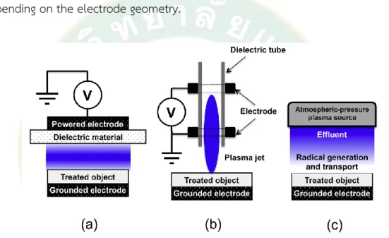
Figure 7 Atmospheric-pressure plasma exposure in biomedical applications; (a) DBD, (b) plasma jet and (c) plasma effluent downstream (Setsuhara, 2016).
APP-irradiation diagram for biomedical applications are shown in Figure 7.
In DBD shows in Figure 7 (a). DBD known as a barrier discharge or a silent discharge, is one type of discharge when at least one of the electrodes is covered by a dielectric material. Layer of dielectric play a role current limiter and shield the formation of arc discharge or a spark. The energy of electrical coupled into a DBD plasma is generally transferred to the electrons, meanwhile the neutral gas remains near to ambient temperatures. DBD system is the non-equilibrium plasma that can be operated at high pressures. The treated object act as a counter electrode (grounded electrode) and the
gap distance is in range of mm or much less than several cm. Hence is suited to treating flat areas of skin. In the DBD type has been used in a wide range of plasma-medicine applications for example sterilization of living animal or human tissue, blood coagulation, wounds healing and skin treatments. However, in the DBD type is restricted by the size of the gap distance between HV electrode and target, and thus plasma sources generated in open space for expanding the flexibility of applications are required.
Other method of open-space exposure has been realized by the plasma- jet in Figure 7 (b). The atmospheric pressure plasma jets (APPJ) are prone to arcing. An arc is a thermal plasma that forms when a current is heated up through the electrodes creating high energy enough. If the temperature at the electrode is high enough more electrons escaping from atom are created due to thermionic emission, which leads to a very high current at low voltage at gas high temperatures. Arcs are very energetic and need to be evaded in many applications. The most significant sources which create non-arcing atmospheric pressure plasmas are dielectric barrier discharges (DBDs), corona discharges and cold atmospheric pressure plasma jets (CAPPJs) (Hofmann, 2013).Plasma is generated in the discharge tube with barrier discharge sustained in flows of noble gases such as He and Ar gas, when flown along dielectric tubes with cylindrical configurations comprise of electrodes and a dielectric tube. In plasma jet, the ionization wave was generated in the discharge tube blown from the discharge tube toward the object. The different designs of atmospheric pressure plasma jets remain (Schutze et al., 1998). The special feature of all of them is that usually inert gas such as helium or argon is blown through a nozzle, dimension size um or mm. In most cases enclosing this nozzle one electrode is connected to a high voltage source around 0.1 to 100 kV and a grounded electrode. The plasma is then usually generated inside the nozzle between these electrodes and blown out via gas flow. To guard arcing in plasma jets three methods can be implemented either separately or in many combinations. One method is to use the geometry of DBD, with the two electrodes being separated by at least one dielectric barrier, such as glass. The advantage of the dielectric barrier is that the charge, which is generated in the volume of the plasma,
17 will be deposited on the dielectric. This leads to a local reduction of the electrical field, which will lead to a self-quenching of the discharge (Hofmann, 2013).
There are several advantages of plasma jets over DBD discharges. DBD discharges, especially in air, are in most cases filamentary and are created randomly over the surface, while a plasma jet forms in most cases a stable diffuse seeing discharge without the need of the surface as a secondary electrode. This makes it possible to treat rough surfaces homogeneously in either a direct contact or an indirect mode, which is a challenge in a DBD geometry. The common disadvantages of plasma jets are the relatively small size of the discharges and the need of (expensive) rare gases. While air jets have also been developed (Kolb et al., 2008).
The plasma-effluent downstream shows in Figure 7 (c). The object is located in the downstream of APP sources, when the effects of the electric filed in region of the treated object can be ineffective, and the effluent of the APP into the open-space, ambient air is source for generation of reactive species. The reactive species generate by the effluent of the plasma source when transported to the objects for treatments (Setsuhara, 2016). As mentioned before, many different kinds of electrode configurations and voltage waveforms can and are used in different plasma research groups. Finally, these plasma sources are different in many ways such as size, rate of gas flow, electrode, etc. A direct contrast of the different plasma sources is in most cases extremely challenging if not impossible without a lot of detailed information of plasma properties (Hofmann, 2013).
2.2 Bacteria
2.2.1 Bacterial cells
One type of prokaryotes are bacteria. Bacteria are simple single cell organisms without cell nucleus or other membrane- bound organelles, unlike eukaryotes. Ordinarily bacteria have a size of around 100 nm and have many shapes, such as rods, spheres, and spirals. The chromosomal DNA of bacteria is a single loop that forms a deformed structure in the cytoplasm known as the nucleoid. Moreover,
bacteria can also carry plasmids, that are small circular DNA molecules providing supplementary genes that are not important to the bacteria, but can assist in their survival in severe environments which are the reasons that make bacteria resistant to antibiotics. All of bacteria cells have a cell membrane in the form of a lipid bilayer. A barrier between the cell interior and cell membrane is known as cytoplasm, and the external world, and molecules can selectively be transported pass it. Furthermore, the lipid membrane, bacterial cells have a cell wall that prevents the cell from pressure produced by osmotic flow of water into the cell (Laroussi et al., 2012).
2.2.2 Staphylococcus aureus
In the 1880s Staphylococcus aureus (S. aureus) was the first time introduced to the scientific community and S. aureus have potential to cause skin and wound infections. Moreover, S. Aureus is a unique bacterium as it has the potential to adapt to for antibiotics resistance leading to the development of methicillin-resistant Staphylococcus aureus (MRSA) in the 1960s. About 25% of population has S. aureus living in the nasal cavities, which being its major reservoir and the most important source for infection. Transportation of S. aureus is affected by genetic and environmental factors, along with cell-wall lipoteichoic acid, hormonal status, and antimicrobial activity of nasal secretions (Boost et al., 2008). Therefore, S. aureus is one of the most poisonous and also dangerous pathogens for human life. S. aureus is the cause of various deep-seated invasive and toxin-mediated illnesses including superficial infections. The broad range of clinical conditions results from a diversity of extracellular components, as well as surface proteins, capsule, biofilm formation, enzymes, and toxins (Petinaki and Spiliopoulou, 2012). MRSA is a major health pathogen problem worldwide which has significantly increased over the last decade.
2.2.3 Pseudomonas aeruginosa
Pseudomonas aeruginosa (P. aeruginosa) is opportunistic pathogen in most cases of humans and animals, leading to severe infections whereby the immune system is compromised (Fergie et al., 1994), or after long time of antibiotic treatments, injuries and medical process (Strateva and Yordanov, 2009) . P. aeruginosa is
19 opportunistic pathogen in gram-negative type ubiquitously present in water and soil.
It has a natural potential to be resistant to penicillin and aminopenicillins, and the cephalosporin of first and second generations. Moreover, it is normally sensitivity to aminoglycosides, fluoroquinolones, lipopeptides, ureidopenicillins, carboxypenicillins, carbapenems and the third generation of cephalosporin. Furthermore, P. aeruginosa can develop resistance to against any antibiotic faster than that we are able to develop antibiotic. (Strateva and Yordanov, 2009).
2.3 Mechanisms of CAPP to bactericidal
The fact that RONS are created by cells and released extracellularly in inflammatory response to infections, tumors, and wounds supports the claim that RONS must be the main active mechanism in CAPP. That is to say RONS are believed to be a part of an intracellular signaling pathway. This class of RONS are called primary species and they are well controlled in the cell and their reactions with biomolecules are reversible. The signaling mechanisms consist of modifications of the intracellular redox state and proteins in signaling pathways by oxidation. One more class of RONS are secondary species and they are toxic and cause irreversible damage by their reactions with biomolecules. The primary RONS comprise of superoxide (O2-), hydrogen peroxide (H2O2), and nitric oxide (NO), and the secondary RONS include hydroxyl (OH) and peroxynitrite (ONOO-). The secondary species only generate in the cell in case primary species react with one another or with a transition metal. The formation of hydroxyl from hydrogen peroxide is catalyzed by iron ions and known as the Fenton reaction. Interestingly, a reaction lead to oxidative damage to pathogen membranes upon immune response, known as lipid peroxidation, the cleavage of lipid peroxides is catalyzed by ferrous ions, which is similar to the Fenton reaction. Another way of forming hydroxyl is through the Haber-Weiss reaction, where O2 is transformed into H2O2 which is then transformed to OH. Peroxynitrite is formed in a reaction between superoxide and nitrogen oxide. The reaction between peroxynitrate and amino acids leads to the formation of nitrated amino acids which causes oxidative damage to proteins (Kong et al., 2009). Therefore, RONS generated in CAPP can both act as an
immune response (primary RONS) and a highly reactive damaging species (secondary RONS).
RONS in CAPP have been reported to have oxidative effects on the outer structures of the cells, that is on the membrane and the cell wall, affecting their structural integrity. Ozone (O3) interferes with cellular respiration, OH (hydroxyl radical) compromises the function of unsaturated fatty acids in the lipid membrane, H2O2
(hydrogen peroxide) has oxidative effect on proteins, lipids, and DNA, and atomic oxygen, metastable oxygen molecules, and superoxide (O2-) can oxidize proteins (Laroussi et al., 2012) . However, the way RONS affect cells depends on their concentration. For example, H2O2 (hydrogen peroxide) can activate cell proliferation or restrain it and induce cell apoptosis and Nitric oxide (NO) which play an important role as an anti-inflammatory agent at low concentration, but at higher concentration it can damage cells (Kong et al., 2009) . When the concentration of RONS are overwhelming to the cell, it is said to be in a state of oxidative stress, damaging lipids, protein, and DNA (Zhang et al., 2016).
As to the metabolic part, ATP (Adenosine triphosphate), the molecule used as the energy currency for life, is the descriptor of the metabolic activity in cells and can be used to observe how bacterial cells in form biofilm react to the stress that follows a CAPP exposure. For short exposure times, bacteria have been shown to try to handle with the stress by either increasing respiration and consequently the ATP production or to uncouple the ATP production from respiration (Joaquin et al., 2009). In a liquid condition, RONS have been shown potential to cause acidification and therefore inactivation of suspended bacteria (Suschek and Opländer, 2016).
Heinlin et al., study in Plasma medicine: possible applications in dermatology.
Experiments shown CAPP have efficient, contact-free and painless disinfection, even in microscopic view without damage to healthy tissue. Plasma have affected the biochemical processes and offer new probability for the selective application of individually designable medical active substances application. For dermatology, new ways are being opened for wound healing, tissue regeneration, therapy of skin disinfection and probably other pathogen (Heinlin et al., 2010).
21 Daeschlein et al., study in Skin decontamination by low- temperature atmospheric pressure plasma jet and dielectric barrier discharge plasma. The APPJ and DBD have a high efficiency in eradicating physiological (PF) and artificially (AF) from the fingertips of healthy participant. Moreover, in the investigation plasma-resistant isolates were not found. CAP has shown to have potential for inactivation skin disease. For the purpose of hand hygiene, the times of plasma exposure should be decreased significantly by technical means (Daeschlein et al., 2012).
Lin et al., study in Ar/O2 Argon-Based Round Atmospheric-Pressure Plasma Jet on Sterilizing Bacteria and Endospores. A round argon-based non-thermal atmospheric- pressure plasma jet (APPJ) for sterilization application was developed and characterized. The APPJ temperature at the tube outlet is lower than 37 °C, so as not to induce damage to normal tissue. The absorbed power of plasma is around 2.7 W.
The OES data shown that APPJ has high intensity of hydroxyl radicals (OH) in glow discharge region. Additionally, this device was applied to eradicate the bacteria (E. coli and B. subtilis) and endospore (B. subtilis endospore). Furthermore, when feeding additional oxygen (0.04%) into working gas it can not only increase the OH radicals but also accelerate the killing of bacteria and endospore (Lin et al., 2016).
Mohd Nasir et al., study in Cold plasma inactivation of chronic wound bacteria.
The researchers used two types of plasma systems to generate cold plasma: a parallel plate dielectric barrier discharge (DBD) and a capillary-guided corona discharge.
Parameters for applied voltage, discharge frequency, exposure time and flow rate of the carrier gas affects the cold plasma chemistry and thus change the composition and concentration of plasma species to reaction with the target sample. Chronic wounds failure to heal, often contaminated by multidrug resistant pathogen, make them resistant to healing. Methicillin-resistant Staphylococcus aureus (MRSA) and P.
aeruginosa are the two major bacteria in infection and clinical non-infection wounds.
The efficiency of cold plasma generated by the two different designs are based on eradication of three different isolates of MRSA and four isolates of P. aeruginosa (Mohd Nasir et al., 2016).
Mai-Prochnow et al., study in Gram-positive and Gram-negative bacteria differ in their sensitivity to cold plasma. The results showed CAP efficiency is direct relative
to the thickness of the cell wall of bacteria in various species. Biofilms of Gram-positive B. subtilis cell wall are around 55.4 nm, have ability resistance to CAP, with less than one log10 reduction after exposure time 10 min. On the other hand, biofilms of Gram- negative P. aeruginosa cell wall are around 2.4 nm and were almost completely eliminated under the same exposure time conditions. Planktonic cultures of Gram- negative P. libanensis also had a higher log10 reduction than Gram-positive S.
epidermidis. Mixed species biofilms of P. aeruginosa and S. epidermidis showed the same trend of Gram-positive bacteria as having ability to resistant treatment of CAP. In addition, when grown in co-culture, Gram-negative P. aeruginosa have higher resistant to CAP than as a mono-species biofilm. OES spectrum found OH and O in the plasma have ability to break bond structure of cell walls. This study showed cell wall thickness being relative to CAP inactivation times of bacteria, but cell membranes and biofilm matrix have also a likely role (Mai-Prochnow et al., 2016).
Kang et al., study in Portable microwave air plasma device for wound healing.
A portable microwave air plasma was developed for safety and effective wound healing process. Generated plasma by a fixed microwave power and two type of gas flows. The result shows the rate of the two air flows define the stability of the plasma jet, gas temperature and including control the concentrations of the reactive species in plasma. Two different system, the NO and ozone system, were identified as acceptable for wound healing without damage from thermal and toxicity. These systems show the same plasma characteristics excluding different concentration of NO and ozone. Both systems show a more than double faster in wound healing process when compared with the untreated case. Speedier potential healing process especially with ozone safety make the NO abundant system the best operation condition for wound healing. The performance of the developed device was monitored by a one- hour continuous generated plasma with a 24 V battery (Kang et al., 2015).