A STUDY TO DETERMINE THE PREVALENCE OF SIGNS AND SYMPTOMS OF CARPAL TUNNEL SYNDROME AND DE QUERVAINS TENOSYNOVITIS IN GARMENT WORKERS IN THE eTHEKWINI DISTRICT OF
KWA-ZULU NATAL
SUBMITTED IN PART FULFILMENT FOR MASTERS DEGREE IN HAND REHABILITATION
RESEARCHER: PRABASHNI PILLAY
(B. PHYSIOTHERAPY)
SUPERVISOR: MRS P. RANGIAH
(B. PHYSIOTHERAPY; HED; M PHYS T)
Author’s Declaration
I, Miss Prabashni Pillay declare that ‘A Study to Determine the Prevalence of Signs and Symptoms of Carpal Tunnel Syndrome and de Quervains Tenosynovitis in Garment Workers,’ is my own work and that all sources that were used or quoted have been indicated by means of complete references. This study has not been submitted in any form to another university or institution.
______________________ __________________________
Miss P. Pillay Date
Acknowledgements
• Department of Occupational Therapy of University of Kwa-zulu Natal
• Managing Directors and all garment workers
• Mrs P. Rangiah, my supervisor, who offered her time and expertise willingly
• Mrs S.D. Naidoo, my manager, for her patience and motivation throughout my research
• Mrs P. Pillay, my mum, a garment worker for 36 years, who dedicated her time and knowledge in helping me understand the garment industry
• My family, friends and colleagues for their support and assistance
Glossary and List of Abbreviations
PREVALENCE: that which exists or is very common at a particular time or in a particular place.
INCIDENCE: the extent to which something happens or has an effect.
CARPAL TUNNEL SYNDROME: Nerve entrapment involving compression of the median nerve at the wrist causing sensory symptoms typically involving the thumb, index, middle and radial half of the ring finger.
De QUERVAINS TENOSYNOVITIS: Inflammation of the tendon sheath of the thumb attributed to excessive friction between the abductor pollicis longus and extensor pollicis brevis tendons and their common sheath, usually caused by twisting and forceful gripping motions with the hands.
MEDIAN NERVE: A major nerve of the hand that dominates the thumb, index and ring finger.
CARPAL TUNNEL: The osseofibrous passage for the median nerve and the flexor tendons, enclosed by the flexor retinaculum and carpal bones of the hand.
REPETITIVE STRAIN INJURIES (RSI’s), CUMULATIVE TRAUMA DISORDER (CTD):
Synonymous terms for disorders caused by prolonged, repetitive tasks.
MFL: Manitoba Federation of Labour CM: centimetres
%: percent
UKZN: University of Kwa-Zulu Natal
OSHA: Occupational Safety and Health Administration AAOS: American Academy of Orthopaedic Surgeons
ABSTRACT
Introduction: Garment work is repetitive and detailed and requires constant use of the hands. It is no surprise that garment workers are at high risk for developing repetitive strain injuries (RSI’s) (MFL Occupational Health Centre, 1999). Work-related upper limb disorders, popularly known as RSI’s, affect over 370,000 people in Great Britain with 86,000 new cases recorded in 2010.
This costs employers almost £300 million in lost working time, sick pay and administration (The Chartered Society of Physiotherapy, 2007). There is however no statistics documented on RSI’s among garment workers found for South Africa. Aim: To determine the prevalence of signs and symptoms of carpal tunnel syndrome and de Quervains tenosynovitis. Method: A study using quantitative data was used. A validated questionnaire consisting of open-ended and closed questions was utilized. Data was collected from two hundred subjects of varying age, gender and ethnic group. Information on signs and symptoms and possible risk factors of RSI’s were obtained. The visual analogue scale was used to assess pain, a goniometer to measure active range of movement, the Phalens test, Reverse Phalens test and Finkelsteins test was used to assess the signs and symptoms of the two occupational repetitive strain disorders. Data analysis:
All data was captured and analysed using the Statistical Package for Social Sciences (SPSS version 15). Descriptive statistics such as mean, standard deviation, proportions, median, mode and interquartile range was used to summarize the data. Pearson’s Chi Square tests and Fishers Exact tests were used to test for association between two categorical variables. Independent Samples t-tests were used for the difference in age distribution between participants that presented with carpal tunnel syndrome and de Quervains tenosynovitis and of those who did not present with them. The level of significance was set at 0.05. Bar graphs, tables and pie charts were used to depict the results. Results/Discussion: The results of this study indicated that 59%
of participants presented with signs and symptoms of de Quervains tenosynovitis and 63% of participants presented with signs and symptoms of carpal tunnel syndrome. The prevalence of carpal tunnel syndrome and de Quervains tenosynovitis was 42% and 43% respectively among garment workers in the eThekwini district. In addition, 100% of participants stated that they work under the following conditions, applying weight through the arms, repeated movement, work with their arms in unsupported positions, fast hand movements and holding or grasping for more than 2 hours continuously per day. Seventy two and a half percent of participants stated that their work entailed using vibratory tools for prolonged hours. Pearson’s Chi Square tests
showed no association of use of vibratory tools to de Quervains tenosynovitis (P=0.666) or to carpal tunnel syndrome. This is inconsistent with the findings of the study completed by Leclerc et al. (1998) who stated that different dimensions of exposure to physical workload are widely recognised as risk factors. These risk factors include rapid hand motions, repetitive bending and twisting of the hands and the wrist, fast work pace, repetitive grasping with the fingers, mechanical stress at the base of the palm and the palm and the use of vibratory tools (Leclerc et al. 1998). Conclusion: This study has identified the prevalence of signs and symptoms of carpal tunnel syndrome and de Quervains tenosynovitis among garment workers. It has also shown that a significant percentage of garment workers presented with symptoms of burning, tingling, itching and numbness in their hands as well as feelings of swollen and ‘useless’ hands. A significant number presented with functional limitations to certain activities of daily living suggestive of the presence of carpal tunnel syndrome. De Quervains tenosynovitis was indicated when a significant number of participants presented with pain, tenderness or swelling over the radial aspect of the wrist as well as functional limitations to certain activities of daily living.
Table of Contents
Page No.
Chapter 1 Introduction
1.1 Background and rationale 1-3
1.2. Aims 4
1.3. Objectives 4
1.4. Significance 4
Chapter 2
Literature Review
2.1. Introduction 5
2.2. Prevalence of repetitive strain injuries 5-7
2.3. Causative factors of repetitive strain injuries 7-9
2.4. Carpal tunnel syndrome
2.4.1. Definition 10
2.4.2. Signs and symptoms 10-11
2.4.3. Prevalence 11
2.4.4. Diagnosis 11-15
2.4.4.1. The Goniometer for the measurement of range of movement 15-16 2.4.4.2. The Dynamometer for measurement of grip strength of the hand 16
2.4.4.3. Specific tests to reproduce carpal tunnel symptoms 16
2.4.4.4. Electro-diagnostic tests 16-17
2.4.5. Management 17
2.4.5.1. Non-operative treatments 17-18
2.4.5.2. Surgery 18-19
2.4.6. Prevention of carpal tunnel syndrome 19
2.4.7. Alternative research related to carpal tunnel syndrome 19-21 2.5. De Quervains tenosynovitis
2.5.1. Definition 21
2.5.2. Anatomy 21-22
2.5.3. Pathology 22
2.5.4. Causes 22-23
2.5.5. Signs and symptoms 23
2.5.6. Diagnosis 23-24
2.5.7. Treatment 24-25
2.6. Conclusion 25
Chapter 3
Methodology
3.1. Research design 26
3.2. Sample
3.2.1. Selected garment factories in eThekwini 26
3.2.2. Population 26
3.3 Inclusion and exclusion criteria
3.3.1. Inclusion criteria 26
3.3.2. Exclusion criteria 26-27
3.4. Data gathering instruments 27-29
3.4.1. Questionnaire 27
3.4.2. Pain 28
3.4.3. Sensation 28
3.4.4. Range of motion 28
3.4.5. Grip strength 28-29
3.4.6. Clinical tests
3.4.6.1. Phalens test 29
3.4.6.2. Reverse Phalens test 29
3.4.6.3. Finkelsteins test 29
3.5. Procedures 29-35
3.6. Ethical considerations 35
3.7. Data analysis 36
Chapter 4 Results
4.1. Introduction 37
4.2. Socio-demographic characteristics of the participants 37
4.2.1. Demographic details of the participants 37
4.2.2. Participants hobbies 38
4.2.3. Participants sports activities 38
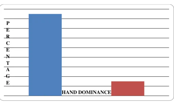
4.2.4. Participants hand dominance 39
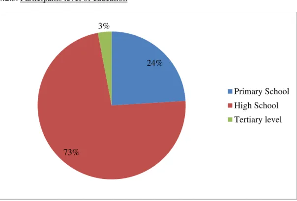
4.2.5. Participants level of education 39-40
4.3. Present and past medical history 40
4.4. Participants occupational details 40
4.4.1. Participants employment history 41
4.4.2. Training experience 41
4.4.3. Participants hours of work including break 41-43 4.4.4. Participants total number of hours of work per day 43 4.4.5. Number of garments produced per hour by all garment workers 44
4.4.6. Participants work setting 44-45
4.4.7. Health and safety in the work place 45
4.5. Participants signs and symptoms 45 4.5.1. Pain
4.5.1.1. Participants pain perception using the visual analogue scale (VAS) 45-46
4.5.1.1. Participants location of pain 47
4.5.2. Sensation 47-48
4.5.3. Range of movement 48
4.5.4. Functional abilities
4.5.4.1. Participants performance on specific activities of daily living
relevant to carpal tunnel syndrome 48-49
4.5.4.2. Participants performance on specific activities of daily living relevant to de Quervains tenosynovitis 49-50 4.5.5. Carpal tunnel syndrome
4.5.5.1. Symptoms of carpal tunnel syndrome 50-51
4.5.5.2. Symptoms post Phalens test 51
4.5.5.3. Symptoms post Reverse Phalens test 52
4.5.5.4. Phalens test 53
4.5.5.5. Reverse Phalens test 53-54
4.5.6. De Quervains tenosynovitis
4.5.6.1. Symptoms de Quervains tenosynovitis 54
4.5.6.2. Symptoms post Finkelsteins test 55
4.5.6.3. Finkelsteins test 55-56
4.5.6.4. Bilateral carpal tunnel syndrome and de Quervains tenosynovitis 56 4.5.6.5. Carpal tunnel syndrome and de Quervains tenosynovitis 57 Chapter 5
Discussion
5.1. Socio-demographic characteristics of the participants 59-60 5.2. Prevalence of signs and symptoms of carpal tunnel syndrome and de Quervains
tenosynovitis 60-62
5.3. External risk factors 62-65
5.4. Functional limitations when performing certain activities of daily living 65-66
Chapter 6
Conclusion, limitations and recommendations
6.1. Introduction 67
6.2. Conclusion 67
6.3. Limitations 67-68
6.4. Recommendations for further studies 68
References 69-73
List of Figures
Page No.
Figure 1: Compression of the median nerve (Bartholet, 2001) 10 Figure 2: De Quervains tenosynovitis of the first extensor compartment
(Griffin, 2005) 21
Figure 3: The mucous sheaths on the posterior aspect of the hand
(Kasdan, 1999) 22
Figure 4: Finkelsteins test
(American Academy of Orthopaedic Surgeons, 2007) 23
Figure 5: Approximate dermatomes and axial lines for the right upper limb
(Tomberlin and Saunders, 2001) 31
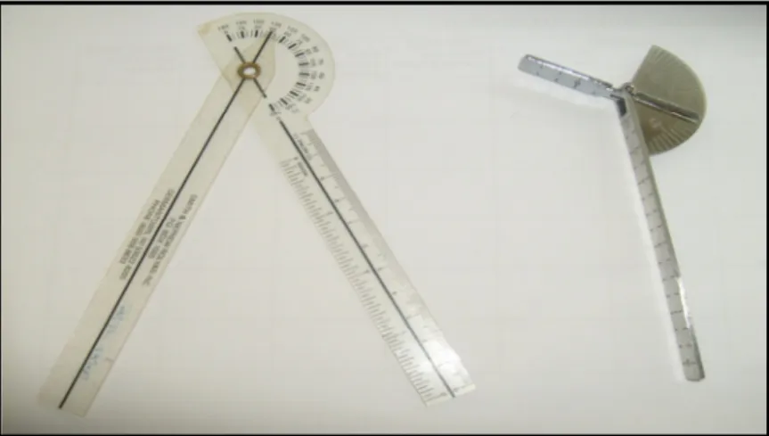
Figure 6: The wrist and digit goniometer 32
Figure 7: Goniometry of wrist extension 32
Figure 8: Goniometry of wrist flexion 32
Figure 9: The Phalens test 34
Figure 10: The Reverse Phalens test being performed by a subject to try and reproduce symptoms of carpal tunnel syndrome 34 Figure 11: The Finkelsteins test in order to reproduce symptoms of de Quervains
tenosynovitis (Kasdan, 1999) 35
Figure 12: The Finkelsteins test in order to reproduce symptoms of de Quervains
tenosynovitis (Kasdan, 1999) 35
Figure 13: Bar graph illustrating participants’ hobbies 38 Figure 14: Bar graph illustrating participants’ hand dominance 39
Page No.
Figure 15: Pie chart illustrating participants’ highest level of education 39 Figure 16: Bar graph illustrating the number of years participants’ worked
in the current position 41
Figure 17: Pie chart illustrating the number of hours worked per day 43 Figure 18: Bar graph illustrating the number of garments produced per hour 44 Figure 19: Bar graph illustrating participants’ sensory deficits 47 Figure 20: Bar graph illustrating participants’ performance on activities of daily
living relevant to carpal tunnel syndrome 48
Figure 21: Bar graph illustrating participants’ performance on specific activities
of daily living relevant to de Quervains tenosynovitis 49
Figure 22: Bar graph illustrating participants’ symptoms of carpal tunnel syndrome 50 Figure 23: Bar graph illustrating participants’ symptoms post Phalens test 51 Figure 24: Bar graph illustrating participants’ symptoms post Reverse Phalens test 52 Figure 25: Bar graph illustrating participants’ results of carpal tunnel syndrome
post Phalens test 53
Figure 26: Bar graph illustrating participants’ results of carpal tunnel syndrome
post Reverse Phalens Test 53
Figure 27: Bar graph illustrating participants’ symptoms of de Quervains
tenosynovitis 54
Figure 28: Bar graph illustrating participants’ symptoms post Finkelsteins test 55 Figure 29: Bar graph illustrating participants’ results of de Quervains
tenosynovitis post Finkelsteins test 55
Page No.
Figure 30: Bar graph illustrating participants’ results of bilateral carpal tunnel
syndrome and de Quervains tenosynovitis 56
Figure 31: Bar graph illustrating participants’ results of carpal tunnel syndrome and
de Quervains tenosynovitis 57
List of Tables
Page No.
Table 1: Prevalence of musculoskeletal disorders in selected occupations
(The Chartered Society of Physiotherapy, 2007) 5-6
Table 2: Normal values for measurement of range of motion of the wrist and thumb
(Trombly and Radomski, 2001) 15
Table 3: Demographic details of participants 37
Table 4: Participants’ role in manufacturing the garment 40 Table 5: Working hours with breaks and activity during breaks (morning session) 42 Table 6: Working hours with breaks and activity during breaks
(mid – morning session) 42
Table 7: Working hours with breaks and activities during breaks (afternoon session) 43 Table 8: Percentage of participants’ working under the following setting 45
Table 9: Pain using the visual analogue scale 46
Table 10: Visual analogue scale readings versus norms 46
Table 11: Location of pain 47
List of Appendices
Page No.
1a. Letter to SNT Fashions 74-75
1b. Letter to Grand Uniform 76-77 1c. Letter to Image Embroidery 78-79 1d. Letter to HIS Clothing 80-81 1e. Letter to TCS Cutting Services 82-83 1f. Letter to RS Fashions 84-85 2a. Consent form 86-87 2b. Consent form (Zulu) 88-89 3a. Questionnaire 90-101
3b. Questionnaire (Zulu) 102-114
4. Ethical clearance 115
5a. Information and exercise booklet – Carpal tunnel syndrome 116-118 5b. Information and exercise booklet – Carpal tunnel syndrome (Zulu) 119-121 6a. Information and exercise booklet – de Quervains tenosynovitis 122-124 6b. Information and exercise booklet – de Quervains tenosynovitis (Zulu) 125-127
Chapter 1 Introduction 1.1. Background and rationale
Carpal tunnel syndrome is the most common nerve compression disorder of the upper extremity. It affects 1% of the general American population and 5% of the working population who are subjected to repetitive use of their hands and wrists in daily living (Concannon et al. 2000). According to Foye (2010), de Quervains tenosynovitis is a common condition of the hand characterized by a localized swelling at the base of the thumb and thickening of the fibrous sheath or retinaculum. De Quervains tenosynovitis is relatively prevalent especially among individuals who use their hands to perform repetitive activities.
Following a thorough search there has been no statistics on the prevalence of carpal tunnel syndrome and de Quervains tenosynovitis in garment workers in South Africa therefore it has become evident that a gap exists for this country regarding these conditions and there is a necessity to obtain data regarding the prevalence of repetitive strain injuries to the hand.
The garment industry is a booming and vibrant industry in South Africa, with South Africa competing amongst various famous brand names. Due to increasing fashion trends many chain stores in South Africa import their garments from other countries. However, due to the great demand of clothing in South Africa, a large number of clothing stores distribute their garments to surrounding manufacturing companies. There are numerous garment manufacturing factories in the eThekwini District of Kwa-Zulu Natal. Large scale manufacturing garment factories have approximately one to two hundred garment workers and small scale factories consist of less than fifty garment workers.
The garment manufacturing industry in Kwa-Zulu Natal is known to be manned by the unskilled industry of the minimally educated or uneducated people. It is noted by many people as one of the most common industries to obtain employment as no experience or qualification is required. There is no training or medical screening offered to employees prior to employment. Most factories rely on a first aid kit if occupational injuries occur. According to recent anecdotal media coverage surrounding the garment industry in South Africa this statement is true as several factories no longer belong to the Bargaining Council, a union that offers support to the garment workers’, as this trade is slowly becoming extinct due to the importing of garments from other countries. Factory owners are now subjected to rely on
minimal services in order to sustain their businesses. The work environment in the majority of manufacturing factories is unsafe and unhealthy (Parimalam et al. 2006), namely, poorly designed workstations, unsuitable furniture, lack of ventilation, inappropriate lighting, excessive noise, insufficient protection from dangerous chemicals, insufficient safety measures in fire emergencies and lack of personal protective equipment. Employees working in such a poor or substandard environment are more prone to occupational injuries (Parimalam et al. 2006).
Are the garment workers in South Africa at risk of developing upper extremity repetitive strain injuries? Are they able to identify early signs and symptoms of repetitive strain injuries and seek help?
Repetitive strain injuries (RSI) are defined as cumulative trauma disorders resulting from prolonged repetitive, forceful or inappropriate movements of the hands. These movements result in damage to muscles, tendons and nerves (Nainzadeh et al. 1999). Occupational repetitive strain injury is becoming a worldwide concern and it is especially serious when it affects our economy. The Chartered Society of Physiotherapy (2007) stated that there were 86 000 new cases of work related upper limb disorders recorded in 2006 in Great Britain.
This cost employers in Great Britain £300 million in lost working time, sick pay and administration. A plan obviously needs to be formulated before the effects of these hand conditions become irreversible.
Occupation-related injuries to the upper limb are increasing daily throughout the world (Leung et al. 2000).Hong Kong, like other cities, has reported a high incidence of occupational hand injury. It was noted that in one month in a 1500 bed hospital, 50% of the injuries were industrial injuries to the hand (Leung et al. 2000). Many of these disorders are suspected of being caused by physical work activities, for example, jobs that involve repetitive movements, heavy physical work and awkward postures (Palmer et al. 2009).
Furthermore, according to the Occupational Safety and Health Administration (OSHA), poor ergonomics in the workplace is also known to be a causative factor of these injuries (Occupational Safety and Health Administration, 2000). The Occupational Safety and Health Administration is an organization that is a division of the United States Department of Labour. The Occupational Safety and Health Administration’s administrator is answerable to the Secretary of Labour who is a member of the cabinet of the President of the United States of America. The function of the Occupational Safety and Health Administration is to ensure
safe and healthy working conditions for working citizens by establishing and implementing standards and by providing training, outreach, education and assistance.
Parimalam et al. (2006) stated that the work environment in the garment manufacturing units in Madurai City is unhealthy and unsafe for the workers, resulting in several health problems.
Ergonomic interventions have been suggested which will eventually help to improve the work environment and also to overcome the health problems (Parimalam et al. 2006). This gives researchers an insight that a problem exists and further research needs to be conducted in order to assist garment workers with possible solutions to promote good health. According to Ranney et al. (1995), following a physical assessment of the 146 female employees in highly repetitive job industries, 54% were found to have musculoskeletal disorders of the upper limb. Carpal tunnel syndrome was the most common form of neuritis with 16 people affected, 7 of whom presented bilaterally with the condition. De Quervains tenosynovitis and wrist flexor tendinitis were the most commonly found disorders in the distal forearm.
A thorough literature search on repetitive strain injuries in South Africa has revealed that there have been no studies on the prevalence of repetitive strain injuries, namely, carpal tunnel syndrome and de Quervains tenosynovitis among garment workers and the possible risk factors associated with it. The choice of conditions for this study was selected as minimum studies and no statistics were obtained on carpal tunnel syndrome and de Quervains tenosynovitis among garment workers in South Africa. Garment production involves the performance of monotonous, highly repetitive and high speed tasks often requiring non- neutral and awkward postures. These exposures place garment workers at risk of work- related musculoskeletal disorders (Herbert and Plattus, 2010). The most common repetitive strain injuries are carpal tunnel syndrome, de Quervains tenosynovitis, epicondylitis and rotator cuff tendinitis. These upper extremity conditions can be clinically diagnosed using specific tests and identifying symptoms thereof (O’Neil et al. 2001).
1.2. Aims
The aim of this study was to determine the prevalence of signs and symptoms of two common occupational repetitive strain disorders of the hand in garment workers, namely, carpal tunnel syndrome and de Quervains tenosynovitis.
1.3. Objectives
The objectives of this study are:
1. To compare the prevalence of signs and symptoms of carpal tunnel syndrome to that of de Quervains tenosynovitis.
2. To determine if subjects’ present with typical signs and symptoms of carpal tunnel syndrome and de Quervains tenosynovitis.
3. To identify possible external risk factors for carpal tunnel syndrome and de Quervains tenosynovitis.
4. To determine whether garment workers present with any functional limitations when performing specific activities of daily living.
1.4. Significance
This study will provide researchers, industrial workers, garment workers and employers with insight into the proportion of clothing workers’ presenting with signs and symptoms of carpal tunnel syndrome and de Quervains tenosynovitis. The study was intended to bring about awareness amongst garment workers about overuse injuries. It has also reflected the need for intervention with regards to workplace modifications and treatment of the conditions in order to promote a safe, pain free, healthy and productive working environment.
Chapter 2 Literature Review 2.1. Introduction
Cumulative trauma disorders due to performance of repetitive tasks account for more than fifty percent of all occupational illnesses in the United States today (Rempel et al. 1992).
These repetitive movements result in damage to the muscles, tendons and nerves. Repetitive strain injuries are also referred to as repetitive stress injuries, repetitive motion disorders and occupational overuse injuries. They may be referred to as well-defined disorders such as carpal tunnel syndrome (CTS) and tendinitis (Nainzadeh et al. 1999).
A variety of primary and secondary sources (textbooks and journal articles) was consulted.
The literature search was dated from 1980 - 2010. Google Scholar, UKZN Online Libraries, Science Direct, PubMed, and Medline online databases were searched using the following keywords: repetitive strain injuries, hand inflammatory conditions, overuse injuries in the workplace, carpal tunnel syndrome and de Quervains tenosynovitis.
2.2. Prevalence of repetitive strain injuries
Table 1 below illustrates the estimated prevalence and rates (%) in Britain of self-reported musculoskeletal disorders mainly affecting the upper limbs or neck caused or made worse by current or most recent jobs for people working in the last 12 months (The Chartered Society of Physiotherapy, 2007).
Table 1: Prevalence of musculoskeletal disorders in selected occupations in Britain (The Chartered Society of Physiotherapy, 2007)
Occupation description Rate per 100 employed in the last 12 months (%)
Prevalence
Process, plant and machine operatives 1.1 24,000
Skilled trades occupations 0.91 30,000
Associate professional and technical occupations
0.76 30,000
Personal service occupations 0.76 17,000
Sales and customer service occupations
0.50 12,000
Administrative and secretarial occupations
0.48 18,000
Elementary occupations 0.45 16,000
Managers and senior officials 0.36 15,000
Professional occupations 0.32 11,000
All occupations 0.60 173,000
The annual prevalence in Britain is the estimated number of people with a work-related illness at any time during the 12-month period. It includes the full range of illnesses from long standing to new cases. The rate is the prevalence estimate divided by the population at risk of having a work-related illness. The Health and Safety Executive (HSE) in Britain reports that for the major occupational groups where the sample numbers were large enough to provide reliable estimates, process, plant and machine operatives and skilled trade occupations carried rates which were statistically significantly higher than the overall rate 0.60% (The Chartered Society of Physiotherapy, 2007). Occupational groups with statistically significantly higher rates were: health and social welfare associate professionals;
process, plant and machine operatives and skilled construction and building trades.
Occupational groups with statistically significant lower rates were professional occupations, managers and senior officials (The Chartered Society of Physiotherapy, 2007).
The United States Bureau of Labour Statistics estimates that more than 332 000 cases of disorders were caused by repeated trauma in 1994. From 1984-1994, the incidence rate increased from 5.1 to 39 cases per 10 000 full time workers. In the workplace, upper extremity repetitive strain injury (RSI) does outnumber lower extremity injuries. In 1989, the total United States Workers’ Compensation costs for upper extremity cumulative trauma
injuries which is a disorder caused by prolonged, repetitive tasks was estimated to be 563 million dollars. Workers considered at high risk for repetitive strain injuries include garment workers, construction workers, meat processors and grocery checkers (Nainzadeh et al.
1999). Following a thorough search for prevalence of repetitive strain injury amongst garment workers in South Africa, it has been established that there has been no studies conducted regarding this area.
2.3. Causative factors of repetitive strain injuries
Repetitive strain injury (RSI) affects a large percentage of the workforce. People develop repetitive strain injuries because they do not know how to or are unable to protect their muscles, nerves and tendons. The workstation may not be set up properly; they may have never been trained to use their hands correctly, or they may be unable to pace themselves (Nainzadeh et al. 1999). The epidemiological literature indicates that the greater the level of exposure to a single risk factor or combination of factors leads to a greater the risk of having a work related musculoskeletal disorder. The literature by the Occupational Safety and Health Administration also indicates that an important factor is the time between each episode of exposure. With adequate time to recover or adapt, and particularly when lower forces are involved, there may be less harm to the body from repeated exposures. The intensity as well as the extended length of the exposure to forceful, repetitive work plays a substantial role in the risk of work related musculoskeletal disorders in many traditional occupational settings (Occupational Safety and Health Administration, 2000).
In workplaces with high rates of work-related musculoskeletal disorders there is little scientific evidence that the principal reason for the excess number of injuries or illnesses is a result of the workers’ psychological reaction to their workplace. However, there is evidence particularly in office settings, suggesting that both physical and psychosocial factors may be important contributors to musculoskeletal disorders (Occupational Safety and Health Administration, 2000). Repetitive strain disorders related to an employee’s occupation occur when the physical capabilities of the employee do not complement the physical requirements of the job. Prolonged exposure to ergonomic risk factors can lead to musculoskeletal disorders (Occupational Safety and Health Administration, 2000).
Conditions that are likely to cause musculoskeletal problems include the following:
• Exerting excessive force in repetition on joints and structures can eventually cause wear and tear of the joints and structures involved thereby causing pain, muscle spasm and inflammation to surrounding structures which will lead to decrease in range of motion of joints and hinder daily activities;
• Excessive repetition of movements can increase blood flow to the area for example, the hands, and cause inflammation thus resulting in tendonitis which could lead to compression of nerves;
• Inappropriate postures, or unsupported positions for prolonged periods can cause compression of nerves, adopting poor postures thus causing musculoskeletal problems;
• Static postures, or positions that an employee must maintain for long periods of time during working hours, can restrict blood flow to other areas of the body thereby causing damage to muscle. These muscles will be restricted of blood flow as well as oxygen for muscles and organs as well as nerves and other structures to function adequately;
• Motion, such as increased speed or acceleration when bending or twisting, can increase the amount of force exerted on the body thus causing strain to muscles which can lead to an inflammatory process;
• Compression of structures in the hand, from grasping sharp edges like tool handles, can concentrate force on the joints of the hand, reduce blood flow and nerve transmission, and damage tendons and tendon sheaths;
• Inadequate recovery time due to overtime, lack of breaks, and failure to vary tasks may leave insufficient time for tissue repair. Tissues need sufficient time to recover from the constant stresses of work practices and can therefore be damaged if healing is delayed or incomplete;
• Excessive vibration, usually from vibrating tools, can decrease blood flow, cause compression of nerves, and contribute to muscle fatigue. Muscle fatigue is due to firm, constant grasping of these tools which are continuously vibrating causing the involved muscles and surrounding structures to be in motion.
• Working in cold temperatures can adversely affect a worker’s co-ordination and manual dexterity and cause a worker to use more force than necessary to perform a task. Cold impairs the performance of complex mental tasks. Manual tasks are also impaired because the sensitivity and dexterity of fingers are reduced in the cold. At even lower temperatures, the cold affects the deeper muscles resulting in reduced muscular strength and stiff joints.
These risk factors, either alone or in combination are likely to subject employees to numerous repetitive twisting, forceful, or flexing motions during a typical workday. To contribute to musculoskeletal disorders these risk factors must be present for a sufficient duration, frequency or magnitude (Occupational Safety and Health Administration, 2000).
2.4. Carpal tunnel syndrome 2.4.1. Definition
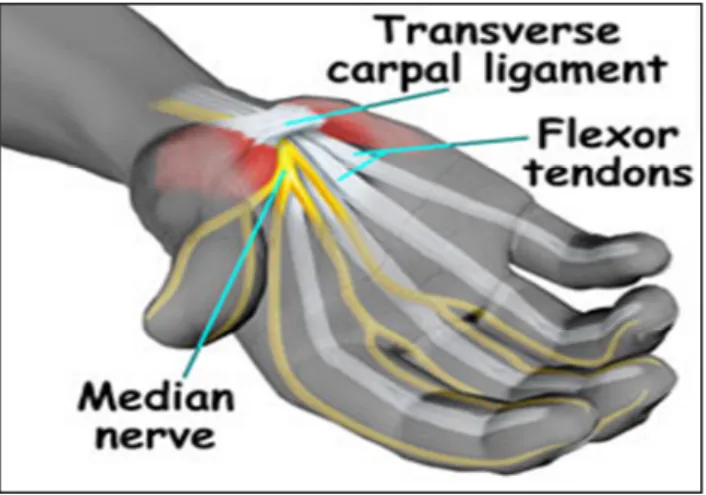
Carpal tunnel syndrome is defined as compression of the median nerve as it passes through the carpal tunnel in the wrist (Beers and Berkow, 1999). The carpal tunnel is a space located posterior between the carpal bones of the wrist (from left to right: trapezium, trapezoid, capitates and hamate), forming the hard ‘carpal floor’ and anterior where the transverse ligament forms a fibrous sheath or the ‘carpal roof’. The tunnel is filled with the flexor tendons of the hand and the median nerve. The median nerve originates from numerous spinal nerves from C5 to T1. It has both sensory and motor functions of the thumb, first, second and lateral aspect of the third digits. Consequently, it is vital for not only grip but also for sensory inputs related to hand function (Scanlon and Maffei, 2009).
Figure 1.Compression of the median nerve (Bartholet, 2001) 2.4.2. Signs and symptoms
Symptoms usually start gradually, with frequent burning, tingling, itching and numbness in the palm of the hand, thumb, first, second and lateral aspect of the third digits, especially the thumb and the index and middle fingers. Some carpal tunnel subjects state that their fingers feel “useless and swollen”, even though little or no swelling is apparent. The affected individual usually first experiences sensory changes associated with carpal tunnel syndrome at night. A person with carpal tunnel syndrome may wake up feeling the need to "shake out"
the hand or wrist. As symptoms worsen, people might feel tingling during the day (National Institute of Health, 2002). Decreased muscle strength in the hand is a late sign and is common for those with moderate to severe carpal tunnel syndrome. Weakness in the hand may make it
difficult to form a fist, grasp small objects, or perform other manual tasks. In chronic and/or untreated cases, the muscles of the thenar eminence may waste away (Scanlon and Maffei, 2009).
2.4.3. Prevalence
Women are three times more likely than men to develop carpal tunnel syndrome, perhaps because the carpal tunnel itself may be smaller in women than in men (National Institute of Health, 2002). The dominant hand is usually affected first and produces the most pain.
Persons with diabetes mellitus or other metabolic disorders that directly affect the body's nerves and make them more susceptible to compression are also at high risk (National Institute of Health, 2002). Neuropathies are the most common complication of diabetes mellitus affecting up to 50% with type 1 and 2 diabetes mellitus (Lin et al. 2011). The diabetes association is due to the fact that when blood glucose levels are elevated, the proteins in the tendons of the carpal tunnel become glycosylated; that is, glucose attaches to the tendon proteins, inflaming them and forming an organic adhesive that makes the tendons less capable to slide without restraint. If one is susceptible to carpal tunnel syndrome because of diabetes or other conditions, the condition may be brought out or exacerbated by repeated forceful flexing of the hands and wrists (Wartburg, 2007).
The risk of developing carpal tunnel syndrome is not confined to people in a single industry or job, but is especially common in those performing assembly line works - manufacturing, sewing, cleaning, and meat, poultry, or fish packaging (National Institute of Health, 2002).
During 1998, an estimated three of every 10,000 workers in the United States lost time from work because of carpal tunnel syndrome. Half of these workers missed more than 10 days of work. The average lifetime cost of carpal tunnel syndrome, including medical bills and lost time from work, is estimated to be about $30,000 for each injured worker (National Institute of Health, 2002).
2.4.4. Diagnosis
Early diagnosis and treatment are important to avoid permanent damage to the median nerve (Tuen, 2007). A clinical assessment is separated into a history and physical examination.
During the history examination carpal tunnel syndrome patients’ often describe diffuse, poorly localized aching that can involve the entire hand and forearm. Many patients report that the hand “falls asleep” with the exception of the little finger. Some patients also describe
weakness, clumsiness, dry skin, coldness, swelling and or changes in the hand. Symptoms are more common during activities requiring the wrist to adopt a flexion or extension posture and discomfort is aggravated by driving, holding a phone, book or a newspaper. Patients often note that symptoms are relieved partially by changes in the hand posture or ‘shaking the hand’ (Tuen, 2007). During the physical examination, provocative tests such as the Tinel sign where paresthesias are provoked by tapping over the median nerve at the wrist, Phalen maneuver where while holding the wrist flexed, paresthesia occurs within 1-2 minutes and direct compression tests are useful. During the motor examination the hand is inspected and examined for muscle atrophy (Tuen, 2007). Dry skin may be visible on digits 1, 2 and 3. The strength of thumb abduction and opposition is tested. Two-point discrimination may be affected before pain and temperature sensation. Even in severe cases of carpal tunnel syndrome, sensation over the thenar area usually is spared, as it is innervated by the palmar cutaneous sensory branch (a median nerve branch that arises proximal to it but does not pass through the carpal tunnel) (Tuen, 2007).
Pain in the hand and wrist is often the main reason patients seek help for their condition. It is often the most difficult symptom to describe and is known to many as subjective during assessment. Cork et al. (2004) stated that the visual analogue scale is a common tool used to measure pain. A survey was performed to determine if the simple Verbal Rating Scale (VRS) could be substituted for the Visual Analogue Scale (VAS) to measure pain intensity in chronic pain patients. Eighty-five (85) chronic pain patients were surveyed using both VAS and VRS. Pearson correlation coefficient(r = 0.906) and p value (< 0.0001) showed excellent correlation between the two and thus the VRS can be used as an alternative. Bijur et al.
(2001) stated that the reliability of the visual analogue scale used to assess pain needed to be researched. A convenience sample of adults presenting with acute pain were selected.
Reliability of the visual analogue scale for acute pain measurement as assessed by intraclass correlation coefficients appeared to be high. Ninety percent of the pain ratings were reproducible within a short period of time. This data suggests that the visual analogue scale is a sufficiently reliable tool that can be used to measure pain.
The purpose of sensation tests is to detect sensory impairment, determine which sensation is affected and determine the severity of the impairment (Lundy-Ekman, 1998). Tomberlin and Saunders (2001) stated that the neurological examination is an important part of a clinician’s objective examination. The neurological part of the neuromusculoskeletal evaluation consists of a series of tests such as light touch testing, tactile thresholds (Semmes Weinstein
Monofilament), two-point discrimination, bilateral simultaneous touch and sensation to temperature to determine if the patient’s problem is caused by spinal nerve root involvement, peripheral nerve pathology or a central nervous system lesion (Lundy-Ekman, 1998).
It was stated by Tomberlin and Saunders (2001) that a neurological examination should be carried out during the initial evaluation for any patient who describes the following symptoms:
- Pain following a nerve path - Numbness (loss of sensation) - Paresthesia (abnormal sensation)
- Weakness (decrease or loss of muscle function)
Byl et al. (1996) stated that repetitive strain injuries are reaching epidemic levels among workers who perform heavy schedules of rapid alternating movements. The purpose of the study was to determine if patients with repetitive strain injury demonstrated degraded sensory motor performance with their hands. It was suggested that when treating patients with repetitive strain injury, discriminative sensory motor skills must be carefully assessed and may need to be addressed as part of an effective treatment program.
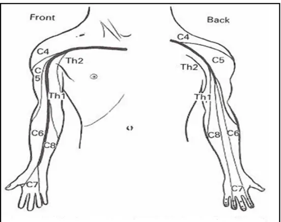
A dermatome refers to the area of the skin innervated by the sensory axons within each segmental nerve (Trombly and Radomski, 2001). Cutaneous nerve fields are the areas of skin innervated by peripheral nerves. Light touch sensation is tested manually by descending along the skin with an object such as a cotton ball. The patient should be asked to report if the sensation from the cotton ball is similar bilaterally. The examination should be done with the patient’s eyes closed and differences should be noted when comparing one limb to the other.
The testing should be done according to dermatomes from the proximal part of the limb to the distal part. These findings can be used as the basis for reassessment on follow up examination and may help determine if the condition is worsening or improving (Tomberlin and Saunders, 2001).
The range of motion examination consists of active, passive and accessory movement tests.
Active and passive motions are types of physiological movement. Physiological motion is movement in standard planes. Accessory movement is the small movement within the joints
and surrounding tissues that is necessary for normal physiological movements. The range of motion examination helps determine the extent to which they are involved. When performing any movement test – active, passive or accessory – the clinician should ask two main questions:
a. What is the effect of the test on the patient’s symptoms?
b. Is the amount of movement normal, hypomobile or hypermobile?
The findings of the range of motion examination are significant if the amount of movement assessed is abnormal when compared to normal values of range of movement as seen in Table 2 below (Tomberlin and Saunders, 2001).
Active motion provides general information about the patient’s functional ability. This includes the patient’s willingness and ability to move the joint. Active movement assesses function and movements in functional patterns rather than straight planar motion yields more information. During active movement testing, the range of motion obtained when the patient is asked to perform a movement should be documented. The quality of movement and symptoms associated with movement should also be noted (Tomberlin and Saunders, 2001).
Passive movement tests differentiate contractile and non-contractile structures. These tests determine if the joint range is restricted, excessive or normal. If the passive movement is greater than the active movement, this indicates that the contractile tissue is at least partly responsible for the patient’s symptoms. If active movement is greater than passive movement, the patient is unable to relax enough to let the clinician complete the testing (Tomberlin and Saunders, 2001).
Table 2: Normal values for measurement of range of motion of the wrist and thumb (Trombly and Radomski, 2001)
MOVEMENTS NORMAL VALUES
IN DEGREES (°)
WRIST: FLEXION 0-80
EXTENSION 0-70
RADIAL DEVIATION 0-20
ULNA DEVIATION 0-30
THUMB: METARCARPAL PHALANGEAL JOINT:
FLEXION 0-50
EXTENSION 50-0
ABDUCTION 0-50
ADDUCTION 0-40
OPPOSITION IN CENTIMETERS (RULER) NO NORM
INTERPHALANGEAL JOINT: FLEXION 0-90
EXTENSION 90-0
DIGITS 2,3,4: METARCARPAL PHALANGEAL JOINT:
FLEXION 0-90
ABDUCTION NO NORM
ADDUCTION NO NORM
PROXIMAL INTERPHALNGEAL JOINT:
FLEXION –EXTENSION 0-100
DISTAL INTERPHALNGEAL JOINT:
FLEXION-EXTENSION 0-90
2.4.4.1. The Goniometer for measurement of range of motion
Trombly and Radomski (2001) stated that a goniometer is used to asses joint motion. In order to ensure reliability, the therapist must place the axis and arms appropriately. In addition to goniometer placement, for the most reliable and accurate results, every effort should be made to make the patient physically and emotionally comfortable by talking to the patient and describing the procedure that is to follow. For the most reliable pretest-posttest information, the same tester should use the same goniometer at the same time of day (Trombly and Radomski, 2001). The method of recording and measuring joint range of motion has followed
the procedures published by the American Academy of Orthopaedic Surgeons. This method is easily understood by most clinicians ensuring the greatest face and content validity (Lehman and Abreu, 1989).
2.4.4.2. The Dynamometer for measurement of grip strength of the hand
The hand held dynamometer is a common tool used to assess power grip in the hand (Tomberlin and Saunders, 2001). Barnes (2007) stated that grip strength is often used clinically as an indicator of hand function and as a quick and effective outcome measure for rehabilitation. In a study by Molenaar et al. (2008), the reliability of the Lode Dynamometer and the Martin vigorimeter was determined. It was found that both the Lode dynamometer and the Martin vigorimeterare reliable instruments with which to measure the grip strength.
The Lode Dynamometer also had a better test retest reliability (Molenaar et al. 2001).
Bellace et al. (2001) conducted a study to evaluate the reliability and validity of the Jamar dynamometer. It was found established that the Jamar dynamometer was found to be highly reliable and valid for measuring hand grip strength.
2.4.4.3. Specifics tests to reproduce carpal tunnel symptoms
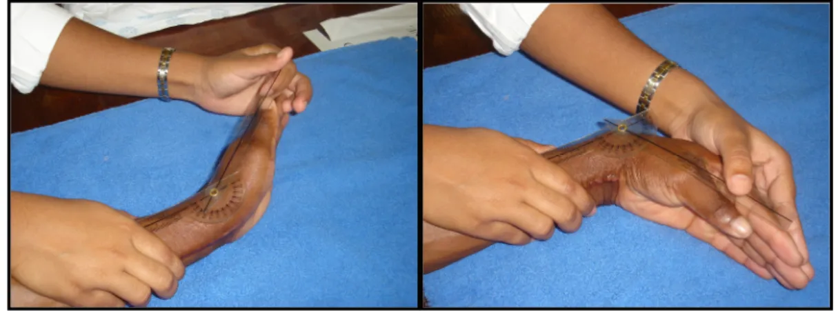
Physicians use specific tests to try to reproduce the symptoms of carpal tunnel syndrome. In the Tinel test, the clinician taps lightly over the site of the median nerve at the distal wrist crease. Development of tingling or discomfort in the fingers supplied by the median nerve constitutes a positive sign (Aroori and Spence, 2007). Phalen and Kendrick described the Phalens test in 1957. Flexion of the wrist causes compression of the nerve between the transverse carpal ligament and the flexor tendons in the carpal tunnel, causing paresthesia in the median nerve distribution reproducing the patients’ symptoms. The Phalens or wrist- flexion test involves having the patient hold his or her forearms upright by pointing the fingers down and pressing the backs of the hands together. The presence of carpal tunnel syndrome is suggested if one or more symptoms, such as tingling or increasing numbness, are felt in the fingers within 1 minute. (Aroori and Spence, 2007).
2.4.4.4. Electro-diagnostic tests
Electro-diagnostic study is a useful adjunct to clinical assessment but does not supplant the necessity for a thorough history and physical examination (Brotzman and Wilk, 1999). Nerve conduction studies measure the sensory and motor transmission velocity in the median nerve at the level of the wrist. The sensory component of the median nerve is affected much earlier
than the motor component and in early stages of carpal tunnel syndrome there is usually a delay in the sensory nerve conduction velocity. Sensory nerve conduction delay is determined by placing an electrode near the base of the ring finger following which the median nerve is stimulated 13cm proximal to the recording of the electrode. The antidromic sensory potentials are recorded and measured. The motor nerve conduction velocity from elbow to wrist is measured using surface electrodes (Aroori and Spence, 2007). In electromyography, a fine needle is inserted into a muscle; the results of the electrical activity are viewed on a screen which determines the severity of damage to the median nerve. Ultrasound imaging can show impaired movement of the median nerve. Magnetic resonance imaging (MRI) can show the anatomy of the wrist but to date has not been especially useful in diagnosing carpal tunnel syndrome (National Institute of Health, 2002).
2.4.5. Management
Treatments for carpal tunnel syndrome should begin as early as possible and managed in conjunction with a multidisciplinary team under a doctor's direction. It is imperative that physiotherapists (even though they are considered first line practitioners and are trained to diagnose and manage these hand conditions) are provided with sufficient information of the severity of the condition as well as the plan of treatment offered by the doctor. The examination including tests performed by the doctor will assist the therapist in devising an effective treatment plan including taking proper precautions not to exacerbate the symptoms.
According to the National Institute of Health (2002), underlying causes such as diabetes or arthritis should be medically treated first. Initial treatment generally involves resting the affected hand and wrist for at least 2 weeks, avoiding activities that may worsen symptoms, and immobilizing the wrist in a splint to avoid further damage from twisting or bending. If there is inflammation, applying cool packs can help reduce swelling (National Institute of Health, 2002).
2.4.5.1. Non-operative treatments These may include:
• The use of a splint, placing the wrist in a neutral position, worn at night. The splint can be worn during the day if the patient’s job allows for it (Brotzman and Wilk, 1999).
• The ability to modify activities for example, discontinuing the use of vibratory tools or placing a support under unsupported arms when sitting in front of the computer (Brotzman and Wilk, 1999).
• A cortisone injection into the carpal tunnel but not into the median nerve. Brotzman and Wilk (1999) stated that studies have shown that less than 25% of patients who took a cortisone injection in to the carpal tunnel became symptom free 18 months following the injection. It was also stated that 80% of patients had temporary relief with cortisone injection and splinting.
• Non-steroidal anti-inflammatory drugs (NSAID’s) can be used to control inflammation (Brotzman and Wilk, 1999).
• Any underlying systemic disease such as diabetes, rheumatoid arthritis or hypothyroidism must be controlled (Brotzman and Wilk, 1999).
2.4.5.2. Surgery
Indications for surgical treatment of carpal tunnel syndrome include: thenar atrophy or weakness, loss of sensation, fibrillation potentials on electro-myelograms and if symptoms persist for more than a year regardless of appropriate conservative measures. The goals of carpal tunnel release are decompression of the median nerve, improvement of excursion and prevention of progressive nerve damage (Brotzman and Wilk, 1999). Surgery consists of division of the transverse carpal ligament. This reduces the pressure on the median nerve by increasing the space in the carpal tunnel. Two types of surgical approaches are used for the treatment of carpal tunnel syndrome; open and endoscopic release. Open carpal tunnel release is the traditional option and still the recommended method of surgical treatment for carpal tunnel syndrome. It was first performed by Herbert Galloway in 1924. The conventional open release carpal tunnel syndrome uses a curved longitudinal inter-thenar incision, approximately 4-5cm in length. It involves opening the subcutaneous tissue, superficial fascia and transverse carpal ligament and 2-3cm of distal forearm fascia under direct vision (Aroori and Spence, 2007).
Endoscopic surgery may allow faster functional recovery and less postoperative discomfort than traditional open release surgery. The surgeon makes two incisions (about 1.25 cm each) in the wrist and palm, inserts a camera attached to a tube, observes the tissue on a screen, and
severs the carpal ligament. This two-portal endoscopic surgery, generally performed under local anesthesia, is effective and minimizes scarring and scar tenderness. One-portal endoscopic surgery for carpal tunnel syndrome is also available (National Institute of Health, 2002).
Although symptoms may be relieved immediately after surgery, full recovery from carpal tunnel surgery can take months. Some patients may have infection, nerve damage, stiffness, and pain at the scar. Occasionally the wrist loses strength because the carpal ligament is cut.
Patients should undergo physiotherapy after surgery to restore wrist strength. Some patients may need to adjust job duties or even change jobs after recovery from surgery. Recurrence of carpal tunnel syndrome following treatment is rare. The majority of patients recover completely (National Institute of Health, 2002).
2.4.6. Prevention of carpal tunnel syndrome
Protecting employees from occupational cumulative trauma disorders like carpal tunnel syndrome poses two challenges: identifying work-related risk factors and instituting appropriate modifications of workstations, tools, work organization and tasks. Ergonomics is rapidly advancing the ability to meet these demands. With training in anatomy, physiology, engineering, psychology and biomechanics, ergonomists are specialists in evaluating work tasks and sites and designing more efficient and safer work environments. Once high risk activities have been identified, ergonomic principles may be used to develop modifications to reduce or eliminate carpal tunnel syndrome (Rempel et al. 1992). The United States Occupational Health and Safety Administration have recently developed guidelines to prevent and reduce work-related cumulative trauma disorders. These guidelines recommend that employees involved in manual handling should minimize the distance between the load and the body, lift loads from knuckle height, keep the travel distance for the lift to less than 10 feet, minimizing twisting and ensuring good handles for grasping tools (Occupational Health and Safety Administration, 2000).
2.4.7. Alternative research related to carpal tunnel syndrome
The National Institute of Neurological Disorders and Stroke (NINDS) stated that scientists are studying the chronology of events that occur with carpal tunnel syndrome in order to better understand, treat and prevent this ailment. By determining distinct biomechanical factors related to pain, such as specific joint angles, motions, force, and progression over
time, researchers are finding new ways to limit or prevent carpal tunnel syndrome in the workplace and decrease other costly and disabling occupational illnesses (National Institute of Health, 2002).
Randomized clinical trials in the United States are being designed to evaluate the effectiveness of educational interventions in reducing the incidence of carpal tunnel syndrome and upper extremity cumulative trauma disorders. Data is to be collected from a National Institute for Occupational Safety and Health-sponsored study of carpal tunnel syndrome among construction workers will provide a better understanding of the specific work factors associated with the disorder, furnish pilot data for planning future projects to study its natural history, and assist in developing strategies to prevent its occurrence among construction and other workers. Additional research may discern differences between the relatively new carpal compression test (in which the examiner applies moderate pressure with both thumbs directly on the carpal tunnel and underlying median nerve, at the transverse carpal ligament) and the pressure provocative test (in which a cuff placed at the anterior aspect of the carpal tunnel is inflated, followed by direct pressure on the median nerve) in predicting carpal tunnel syndrome. The use of alternative therapies, such as acupuncture, to prevent and treat this disorder is also being investigated (National Institute of Health, 2002).
Palmer et al. (2006) conducted a systematic literature review on carpal tunnel syndrome and its relation to occupation. The objective of the study was to assess occupational risk factors for carpal tunnel syndrome. This was accomplished by identifying relevant primary research from two major reviews in the 1900’s and supplemented this material by a systematic search of the Medline and Embase biomedical databases from the start of the electronic record to 1 January 2005. Reports were obtained and their bibliographies checked for other relevant publications. From each paper, a standardized set of information on study populations, exposure contrasts and estimates of effects were abstracted. Altogether, thirty eight primary reports were summarized, with analysis based either on a comparison of job titles or of physical activities in the job or both. It was found that regular and prolonged use of hand-held vibratory tools increases the risk of carpal tunnel syndrome two-fold. The balance of evidence on keyboard and computer work did not indicate an important association with carpal tunnel syndrome. Although the papers that were considered had limitations, a substantial and coherent body of evidence supports preventative policies aimed at avoiding highly repetitive wrist-hand work. Limitations mentioned involved potential information bias, limiting their statistical power may not fully have been controlled for confounding and the
possibility that investigations were prompted by the observation of workplace clusters which could have led to unrepresentatively high risk estimates (Palmer et al. 2006).
2.5. De Quervains tenosynovitis 2.5.1. Definition
De Quervains tenosynovitis is defined as stenosing tenosynovitis of the short extensor muscle (extensor pollicis brevis) and the long abductor tendon (abductor pollicis longus) of the thumb (Beers and Berkow, 1999).
2.5.2. Anatomy
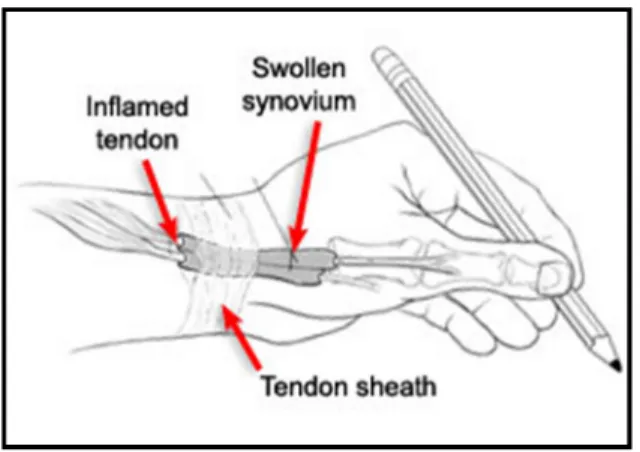
The first dorsal compartment at the wrist includes the tendons of the abductor pollicis longus and the extensor pollicis brevis. Patients with this condition usually report pain at the dorsolateral aspect of the wrist with referral of pain toward the thumb and or the lateral forearm. Inflammation of a tenosynovium is called tenosynovitis. In the case of de Quervains tenosynovitis, the inflammation causes an impingement of the tendons in the tunnel, causing friction on the tendons, further inflaming the tenosynovium and tendons (The Stretching Institute, 1999-2010).
Figure 2. De Quervains tenosynovitis of the first extensor compartment (Griffin, 2005)
Two of the main tendons to the thumb pass through a tunnel (or series of pulleys) located on the dorsolateral aspect of the wrist. Tendons are covered by a slippery thin soft-tissue layer, called synovium. This layer allows the tendons to slide easily through the tunnel. Any swelling of the tendons located near these nerves can put pressure on the nerves. This can
cause wrist pain or numbness in the fingers (American Academy of Orthopedic Surgeons, 2007).
2.5.3. Pathology
Figure 3. The mucous sheaths on the posterior aspect of the hand (Kasdan, 1999) Abductor pollicis longus and extensor pollicis brevis have almost the same function: the movement of the thumb away from the hand in the plane of the hand. The impaired gliding is caused by the thickening of the extensor retinaculum (the thickened part of the general tendon sheath that holds the extensor muscles in place) of the wrist. De Quervain, a Swiss physician, is given credit for first describing this condition with a report of 5 cases in 1895 and 8 additional cases in 1912. On histopathological examination predominant features are degenerative changes (myxoid degeneration, fibrocartilaginous metaplasia and deposition of mucopolysaccharide). Pain is elicited by mechanical impingement between the tendon and fibro-osseous canal resulting in stimulation of nociceptors (Winters et al. 2009). In the first dorsal compartment of the wrist, a tendon sheath encloses the abductor pollicis longus and extensor pollicis brevis tendons at the lateral border of the anatomical snuffbox.
Inflammation at this site commonly is seen in patients who use their hands and thumbs in a repetitive fashion. Thus de Quervains tenosynovitis can result from cumulative microtrauma.
Inflammation also may occur after an episode of acute trauma to the site (Foye et al. 2010).
2.5.4. Causes
This disorder is the most common overuse injury involving the wrist and often occurs in individuals who regularly use a forceful grasp coupled with ulnar deviation of the wrist such as in a tennis serve or squash (Brotzman and Wilk, 1999). Excessive friction between the
tendons of abductor pollicis longus and extensor pollicis brevis in their common sheath causes fibrous thickening of the sheath and stenosis of the osseofibrous tunnel. This excessive friction is caused by repetitive forceful use of the hands during gripping and wringing (Moore and Dalley, 1999). Beers and Berkow (1999) stated that this disorder usually occurs after repeated use (especially in wringing), of the wrist although it is occasionally associated with rheumatoid arthritis.
2.5.5. Signs and symptoms
• Pain may be felt over the radial aspect of the wrist. Pain may appear gradually or suddenly. Pain may also be present in the forearm. The pain is usually worse when the hand and thumb are in use. This is especially true when forcefully grasping objects or twisting the wrist.
• Swelling may be present over the radial aspect of the wrist. This swelling may occur together with a fluid-filled cyst in this region.
• A "catching" or "snapping" sensation may be felt when moving the thumb.
• Pain and swelling may make it difficult to move the thumb and wrist.
• Numbness may be experienced on the posterior aspect of the thumb and index finger.
This is caused as the nerve lying superior to the tendon sheath is irritated (American Academy of Orthopedic Surgeons, 2007).
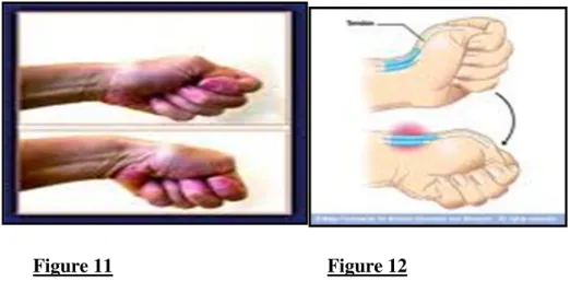
2.5.6. Diagnosis
Figure 4. Finkelsteins test (American Academy of Orthopaedic Surgeons, 2007) The Finkelstein test is conducted by making a fist with the fingers closed over the thumb and the wrist is bent toward the little finger. The Finkelstein test can be quite painful for the person with de Quervains tendinitis. Tenderness directly over the tendons on the thumb side
of the wrist is a common finding with this test (American Academy of Orthopaedic Surgeons, 2007).
Pain can be assessed using the visual analogue scale as in carpal tunnel syndrome.
Neurological examination in de Quervains tenosynovitis follows the same procedure as in carpal tunnel syndrome. Range of movement is assessed using the universal goniometer and using the baseline values for comparison. Strength is evaluated using a hand dynamometer (American Academy of Orthopaedic Surgeons, 2007).
2.5.7. Treatment
Conservative Management
A thumb spica splint is used to immobilize the first dorsal compartment tendons with a commercially available splint or, depending on the patients comfort, a custom molded orthoplast device. The splint maintains the wrist in 15-20 degrees of extension and the thumb in 30 degrees of radial and palmar abduction. The interphalangeal joint is left free, and motion at this joint is encouraged. The patient wears the splint during the day for the first 2 weeks and at night until the next visit to the therapist, generally after 6-8 weeks. Splinting may continue longer, depending on the response to treatment. The splint can be discontinued during the day if symptoms permit and if daily activities are gradually resumed. Work place activities are advanced accordingly (Brotzman and Wilk, 1999).
Other considerations include:
• A corticosteroid sheath injection can be offered to patients with moderate to marked pain or with symptoms lasting more than 3 weeks. The injection should individually distend the abductor pollicis longus and the extensor pollicis brevis sheath.
Discomfort after the injection is variable, and a 2-3 day supply of mild analgesic is recommended
• A systemic non-steroidal anti-inflammatory drug is commonly prescribed for the initial 6-8 weeks of treatment.
• Thumb use is restricted so that the first dorsal compartment tendons are at relative rest. Activities that require prolonged thumb interphalangeal joint flexion, pinch, or repetitive motions are avoided.