ANTIOXIDATIVE AND ANTIDIABETIC EFFECTS OF SOME AFRICAN MEDICINAL PLANTS
BY
AMINU MOHAMMED
Student number: 212562498
MARCH, 2016
ANTIOXIDATIVE AND ANTIDIABETIC EFFECTS OF SOME AFRICAN MEDICINAL PLANTS
BY
AMINU MOHAMMED Student number: 212562498
Submitted in fulfillment of the requirements for the award of Doctor of Philosophy degree in Biochemistry
School of Life Sciences
College of Agriculture, Engineering and Science
As the candidate’s supervisor I have approved this thesis/dissertation for submission.
Signed: ……….. Name: Dr. M.S. Islam Date: ……….
COLLEGE OF AGRICLUTURE, ENGINEERING AND SCIENCE DECLARATION I - PLAGIARISM
I, Aminu Mohammed……….. declare that
1. The research reported in this thesis, except where otherwise indicated, is my original research.
2. This thesis has not been submitted for any degree or examination at any other university.
3. This thesis does not contain other persons’ data, pictures, graphs or other information, unless specifically acknowledged as being sourced from other persons.
4. This thesis does not contain other persons' writing, unless specifically acknowledged as being sourced from other researchers. Where other written sources have been quoted, then:
a. Their words have been re-written but the general information attributed to them has been referenced
b. Where their exact words have been used, then their writing has been placed in italics and inside quotation marks, and referenced
5. This thesis does not contain text, graphics or tables copied and pasted from the Internet, unless specifically acknowledged, and the source being detailed in the thesis and in the references sections.
Signed: ………..
Declaration Plagiarism 22/05/08 FHDR Approved
DECLARATION II
I, Aminu Mohammed hereby declare that the dissertation entitled “Antioxidative and antidiabetic effects of some African medicinal plants” is the result of my own investigation and research and that it has not been submitted in part or in full for any other degree or to any other university. Where use of the work of others was made, it is duly acknowledged in the text.
Student: Mr. Aminu Mohammed Signature ……….
Supervisor: Dr. M.S. Islam Signature ……….
PUBLICATIONS AND PRESENTATIONS
DETAILS OF CONTRIBUTION TO PUBLICATIONS that form part of and/or include research presented in this thesis (include publication in preparation, submitted, in press and published and give details of the contributions of each author to the experimental work and writing of each publication).
In all the publications included in this thesis, I designed the work, performed all the experiments and wrote all the publications. The co-authors contributed by conducting an editorial work, checking the scientific content of the work and the correctness of my statistical analysis of data and interpretation of the findings.
Published and/or accepted papers
Publication 1
Mohammed, A., Koorbanally, N.A., Islam, M.S. (2015). Ethyl acetate fraction of Aframomum melegueta fruit ameliorates pancreatic β-cell dysfunction and major diabetes-related parameters in a type 2 diabetes model of rats. Journal of Ethnopharmacology 175: 518-527.
Publication 2
Mohammed, A., Koorbanally, N.A., Islam, M.S. (2015). Phytochemistry, anti-oxidative and anti-diabetic effects of various parts of Eugenia caryophyllata Thunb. in vitro. Acta Poloniae Pharmaceutica-Drug Research 72: 1201-1215.
Publication 3
Mohammed, A., Koorbanally, N.A., Islam, M.S. (2015). Anti-oxidative activity, phytochemistry, and inhibition of key enzymes linked to type 2 diabetes by various parts of Aframomum melegueta in vitro.
Acta Poloniae Pharmaceutica-Drug Research In press (Accepted on 28 March, 2015).
Publication 4
Mohammed, A., Ibrahim, M.A., Islam, M.S. (2014). African medicinal plants with anti-diabetic potentials:
a review. Planta Medica 80: 354-377.
Publication 5
Mohammed A., Koorbanally N.A., Islam M.S. Anti-diabetic effect of Xylopia aethiopica (Dunal) A. Rich.
(Annonaceae) fruit acetone fraction in a type 2 diabetes model of rats. Journal of Ethnopharmacology 180: 131-139.
Papers are intended to submit:
Publication 6
Mohammed, A., Islam, M.S. Anti-oxidant potential of Aframomum melegueta fruit ethyl acetate fraction in a type 2 diabetes model of rats (in preparation).
Publication 7
Mohammed, A., Koorbanally, N.A., Islam, M.S. Anti-oxidative activity, phytochemistry, and inhibition of key enzymes linked to type 2 diabetes by various parts of Xylopia aethiopica in vitro (in preparation).
Publication 8
Mohammed, A., Koorbanally, N.A., Islam, M.S. Anti-oxidative action and inhibition of key enzymes linked to type 2 diabetes of various solvent fractions from fruit ethanolic extract of Xylopia aethiopica in vitro (in preparation).
Publication 9
Mohammed, A., Koorbanally, N.A., Islam, M.S. In vivo anti-oxidant potential of Xylopia aethiopica fruit acetone fraction in a type 2 diabetes model of rats (in preparation).
Publication 10
Mohammed, A., Koorbanally, N.A., Islam, M.S. Anti-oxidative and anti-diabetic effects of various parts of Capsicum annuum L. (Solanaceae) in vitro (in preparation).
Publication 11
Mohammed, A., Koorbanally, N.A., Islam, M.S. Acetone fraction from Capsicum annuum fruit possesses anti-oxidative effects and inhibits the activities of carbohydrate digesting enzymes in vitro (in preparation).
Publication 12
Mohammed, A., Islam, M.S. Anti-diabetic effects of Capsicum annuum L. fruit acetone fraction in a type 2 diabetes model of rats (in preparation).
Publication 13
Mohammed, A., Islam, M.S. Anti-oxidant action of Capsicum annuum fruit acetone fraction in in a type 2 diabetes model of rats (in preparation).
Publication 14
Mohammed, A., Koorbanally, N.A., Islam, M.S. Anti-diabetic action of some African natural products (Aframomum melegueta, Xylopia aethiopica and Capsicum annuum) in vitro and and isolation of bioactive compounds (in preparation).
Presentions:
Presention 1
Mohammed, A., Islam, M.S. Anti-diabetic effect of acetone fraction from Xylopia aethiopica fruit in a type 2 diabetes model of rats. A poster presentation at the 23rd World Diabetes Congress 2015 (30th November, 2015 to 4th December, 2015) at Vancouver, Canada. I have been awarded one of the 100 prestigious IDF Travel Grants to present the work at the conference.
Presention 2
Mohammed, A., Islam, M.S. Ethyl acetate fraction of Aframomum melegueta fruit ameliorates pancreatic β-cell dysfunction and major diabetes-related parameters in a type 2 diabetes model of rats. A poster presentation at the 15th Congress of International Society for Ethnopharmacology, (5-8 May, 2015) at BeitZaman Hotel & Resort, Petra, Jordan.
Presention 3
Mohammed, A., Islam, M.S. Anti-diabetic effect of Xylopia aethiopica (Dunal) A. Rich. fruit acetone fraction in a type 2 diabetes model of rats. An oral presentation at the College of Agriculture, Engineering and Sciences Research Day (22nd September 2015) at C Block Lecture Theatre Complex, University of KwaZulu-Natal, Pietermaritzburg Campus, Durban 4000, South Africa.
Presention 4
Mohammed, A., Islam, M.S. Anti-diabetic effect of Aframomum melegueta ethyl acetate fraction from fruit in type 2 diabetes model of rats. An oral presentation at the College of Agriculture, Engineering and Sciences Research Day (27th October 2014) at T Block Lecture Theatre Complex, University of KwaZulu- Natal, Westville Campus, Durban 4000, South Africa.
Presention 5
Mohammed, A., Islam, M.S. Anti-oxidative potential and inhibition of key enzymes linked to type-2 diabetes mellitus of various fractions from sweet pepper (Capsicum annuum L.) in vitro. A poster presentation at the School of Life Sciences Research Day (26th May, 2014) at T Block Lecture Theatre Complex, University of KwaZulu-Natal, Westville Campus, Durban 4000, South Africa.
Student: Mr. Aminu Mohammed Signature ……….
ABSTRACT
Three (3) medicinal plants [Aframomum melegueta K. Schum., Xylopia aethiopica (Dunal.) A. Rich. and Capsicum annuum L.] were selected based on their traditional uses in the treatment of diabetes in Africa.
Various crude extracts and fractions from different parts of the plants were screened using several anti- oxidative and anti-diabetic tests in vitro. Most active fractions from each plant were used to examine in vivo anti-diabetic activity in type 2 diabetes (T2D) rat model. Additionally, possible bioactive compounds from most active extracts and fractions were analyzed by using GC-MS, TLC and NMR spectroscopy. The results showed that ethanolic extracts derived from the fruits of the plants demonstrated excellent anti-oxidative and anti-diabetic activities in vitro compared to other extracts from the same or different parts of these plants. After fractionation, ethyl acetate fraction from A. melegueta and acetone fractions from X. aethiopica and C. annuum exhibited strong radical scavenging (IC50: 1-120 µg/mL) activity, inhibition of hemoglobin glycation (IC50: 100-150 µg/mL), α-amylase (IC50: 50-170 µg/mL) and α-glucosidase (IC50: 40-87 µg/mL) activities hence were used for the in vivo study. The GC-MS analysis of the three (3) most active fractions revealed the presence of mostly phenolic compounds of 4-hydroxy-3-methoxyphenyl derivatives. Furthermore, the data of the in vivo study showed that oral intervention of the fractions (150 and 300 mg/kg bw) for 4 weeks demonstrated potent anti-diabetic actions via improving body weight gain, reducing feed and fluid intake and hyperglycemia, improving glucose tolerance ability, insulin sensitivity, amelioration of pancreatic β-cell histology and β-cell functions, improving dyslipidemia in a T2D rat model.
Additionally, the pancreatic histopathological damages and other oxidative damages caused by the induction of diabetes were attenuated to near normal in the liver, kidney, heart and pancreas of the treated animals. The bioassay-guided fractionations lead to the isolation of 3 arylalkanes (6-paradol (1), 6-shagaol (2), and 6- gingerol (3)) and oleanolic acid (4) from A. melegueta fruits, when oleanolic acid (4) was the first to be isolated from A. melegueta. Moreover, 6-gingerol (3) and oleanolic acid (4) were similarly isolated for the first time from X. aethiopica fruits as well. These compounds have exhibited significant inhibitions against the α-amylase and α-glucosidase actions and thus are possible anti-diabetic agents and the anti-diabetic action of A. melegueta and X. aethiopica fruits is attributed to the presence of these compounds. This study also confirmed the use of these plants in African anti-diabetic traditional medicines by traditional healers. However, further clinical study is required to confirm these effects in human subjects.
DEDICATION
To my parent Alhaji Abubakar Mohammed and Hajiya Hauwa’u Aminu for their guidance, support and wisdom.
ACKNOWLEDGEMENTS
First of all, I would like to express my sincere gratitude to Almighty Allah, the Giver of favors for giving me life, time and health up to this time. These are the basis upon which the entire success of this work was built. I also thank him for the gift of my parents who have tirelessly stood by me since childhood.
I must acknowledge the tremendous support of my elegant supervisor Dr. M.S. Islam for his support, time and encouragement despite his tight schedule. His constructive comments, patience, encouragement and motivation inspired me greatly. It is an input I will use forever in my academic and scientific career. The immense contribution and guidance of Prof. N.A. Koorbanally is highly appreciated as well.
To my lovely wife Mrs. Maryam M. Zayyan and my daughter Nana Aisha, no amount of words will explain my sincere and heartily appreciation for your tireless prayers, patience and support. You have been the pillars of my success and I will be forever grateful.
I am greatly indebted to the efforts of technical staff from various units. The staff in the Biomedical Research Unit (BRU), Westville Campus, especially Dr. Linda Bester, Mr. David Mompe and Rita Radebe deserve special mention. The help from Shoohana Singh of histology unit, Department of Physiology is beyond rewarding.
I must appreciate the contributions of the past and present colleagues of Biomedical Lab. Namely:
Dr. Mohammed Auwal Ibrahim, Talent Chipiti, Praglathan Naidoo, Siphiwe Dlamini, Shandre Pillay, Chika Chukwuma, Dr. Ramgopal Mopuri, Nokonwaba Nkondlo, Nomcebo Mchunu and Shireen Nadesan.
You have always been good friends in the lab. So also Mrs. Victoria Awolola of Natural Products Lab, Department of Chemistry, University of KwaZulu-Natal, South Africa, your contribution is highly appreciated.
I am also greatly indebted to the Ahmadu Bello University, Zaria, Nigeriaand Education Trust Fund (ETF) Desk Office-ABU for approving my fellowship at UKZN, South Africa. On that note, I must appreciate the immense contributions from the colleagues of Biochemistry Department, Ahmadu Bello University, Zaria, Nigeria. Prof. I.A. Umar, Prof. H.M. Inuwa, Prof. S. Ibrahim, Dr. A. Salihu, Dr. A.B.
Sallau and Prof. M.K. Atiku of Bayero University Kano, Nigeria deserve a special mention.
Finally, I would like to appreciate the Nigerian Community in South Africa whom has been my family. To mention a few, Dr. Abdullahi A. Yusuf, Dr. Aliyu Babando, Dr. Hamisu Ibrahim, Dr. Halliru Ibrahim, Dr. Ibrahim Abdulkadir, Murtala Isah Bindawa, Shuaibu M. Bala, Salihu Zubairu, Falalu Hamza, Buhari Badamasi, Aliyu Adamu, Jibril Nuhu, Abdulkadir Ibrahim, AbduSalam Yakasai, A. Abdulkadir, Alhaji Zubairu, Umar and Ayodeji Oyenihi among others. They were really encouraging.
TABLE OF CONTENTS
PAGE
DECLERATION I iii
DECLERATION II iv
PUBLICATIONS AND CONFERENCES v
ABSTRACT ix
DEDICATION x
ACKNOWLEDMENTS xi
TABLE OF CONTENTS xii
LIST OF TABLES xix
LIST OF FIGURES xxii
LIST OF ABBREVIATIONS xxviii
CHAPTER I 1
1.0 Introduction and background of the study 1
1.1 Literature review 2
1.2 Diabetes mellitus 2
1.2.1 Types of diabetes mellitus 2
1.2.2 Type 1 diabetes mellitus 2
1.2.3 Type 2 diabetes mellitus 3
1.2.4 Gestational diabetes mellitus 4
1.2.5 Specific types of diabetes due to other causes (secondary type) 5
1.2.6 Prevalence of diabetes mellitus 6
1.2.7 Diabetes associated complications 6
1.2.8 Treatment and management of diabetes mellitus 7
1.2.9 Non-pharmacological therapy 8
1.2.10 Pharmacological therapy 8
1.3 Medicinal plants 10
1.3.1 Medicinal plants in future drug discovery 10
1.3.2 Selection of medicinal plant for drug discovery 11 1.3.3 Collection, authentication and preparation of plant material 12
1.3.4 Preliminary in vitro studies 12
1.3.5 Bioassay-guided fractionation of the active principles 13
1.3.6 In vivo, clinical and toxicological studies 13 1.3.7 Medicinal plants for the treatment of diabetes 14 1.3.8 African medicinal plants with anti-diabetic potentials 14 1.3.9 Bioactive compounds from African medicinal plants with anti-diabetic
potentials
16
1.4 Aframomum melegueta K. Schum. 22
1.4.1 Description 22
1.4.2 Distribution 22
1.4.3 Phytochemistry 22
1.4.4 Ethnobotanical uses 23
1.4.5 Pharmacological importance 23
1.5 Xylopia aethiopica (Dunal) A. Rich 25
1.5.1 Description 25
1.5.2 Distribution 25
1.5.3 Phytochemistry 25
1.5.4 Ethnobotanical uses 25
1.5.5 Pharmacological importance 26
1.6 Capsicum annuum L. 27
1.6.1 Description 27
1.6.2 Distribution 27
1.6.3 Phytochemistry 27
1.6.4 Ethnobotanical uses 28
1.6.5 Pharmacological importance 28
1.7 Statement of the problem 29
1.8 Justification and significance of the research work 30
1.9 Objective of the study 30
1.9.1 General objective 30
1.9.2 Specific objectives 30
CHAPTER 2 32
2.0 Materials and methods 32
2.1 Chemicals and reagents 32
2.2 Equipment 32
2.3 Plant materials 32
2.3.1 Preparation of the plant extracts 33
2.3.2 Fractionation of the crude extracts 33
2.4 In vitro studies 33
2.4.1 Estimation of total polyphenol content 33
2.4.2 Determination of total flavonoid content 34
2.4.3 DPPH radical scavenging activity 34
2.4.4 Ferric (Fe3+) reducing anti-oxidant power assay 34
2.4.5 Inhibition of hemoglobin glycation 35
2.4.6 α-Amylase (E.C. 3.2.1.1) inhibitory effect 35
2.4.7 α-Glucosidase (E.C. 3.2.1.20) inhibitory effect 36
2.4.8 Calculation of IC50 values 36
2.4.9 Mechanism of α-glucosidase and α-amylase inhibitions 36 2.4.10 Gas Chromatography-Mass Spectroscopic (GC-MS) analysis 37
2.5 In vivo studies 37
2.5.1 Experimental animals 37
2.5.2 Animal grouping 37
2.5.3 Induction of Type 2 diabetes (T2D) 38
2.5.4 Intervention period 39
2.5.5 Oral glucose tolerance test (OGTT) 39
2.5.6 Collection and preparation of blood and organs 39
2.5.7 Analytical methods 40
2.5.8 Histopathological examination of pancreatic tissue 40
2.5.9 Determination of reduced glutathione (GSH) 41
2.5.10 Determination of thiobarbituric acid reactive substance (TBARS) concentration as malondialdehyde (MDA) equivalent
41
2.5.11 Determination of catalase activity 41
2.5.12 Determination of superoxide dismutase (SOD) activity 41 2.5.13 Determination of glutathione peroxidase (GPx) activity 42 2.5.14 Determination of glutathione reductase (GR) 42 2.6 Isolation of the bioactive anti-diabetic compounds from the fractions 42
2.6.1 Isolation of the bioactive compounds from the ethyl acetate fraction of ethanolic extract of Aframomum melegueta fruit
43
2.6.2 Isolation of the bioactive compound from the acetone fraction of ethanolic extract of Xylopia aethiopica fruit
43
2.6.3 Isolation of the bioactive compound from the acetone fraction of ethanolic extract of Capsicum annuum fruit
43
2.7 Statistical analysis 44
CHAPTER 3 45
3.0 Anti-diabetic and anti-oxidative actions of various extracts and fractions from different parts of Aframomum melegueta: an in vitro and in vivo approach
45
3.1 Anti-oxidative activity, phytochemistry, and inhibition of key enzymes linked to type 2 diabetes by various parts of Aframomum melegueta in vitro
45
3.1.1 Abstract 45
3.1.2 Introduction 46
3.1.3 Materials and methods 47
3.1.4 Results 47
3.1.5 Discussions 57
3.1.6 Conclusions 60
3.2 Phytochemistry, anti-oxidative and anti-diabetic effects of various solvent fractions from fruit ethanolic extract of Aframomum melegueta in vitro
61
3.2.1 Abstract 61
3.2.2 Introduction 62
3.2.3 Materials and methods 63
3.2.4 Results 63
3.2.5 Discussions 71
3.2.6 Conclusions 73
3.3 Ethyl acetate fraction of Aframomum melegueta fruit ameliorates pancreatic β-cell dysfunction and major diabetes-related parameters in a type 2 diabetes model of rats
74
3.3.1 Abstract 74
3.3.2 Introduction 75
3.3.3 Materials and methods 76
3.3.4 Results 76
3.3.5 Discussions 85
3.3.6 Conclusions 87
3.4 Anti-oxidant potential of Aframomum melegueta fruit ethyl acetate fraction in a type 2 diabetes model of rats
88
3.4.1 Abstract 88
3.4.2 Introduction 88
3.4.3 Materials and methods 90
3.4.4 Results 90
3.4.5 Discussions 95
3.4.6 Conclusions 96
CHAPTER 4 97
4.0 In vitro and in vivo anti-diabetic and anti-oxidative effects of various extracts and fractions from different parts of Xylopia aethiopica
97
4.1 Phytochemistry, anti-oxidative and anti-diabetic effects of extracts from various parts of Xylopia aethiopica in vitro
97
4.1.1 Abstract 97
4.1.2 Introduction 98
4.1.3 Materials and methods 99
4.1.4 Results 99
4.1.5 Discussions 108
4.1.6 Conclusions 110
4.2 Anti-oxidative effect and inhibition of key enzymes linked to type 2 diabetes of various solvent fractions from fruit ethanolic extract of Xylopia aethiopica in vitro
111
4.2.1 Abstract 111
4.2.2 Introduction 112
4.2.3 Materials and methods 112
4.2.4 Results 112
4.2.5 Discussions 120
4.2.6 Conclusions 121
4.3 Anti-diabetic effect of Xylopia aethiopica (Dunal) A. Rich. fruit acetone fraction in a type 2 diabetes model of rats
123
4.3.1 Abstract 123
4.3.2 Introduction 124
4.3.3 Materials and methods 125
4.3.4 Results 125
4.3.5 Discussions 135
4.3.6 Conclusions 137
4.4 In vivo anti-oxidant potential of Xylopia aethiopica fruit acetone fraction in a type 2 diabetes model of rats
138
4.4.1 Abstract 138
4.4.2 Introduction 138
4.4.3 Materials and methods 139
4.4.4 Results 139
4.4.5 Discussions 144
4.4.6 Conclusions 145
CHAPTER 5 146
5.0 In vitro and in vivo anti-diabetic and anti-oxidative effects of various extracts and fractions from the different parts of Capsicum annuum
146
5.1 Anti-oxidative and anti-diabetic effects of various parts of Capsicum annuum L.
(Solanaceae) in vitro
146
5.1.1 Abstract 146
5.1.2 Introduction 147
5.1.3 Materials and methods 148
5.1.4 Results 148
5.1.5 Discussions 158
5.1.6 Conclusions 160
5.2 Acetone fraction from Capsicum annuum fruit possesses anti-oxidative effects and inhibits the activities of carbohydrate digesting enzymes in vitro
161
5.2.1 Abstract 161
5.2.2 Introduction 162
5.2.3 Materials and methods 163
5.2.4 Results 163
5.2.5 Discussions 170
5.2.6 Conclusions 172
5.3 Anti-diabetic effects of Capsicum annuum L. fruit acetone fraction in a type 2 diabetes model of rats
173
5.3.1 Abstract 173
5.3.2 Introduction 174
5.3.3 Materials and methods 175
5.3.4 Results 175
5.3.5 Discussions 184
5.3.6 Conclusions 187
5.4 Anti-oxidant action of Capsicum annuum fruit acetone fraction in in a type 2 diabetes model of rats
188
5.4.1 Abstract 188
5.4.2 Introduction 188
5.4.3 Materials and methods 190
5.4.4 Results 190
5.4.5 Discussions 194
5.4.6 Conclusions 196
CHAPTER 6 197
6.1 Anti-diabetic action of some African natural products (Aframomum melegueta, Xylopia aethiopica and Capsicum annuum) in vitro and and isolation of bioactive compounds
197
6.1.1 Abstract 197
6.1.2 Introduction 198
6.1.3 Materials and methods 200
6.1.4 Results 200
6.1.5 Discussions 204
6.1.6 Conclusions 205
CHAPTER 7 208
7.0 General discussion, conclusion and further research 208
7.1 General discussion 208
7.2 Overall conclusion 214
7.3 Further research 215
References 217
Appendices I (Publications) 251
Appendices II (NMR spectra) 310
LIST OF TABLES
PAGE Table 1.1 Most studied anti-diabetic medicinal plants in the African continent 15 Table 1.2 Bioactive compounds from African medicinal plants with reported activities 17 Table 3.1 Percentage yield, total polyphenol and flavonoid contents of various solvent
extracts of A. melegueta parts
48
Table 3.2 IC50 values of various extracts of A. melegueta parts in different anti-oxidative and anti-diabetic models
50
Table 3.3 Identified compounds from the EtOH fruit and leaf extracts of A. melegueta by GC-MS
57
Table 3.4 Percentage yield, total polyphenol and flavonoid contents of various fractions from ethanolic fruit extract of A. melegueta
65
Table 3.5 IC50 values of various solvent fractions from ethanolic fruit extract of A.
melegueta in different anti-oxidative and anti-diabetic models
65
Table 3.6 Identified compounds from the ethyl acetate fraction from fruit ethanolic extract of A. melegueta by GC-MS
69
Table 3.7 Area under the curve (AUC) of different animal groups at the end of the experimental period
79
Table 3.8 Serum insulin and fructosamine levels, HOMA-IR and HOMA-β scores of different animal groups at the end of the experimental period
80
Table 3.9 Effect of AMEF on liver weights and liver glycogen concentrations in different animal groups as the end of the experimental period
82
Table 3.10 Serum lipid profiles atherogenic and coronary risk indices of different animal groups at the end of the experimental period
83
Table 3.11 Serum ALT, AST, ALP and other biochemical parameters different animal groups at the end of the experimental period
84
Table 4.1 Percentage yield, total polyphenol and flavonoid contents of various solvent extracts of X. aethiopica parts
99
Table 4.2 IC50 values of various solvent extracts of X. aethiopica parts in different anti- oxidative and anti-diabetic models
102
Table 4.3 Identified compounds from the fruit, leaf and stem EtOH extracts of by GC- MS
107
Table 4.4 Percentage yield, total polyphenol and flavonoid contents of various fractions from ethanolic fruit extract of X. aethiopica
113
Table 4.5 IC50 values of various solvent fractions from ethanolic fruit extract of X.
aethiopica in different anti-oxidative and anti-diabetic models
114
Table 4.6 Identified compounds from the acetone fraction from fruit ethanolic extract of X. aethiopica by GC-MS
118
Table 4.7 Area under the curve (AUC) of different animal groups at the end of the experimental period
128
Table 4.8 Effect of XAAF on serum insulin and fructosamine levels and calculated HOMA-IR and HOMA-β scores in different animal groups at the end of the intervention period
130
Table 4.9 Effect of XAAF on liver weights and liver glycogen concentrations in T2D rats 132 Table 4.10 Serum lipid profiles and other biochemical parameters in different animal
groups at the end of the intervention period
133
Table 4.11 Serum biochemical parameters in different animal groups at the end of the intervention period
134
Table 5.1 Percentage yield, total polyphenol and flavonoid contents of various solvent extracts of C. annuum parts
148
Table 5.2 IC50 values of various extracts of C. annuum parts in different anti-oxidative and anti-diabetic models
150
Table 5.3 Identified compounds from the EtOH fruit, leaf and stem extracts of C. annuum by GC-MS
156
Table 5.4 Percentage yield, total polyphenol and flavonoid contents of various fractions from ethanolic fruit extract of C. annuum
163
Table 5.5 IC50 values of various solvent fractions from ethanolic fruit extract of C.
annuum in different anti-oxidative and anti-diabetic models
165
Table 5.6 Identified compounds from the acetone fraction from fruit ethanolic extract of C. annuum by GC-MS
168
Table 5.7 Area under the curve (AUC) of different animal groups at the end of the experimental period
178
Table 5.8 Effect of CAAF on serum insulin and fructosamine levels and calculated HOMA-IR and HOMA-β scores in different animal groups at the end of the intervention period
179
Table 5.9 Effect of CAAF on liver weights and liver glycogen concentrations in T2D rats 181
Table 5.10 Serum lipid profiles and other biochemical parameters in different animal groups at the end of the intervention period
182
Table 5.11 Serum biochemical parameters in different animal groups at the end of the intervention period
183
Table 6.1 IC50 values of bioactive compounds isolated from the ethyl acetate fraction of A. melegueta and acetone fractions of C. annuum and X. aethiopica fruit in anti-diabetic models
201
Table 6.2 Kinetic analysis of α-amylase and α-glucosidase inhibition by compounds isolated from the ethyl acetate fraction of A. melegueta and acetone fractions of C. annuum and X. aethiopica fruit in anti-diabetic models
203
LIST OF FIGURES
PAGE
Figure 1.1 Pathogenesis of type 1 diabetes 3
Figure 1.2 Pathogenesis of type 2 diabetes 4
Figure 1.3 Pathogenesis of gestational diabetes mellitus 5 Figure 1.4 Hyperglycemia-induced oxidative damages in diabetes mellitus 7 Figure 1.5 Target organs/tissues and mode of actions of orally anti-diabetic drugs 10 Figure 1.6 Flow chart for plant-derived drug discovery 11
Figure 1.7 Aframomum melegueta plant 22
Figure 1.8 Xylopia aethiopica plant 25
Figure 1.9 Capsicum annuum plant 27
Figure 2.1 Animal grouping in vivo study 38
Figure 3.1 Ferric reducing power (relative to gallic acid) of fruit and leaf extracts of A. melegueta
49
Figure 3.2 DPPH Radical scavenging activity (%) of fruit, leaf and stem extracts of A. melegueta
51
Figure 3.3 Inhibition of hemoglobin glycosylation (%) of fruit and leaf extracts of A. melegueta
52
Figure 3.4 α-amylase inhibition (%) of fruit and leaf extracts of A. melegueta 53 Figure 3.5 α-glucosidase inhibition (%) of fruit leaf and stem extracts of A.
melegueta
54
Figure 3.6 GC-MS Chromatograms of ethanolic extracts of fruit and leaf of A.
melegueta
55
Figure 3.7 Structures of identified compounds from fruit and leaf of A. melegueta 56 Figure 3.8 Total reducing power (relative to gallic acid) of various fractions from
fruit ethanolic extract of A. melegueta
64
Figure 3.9 DPPH Radical scavenging activity (%) of various fractions from fruit ethanolic extract of A. melegueta
65
Figure 3.10 Inhibition of hemoglobin glycation (%) of various fractions from fruit ethanolic extract of A. melegueta
66
Figure 3.11 α-amylase inhibition (%) of various fractions from fruit ethanolic extract of A. melegueta
66
Figure 3.12 α-glucosidase inhibition (%) of various fractions from fruit ethanolic extract of A. melegueta
67
Figure 3.13 Lineweaver-Burke’s plot of (A) α-glucosidase and (B) α-amylase catalyzed reactions in the presence and in the absence of the ethyl acetate (EtOAc) fraction derived from the A. melegueta fruit ethanolic extract
68
Figure 3.14 GC-MS Chromatogram (A) and the structures of compounds (B) identified from the ethyl acetate fraction from fruit ethanolic extract of A. melegueta
70
Figure 3.15 Mean body weight change in all animal groups during the entire study period
76
Figure 3.16 Mean food and fluid intake of different animal groups during 4-week intervention period
77
Figure 3.17 Weekly NFBG of all animal groups during the entire experimental period
78
Figure 3.18 Oral glucose tolerance test (OGTT) in all animal groups in the last week of the 4-week experimental period
79
Figure 3.19 Histopathological examination of the pancreatic islets of different animal groups at the end of the intervention period
80
Figure 3.20 Serum and organs glutathione contents of all animal groups during the entire study period
90
Figure 3.21 Serum and organs glutathione contents of all animal groups during the entire study period
91
Figure 3.22 Superoxide dismutase (SOD) activities of serum and organs of all animal groups during the entire study period
92
Figure 3.23 Catalase activities of serum and organs of all animal groups during the entire study period
93
Figure 3.24 Glutathione peroxidase activities of serum and organs of all animal groups during the entire study period
94
Figure 3.25 Glutathione reductase activities of serum and organs of all animal groups during the entire study period
94
Figure 4.1 Ferric reducing power (relative to gallic acid) of fruit, leaf and stem extracts of X. aethiopica
100
Figure 4.2 DPPH Radical scavenging activity (%) of fruit, leaf and stem extracts of X. aethiopica
101
Figure 4.3 Inhibition of hemoglobin glycosylation (%) of fruit and leaf extracts of X. aethiopica
103
Figure 4.4 α-Amylase inhibition (%) of fruit and leaf extracts of X. aethiopica 104 Figure 4.5 α-Glucosidase inhibition (%) of fruit and leaf extracts of X. aethiopica 105 Figure 4.6 GC-MS Chromatograms of ethanolic extracts of fruit and leaf extracts
of X. aethiopica
106
Figure 4.7 Total reducing power (relative to gallic acid) of various fractions from fruit ethanolic extract of X. aethiopica
113
Figure 4.8 DPPH Radical scavenging activity (%) of various fractions from fruit ethanolic extract of X. aethiopica
114
Figure 4.9 Inhibition of hemoglobin glycation (%) of various fractions from fruit ethanolic extract of X. aethiopica
115
Figure 4.10 α-Amylase inhibition (%) of various fractions from fruit ethanolic extract of X. aethiopica
115
Figure 4.11 α-Glucosidase inhibition (%) of various fractions from fruit ethanolic extract of X. aethiopica
116
Figure 4.12 Lineweaver-Burke’s plot of α-amylase and α-glucosidase catalyzed reactions in the presence and in the absence of the acetone fraction derived from the X. aethiopica fruit ethanolic extract
117
Figure 4.13 GC-MS Chromatogram of the acetone fraction from fruit ethanolic extract of X. aethiopica
118
Figure 4.14 The structures of compounds identified from the acetone fraction from fruit ethanolic extract of X. aethiopica
119
Figure 4.15 Mean body weight change of all animal groups during the study period
125
Figure 4.16 Food and fluid intake in different animal groups during the experimental period
126
Figure 4.17 Weekly NFBG in all animal groups during the intervention period 127 Figure 4.18 Oral glucose tolerance test (OGTT) in all animal groups at the last
week of the 4-week intervention period
128
Figure 4.19 Histopathological examination of the pancreatic tissues of different animal groups at the end of the intervention period
130
Figure 4.20 Serum and organs thiobarbituric acid reactive substances of all animal groups during the entire study period
140
Figure 4.21 Serum and organs glutathione contents of all animal groups during the entire study period
141
Figure 4.22 Superoxide dismutase (SOD) activities of serum and organs of all animal groups during the entire study period
141
Figure 4.23 Catalase activities of serum and organs of all animal groups during the entire study period
142
Figure 4.24 Glutathione peroxidase activities of serum and organs of all animal groups during the entire study period
143
Figure 4.25 Glutathione reductase activities of serum and organs of all animal groups during the entire study period
143
Figure 5.1 Ferric reducing power (relative to gallic acid) of fruit, leaf and stem extracts of C. annuum
149
Figure 5.2 DPPH Radical scavenging activity (%) of fruit and leaf extracts of C.
annuum
151
Figure 5.3 Inhibition of hemoglobin glycosylation (%) of fruit and leaf extracts of C. annuum
152
Figure 5.4 α-Amylase inhibition (%) of fruit and leaf extracts of C. annuum 153 Figure 5.5 α-Glucosidase inhibition (%) of fruit, leaf and stem (C) extracts of C.
annuum
154
Figure 5.6 GC-MS Chromatograms of ethanolic extracts of fruit and leaf of C.
annuum
155
Figure 5.7 Structures of identified compounds from ethanolic extracts of fruit and leaf of C. annuum
157
Figure 5.8 Total reducing power (relative to gallic acid) of various fractions from fruit ethanolic extract of C. annuum
164
Figure 5.9 DPPH Radical scavenging activity (%) of various fractions from fruit ethanolic extract of C. annuum
164
Figure 5.10 Inhibition of hemoglobin glycation (%) of various fractions from fruit ethanolic extract of C. annuum
165
Figure 5.11 α-Amylase inhibition (%) of various fractions from fruit ethanolic extract of C. annuum
166
Figure 5.12 α-Glucosidase inhibition (%) of various fractions from fruit ethanolic extract of C. annuum
166
Figure 5.13 Lineweaver-Burke’s plot of (A) α-amylase and (B) α-glucosidase catalyzed reactions in the presence and in the absence of the acetone fraction derived from the C. annuum fruit ethanolic extract
167
Figure 5.14 GC-MS chromatogram and structures of identified compounds from acetone fraction from fruit of C. annuum
169
Figure 5.15 Mean body weight change of all animal groups during the study period
176
Figure 5.16 Food and fluid intake in different animal groups during the experimental period
176
Figure 5.17 Weekly NFBG in all animal groups during the intervention period 177 Figure 5.18 OGTT of all animal groups in the last week of the 4-week intervention
period
178
Figure 5.19 Histopathological examination of the pancreatic islets of different animal groups at the end of the intervention period
179
Figure 5.20 Serum and organs thiobarbituric acid reactive substances of all animal groups during the entire study period
190
Figure 5.21 Serum and organs glutathione contents of all animal groups during the entire study period
191
Figure 5.22 Superoxide dismutase (SOD) activities of serum and organs of all animal groups during the entire study period
192
Figure 5.23 Catalase activities of serum and organs of all animal groups during the entire study period
192
Figure 5.24 Glutathione peroxidase activities of serum and organs of all animal groups during the entire study period
193
Figure 5.25 Glutathione reductase activities of serum and organs of all animal groups during the entire study period
194
Figure 6.1 Structures of compounds 1-4 isolated from A. melegueta and X.
aethiopica
200
Figure 6.2 α-Amylase Inhibition (%) of compounds isolated from A. melegueta, X. aethiopica and C. annuum fruits
201
Figure 6.3 α-Glucosidase Inhibition (%) of compounds isolated from A.
melegueta, X. aethiopica and C. annuum fruits
202
Figure 6.4 Lineweaver-Burke plot for α-amylase in the absence and presence of the inhibitors (bioactive compounds)
204
Figure 6.5 Lineweaver-Burke plot for α-glucosidase in the absence and presence of the inhibitors (bioactive compounds)
205
LIST OF ABBREVIATIONS
ADA American diabetes association
AI Atherogenic index
ALP Alkaline phosphatase ALT Alanine transaminase
AMEF Aframomum melegueta ethyl acetate fraction AST Aspartate transaminase
AUC Area under the curve BRU Biomedical Resource Unit
CAAF Capsicum annuum acetone fraction CK-MB Creatine kinase
CRI Coronary artery risk index
DAMH Diabetic Aframomum melegueta high dose DAML Diabetic Aframomum melegueta low dose DBC Diabetic control
DCAH Diabetic Capsicum annuum high dose DCAL Diabetic Capsicum annuum low dose DETAPAC Diethylenetriaminepentaacetic acid
DM Diabetes mellitus
DMF Diabetic metformin DMSO Dimethyl sulfoxide DNS Dinitrosalicylic acid DPP-4 Dipeptidyl peptidase-4
DPPH 1, 1-Diphenyl-2-picrylhydrazyl radical DXAH Diabetic Xylopia aethiopica high dose DXAL Diabetic Xylopia aethiopica low dose EMA European medicines agency
ELISA Enzyme-linked immunosorbent assay EtOAc Ethyl acetate
EtOH Ethanolic
FBG Fasting blood glucose
FDA Food and drug administration FRAP Ferric reducing anti-oxidant power
GADA Glutamic acid decarboxylase antibodies GAE Gallic acid equivalent
GC-MS Gas chromatography-mass spectroscopic GDM Gestational diabetes mellitus
GLUT 2 Glucose transporter type 2 GLUT 4 Glucose transporter type 4 GLP Glucagon-like peptide GPx Glutathione peroxidase GR Glutathione reductase
HOMA-β Homeostatic model assessment-β cell function HOMA-IR Homeostatic model assessment-insulin resistance IDDM Insulin dependent diabetes mellitus
IDF International Diabetes Federation IFG Impaired fasting glucose
IGT Impaired glucose tolerance LDH Lactate dehydrogenase MDA Malondialdehyde
MODY Maturity-onset diabetes of the young NAMH Normal Aframomum melegueta high dose
NC Normal control
NCAH Normal Capsicum annuum high dose NFBG Non-fasting blood glucose
NIDDM Non-insulin dependent diabetes mellitus NMR Nuclear magnetic resonance spectroscopy pNPG p-Nitropheynyl glucopyranoside
NXAH Normal Xylopia aethiopica high dose OGTT Oral glucose tolerance test
PPAR Peroxisome proliferator activated receptor QE Quercetin equivalent
ROS Reactive oxygen species SOD Superoxide dismutase STZ Streptozotocin
TBARS Thiobarbituric acid reactive substance T1D Type 1 diabetes
T2D Type 2 diabetes
TLC Thin layer chromatography WHO World health organization
XAAF Xylopia aethiopica acetone fraction
CHAPTER 1
Introduction and Literature Review 1.0 Introduction and background of the study
Diabetes mellitus (DM) is a group of complex and chronic metabolic disorders with diverse multiple etiologies. It is characterized by high blood glucose (hyperglycemia) resulting from malfunction in insulin secretion and/or insulin action, both leading to impair metabolism of carbohydrates, lipids and proteins (ADA, 2015). The alterations in the utilization of complex biomolecules by the most affected tissues (liver, muscle and adipose tissue) due to hyperglycemia initiate a sequence of oxidative processes that cause dysfunction and failure of other organs in the body.
Long-term complications may affect the organs such as kidneys, eyes, nerves, heart and blood vessels, and in absence of effective treatment result into death (ADA, 2015; Surampud et al. 2009; Maritim et al. 2003).
At present, different approaches are used to control diabetes using modern synthetic anti-diabetic drugs, insulin injection and life style modification. The synthetic anti-diabetic drugs include sulphonylureas, glucosidase inhibitors, dipeptidyl peptidase-4 (DPP-4) inhibitors and biguanide.
However, these synthetic drugs have characteristic profiles of serious side effects, which include hypoglycemia, weight gain, gastrointestinal discomfort and nausea, liver and heart failure, and diarrhea (Hung et al. 2012; Michael et al. 2005). This is in addition to being rather costly and not affordable by the majority of people in developing countries especially for African populations. These limitations coupled with an exponential increase in the prevalence of diabetes motivate researchers to scientifically validate the folkloric use of a number of medicinal plants and/or their isolated bioactive compounds as possible alternative therapies for diabetes. The prime target for such research is to pave the way for the development of newer plant-derived anti-diabetic compounds that could be used to ameliorate the diabetes associated complications. This can subsequently be standardized and be used as drug for the treatment of the DM.
Furthermore, in many continents such as Africa, herbs and natural products form an integral component of the health care delivery system (Cragg and Newman, 2013). This has been further supported by the World Health Organization (WHO) report that 80% of the population in Africa depends almost entirely on traditional medicines, herbal medicines in particular, for their primary health care needs (WHO, 2001). This is attributed to the proven effectiveness of the plant-based therapies as well as the availability of these medicinal plants. Because, the African continent accounts for about 25% of the total number of higher plants in the world where more than 5400 medicinal plants were reported to have over 16300 medicinal uses (van Wyk et al. 2008). Fortunately, some plant products either in the form of crude extracts, fractions or isolated compounds have been screened or investigated for possible anti-diabetic remedy in Africa (Mohammed et al. 2014). However, the number of plants
and/or isolated bioactive compounds with potential anti-diabetic actions is very limited and many of their anti-diabetic effects have not yet been scientifically validated.
1.1 Literature Review
1.2 Diabetes Mellitus
Diabetes mellitus (DM) is a disorder that causes elevation of blood glucose, otherwise known as hyperglycemia (fasting blood glucose level: ≥126 mg/dL or 7.0 mmol/L; or postprandial hyperglycemia:
≥200 mg/dL or 11.1 mmol/L) due to either decrease in insulin secretion and/or insulin sensitivity of target tissues (Panini, 2013; ADA, 2015).
1.2.1 Types of diabetes mellitus
Originally, diabetes has been classified into two major classes: (1) type 1 or insulin dependent diabetes mellitus (IDDM) and (2) type 2 or non-insulin dependent diabetes mellitus (NIDDM) (WHO, 1980). However, rapidly changing pathogenesis of diabetes has been taken into account for the new classification of DM. The recent classification by the American Diabetes Association (ADA), diabetes is categorized into four types: type 1 diabetes, type 2 diabetes, gestational diabetes and the secondary form of diabetes which encompasses all types of diabetes due to other causes, for instance, monogenic diabetes syndromes, diseases of the exocrine pancreas and drug- or chemical-associated diabetes (ADA, 2015).
1.2.2 Type 1 diabetes mellitus
This form of diabetes is due to autoimmune-mediated destruction of the pancreatic β-cells as a result of production of humoral auto antibodies (ADA, 2015; Canivell and Gomis, 2014). Although the cause of type 1 diabetes (T1D) remains elusive, it is strongly linked to interplay between genetic predisposition and environmental factors that possibly triggers an autoimmune destruction of the pancreatic β-cells leading to absolute insulin deficiency (Patterson et al. 2014). The environmental factors include infectious agents such as viruses (coxsackie B virus, rubella virus) and food toxins. The destruction of pancreatic β-cells is gradual and variable, being rapid in infants and children and slower in adults (Joslin and Kahn, 2005). The mechanism involves on selective destruction of pancreatic β-cells in T1D is poorly understood due to the dissimilarities of pancreatic lesions (Ozougwu et al. 2013). The proposed mechanism involves the infiltration of lymphocytes (innate immune cells) or insulitis due to co-interaction of genetic and environmental factors. The infiltration of innate immune cells produces cytokines such as glutamic acid decarboxylase antibodies (GAD-65), islet cell antibodies (ICA512A/ICA) and insulin antibodies (IAA), which promote pancreatic β-cell apoptosis and increase
infiltration of islet reactive T cells that ultimately attack and destroys pancreatic β-cells (Szablewski, 2014).
Similarly, other form of T1D categorized as ʺidiopathic diabetesʺ which includes all forms of T1D with no known etiology and is mostly found among individuals from Asian or African regions.
Individuals with this type of diabetes demonstrate no evidence of autoimmunity and exhibit insulinopenia and are prone to ketoacidosis (ADA, 2015; Canivell and Gomis, 2014). Apart from above, a brief summary of the pathogenesis of T1D is presented in Figure 1.1.
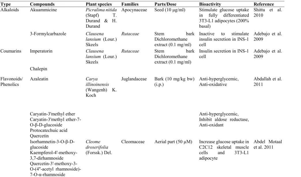
Figure 1.1: Pathogenesis of type 1 diabetes (copied without permission from Atkinson and Eisenbarth, 2001). FPIP, first phase of insulin response; GADA, glutamic acid decarboxylase antibodies; ICA512A/ICA, islet cell antibodies; IAA, insulin antibodies.
1.2.3 Type 2 diabetes mellitus
Type 2 diabetes (T2D) is a heterogeneous disorder characterized by insulin resistance and partially dysfunctional pancreatic β-cells which cannot properly secrete insulin in response to hyperglycemia (Hui et al. 2007). It is the most prevalent type of diabetes, accounting for more than 90%
of all reported diabetes cases in the world (IDF, 2014). The insulin deficiency is relative rather than absolute and usually no insulin treatment (unless special cases) is required for T2D (ADA, 2015). The pathogenesis of insulin resistance in T2D is complex and involves genetic (defect on insulin and its receptor genes etc.) and environmental (obesity, sedentary life, age and physical inactivity) factors (Tuomilehto et al. 2001). Furthermore, inadequate insulin secretion by pancreatic β-cell in type 2 diabetic individuals disrupts the regulation of hepatic gluconeogesis, muscles glucose uptake and lipolysis in adipose tissues (Gastaldelli, 2011). The consequence is postprandial hyperglycemia which results in to T2D. The summary of the pathogenesis for T2D is presented in Figure 1.2.
Figure 1.2: Pathogenesis of type 2 diabetes (copied without permission from Caballero, 2005).
1.2.4 Gestational diabetes mellitus
Gestational diabetes mellitus (GDM) have been defined as heterogeneous group of disorders associated with any glucose intolerance diagnosed usually in the third trimester of the pregnancy (Ashwal and Hod, 2015) when in many cases disorder may improve or disappear after the delivery of baby. Similarly, it has been reported that about 3% to 65% of women with a history of GDM are at high risk of developing T2D in the later part their lives (Lee et al. 2008). In genetic predisposed women (during pregnancy), the alterations in glucose metabolism may lead to mix insulin resistance and impaired insulin secretion (Whitelaw and Gayle, 2010). This condition usually exaggerates as the pregnancy period increases which ultimately result in to hyperglycemia. The pathogenesis of GDM is summarized in Figure 1.3.
Figure 1.3: Pathogenesis of gestational diabetes mellitus (GDM) (prepaid based on Whitelaw and Gayle (2010) report).
1.2.5 Specific types of diabetes due to other causes (secondary type)
This is further sub-divided in to the following;a) Maturity-onset diabetes of the young (MODY)
This form of diabetes is due to the defect on DNA methylation of genes for pancreatic β-cell development (Thomas and Philipson, 2015). It is mainly categorized into neonatal diabetes and maturity- onset diabetes of the young (MODY). The onset of elevated blood glucose as a result of pancreatic β- cell failure usually occurs at an early stage of life (within the first 6 months). Furthermore, in MODY, there is a defect in insulin secretion with less or no reported alterations in insulin function (ADA, 2015;
Thomas and Philipson, 2015).
b) Cystic fibrosis related diabetes
The cystic fibrosis–related diabetes is frequently occurs in individuals with cystic fibrosis. The disorder is due to impair insulin secretion as a result of partial fibrotic reduction of the pancreatic β-cell mass (ADA, 2015). Insulin sensitivity is usually normal or may be partially impaired in this kind of diabetes (Ode and Moran, 2013). Other disorders associated with pancreas include chronic pancreatitis, hereditary hemochromatosis and pancreatic neoplasia.
c) Drug associated diabetes
The medications of some diseases were reported to interfere with complex metabolic processes, thereby alters the secretion and/or function of insulin in physiological system (Thomas and Philipson, 2015). This may likely triggers diabetes or insulin resistance in susceptible individuals. For instance, glucocorticoids were reported to cause insulin resistance, hyperglycemia, and do interfere with some stages in the insulin-signaling pathways via multiple mechanisms (Ferris and Kahn, 2012). Antiretroviral drugs (HIV protease inhibitors) were also reported to induce insulin resistance and impaired glucose tolerance by inhibiting glucose disposal via GLUT4 in cellular system (Koster et al. 2003). Hence, it is clear that the pathogenesis of diabetes changing rapidly to create newer type of diabetes which directly affecting the total prevalence of diabetes mellitus.
1.2.6 Prevalence of diabetes mellitus
The global prevalence of DM is increasing exponentially. Recent data from the International Diabetes Federation (IDF) indicates that DM affects over 387 million people globally and this figure is likely to rise to 592 million by 2035 (IDF, 2014; Guariguata et al. 2014). The disease affects nearly 8.3%
of adult (20-79 years) which are mostly live in the low- and middle-income countries. In Africa, more than 22 million people have diabetes, accounting for about 5.1% of adults (mostly < 60 years) in the region (Peer et al. 2014; IDF, 2014). In addition, multiple factors contribute to this rising prevalence of diabetes including population growth, urbanization, dietary change, nutritional transition, physical inactivity and so on (Guariguata et al. 2014; Hirst et al. 2013).
1.2.7 Diabetes associated complications
Although the pathogenesis of T1D and T2D are different, the consequences of their resulting complications are almost similar (van Dijk and Berl, 2004). These complications are categorized as either acute or chronic, which partly or solely depend on the uncontrolled hyperglycemia (Monnier et al. 2006).
Acute complications include ketoacidosis, hyperosmolar non-ketotic coma and hypoglycemia (Fishbein and Palumbo, 1995). The chronic crises comprise of microvascular (retinopathy, neuropathy and nephropathy) and macro vascular (myocardial infarction, atherosclerosis and peripheral vascular diseases) complications (Heydari et al. 2010; Rahman et al. 2007). The over production of reactive oxygen species (ROS) via metabolic processes and due to hyperglycemia have been considered as the major hallmarks of diabetes-associated complications (Pazdro and Burgess, 2010). In chronic uncontrolled hyperglycemia, there is an increased production of ROS and a declined of in vivo anti- oxidant defense system, a term referred as oxidative stress. The ROS are derived from the normal physiological processes and become highly deleterious if the levels increases and not arrested by complex anti-oxidant systems in the body (Chang and Chuang, 2010). Furthermore, major contributors of hyperglycemia-induced oxidative stress include glucose toxicity, protein glycosylation, increased
production of glycation end products and mitochondrial ROS, the polyol, hexosamine and affected protein kinase C pathways (Giacco and Brownlee, 2010; Chang and Chuang, 2010; Chung et al. 2003;
Maritim et al. 2003). The schematic diagram indicating hyperglycemia-induced oxidative damages is presented in Figure 1.4. The mechanisms involve in micro-vascular and macro-vascular complications can be linked to hyperglycemia-induced oxidative stress. For instance, in diabetic retinopathy, the increased oxidative stress due to inability of the cells to convert glucose to sorbitol caused cellular injury and increased the accumulation of advanced glycated end products (Jiang, 2000).
Figure 1.4: Hyperglycemia-induced oxidative damages in diabetes mellitus (copied without permission from Brownlee, 2005).
1.2.8 Treatment and management of diabetes mellitus
The importance of protecting or delaying hyperglycemia cannot be overemphasized. The acute as well as chronic complications of diabetes are the major causes of morbidity and mortality in all types of diabetes (Fowler, 2008). Fortunately, in the last 2 decades, concerted research efforts by diabetologists and other relevant research scientists highlighted two main strategies to control DM which can be used either alone or in combination of the two, depending on the type and severity of diabetic condition. These include pharmacological and non-pharmacological approaches (Stolar et al. 2008).
1.2.9 Non-pharmacological therapy
The non-pharmacological option refers to the use of lifestyle intervention strategies in patients with established risk factors for T2D in order to delay the progression and development of DM. Lifestyle changes such as weight loss, increased physical exercise and some dietary manipulations are effective in preventing and controlling diabetes (Tuomilehto, 2009) and this was demonstrated in a number of randomized controlled clinical trials such as Swedish Malmo feasibility study (Eriksson and Lindgarde, 1991), Chinese Da Qing study (Pan et al. 1997), Finnish diabetes prevention study (Tuomilehto et al.
2001), U.S diabetes prevention program (DPP, 2002), Japanese lifestyle intervention study (Kosaka et al. 2005) and the Indian diabetes prevention program (Ramachandran et al. 2006). It has been reported that lifestyle modification by diabetic patients significantly improved hepatic and muscle insulin sensitivity, muscle glucose uptake and utilization, and the overall glycemic control in addition to decrease lipids and blood pressure levels (Hayes, 2008).
1.2.10 Pharmacological therapy
Pharmacological intervention to prevent diabetes involves the use of drugs or any agents to treat patients with established risk factors. The basic risk factors are impaired glucose tolerance (IGT) or impaired fasting blood glucose (IFG) because approximately 50% of IGT and IFG individuals progress to diabetes (T2D) over their lifetime (De Fronzo and Abdul-Ghani, 2011).
Insulin
It has been well established that insulin therapy reduce hyperglycemia and micro- and macro- vascular complications associated with diabetes in several randomized clinical trials (Holman et al.
2008; UKPDS, 1998; Ohkubo et al. 1995). However, insulin therapy is associated with two major limitations; hypoglycemia and weight gain, arose due to intensive insulin treatment (Swinnen et al.
2009).
Mode of insulin action
Insulin is a polypeptide hormone produced by the pancreatic β-cells of the islets of Langerhans in response to hyperglycemia (Harvey and Ferrier, 2011).The β-cells are freely permeable to glucose via type 2 glucose transporter (GLUT 2) which are immediately phosphorylated to glucose 6-phosphate by glucokinase enzyme. This stimulates the rise in metabolic flux through glycolysis, the citric acid cycle, and the generation of ATP (Murray et al. 2003). Elevation of ATP concentration down regulates the ATP-sensitive K+ channel, leading to depolarization of the pancreatic β-cell membrane. This may eventually increases Ca2+ influx via voltage-sensitive Ca2+ channels and stimulates the exocytosis of insulin (Kieffer et al. 1997). Hence, the level of insulin in the blood is proportionate to that of the blood glucose. Therefore, insulin stimulates glucose transport into adipose tissue and skeletal muscles via type
4 glucose transporter (GLUT 4) which are utilized as metabolic fuel and stored as well (Kieffer et al.
1997).
Conventional oral hypoglycemic drugs
Several classes of pharmacological agents have shown to reduce the risk of developing DM in double blind randomized controlled clinical trials (Phung et al. 2011). These include biguanides (phenformin), α-glycosidase inhibitors (acarbose), sulfonylureas glibenclamide), thiazolidinediones (rosiglitazone) and dipeptidyl peptidase-4 inhibitors (vildagliptin) (Phung et al. 2011; Padwal et al.
2005).
Moreover, these classes of drugs demonstrate their anti-diabetic actions via diverse mechanisms (Figure 1.5). Sulfonylureas, also called insulin secretogogues, improve insulin release from the pancreatic β-cells (Aguilar-Bryan et al. 1995). Biguanides improve sensitivity of insulin to the liver and muscle cells and inhibit hepatic glucose production (De Fronzo, 1999). Some enzyme inhibitors also reduce blood glucose by inhibiting the activities of carbohydrates digestive enzymes (α-amylase and α- glucosidase) located at the intestinal brush border (Krentz et al. 1994). Furthermore, thiozolidinediones also referred as peroxisome proliferator activated receptor (PPAR)-γ agonist which improves insulin sensitivity in the liver, muscle and adipose tissues. They do achieve such effects by binding to PPARγ receptor, which eventually increase the expression of glucose transporters (Kintscher and Law, 2005;
Lehrke and Lazar, 2005). Dipeptidyl peptidase-4 (DPP-4) inhibitors cause reduction on blood glucose levels via inhibiting an enzyme involve in the breakdown of incretin like peptides such as glucagon-like peptide (GLP)-1 and glucose-dependent insulinotropic peptide (GIP) (Hung et al. 2012).
However, these conventional oral drugs stated above have many unwanted adverse effects (hypoglycemia, weight gain, gastrointestinal discomfort, nausea, hypersensitivity, liver and heart failure, and diarrhea) and are not cost effective for the majority of the people, particularly for those who are from the developing countries (Michael et al. 2005). Therefore, safer and more effective treatment options with fewer side effects are required in order to control the progression DM. Thus, with the increasing interest in alternatives to the present conventional oral drugs in the treatment of diabetes, the use of plant- derived products is emerging and receiving much attention by many researchers.
Figure 1.5: Target organs/tissues and mode of actions of orally anti-diabetic drugs (copied without permission from Koutnik, 2013). (DPP4: Dipeptidyl peptidase-4; GLP-1: Glucagon-like peptide).
1.3 Medicinal plants
Medicinal plants are a plants of which one or more parts contain substances that can be used for therapeutic purposes or which are precursors of the useful drugs (Ebadi, 2006; Lewis, 1981). Medicinal plants have formed the basis of health care system worldwide for several years and are still widely used as good sources for the present modern drugs. Recognition of their therapeutic and economic value is emerging and receiving much attention. Plants are important for pharmacological research and drug discovery, not only when bioactive compounds are used directly for treatment of ailments, but also as template for the synthesis of modern conventional drugs (Mendonça-Filho, 2006; Geldenhuys and Mitchell, 2006).
1.3.1 Medicinal plants in future drug discovery
Medicinal plants are natural products endowed with tremendous capacities to treat wide arrays of diseases. The use of plant-based formulation to treat diseases also known as herbal medicine is the oldest form of medicine since around 2600 BCE (Cragg and Newman, 2013; Soladoye et al. 2012). Fortunately, this area has documented the traditional uses of more than 1000 plant-derived formulations which are still being used to treat both communicable and non-communicable diseases. Studies conducted previously have demonstrated that about 80% of isolated compounds, which are used in the modern medicines, are derived from medicinal plants and traditionally these medicinal plants were used for the same or similar purposes (Cragg and Newman, 2013; Farnsworth et al. 1985). For instance, the anti- diabetic agent galegine (a template for synthesis of metformin) was isolated from Galega officinalis L., which plant was used to treat diabetes traditionally (Heinrich, 2010). Similarly, the anti-malarial drug, artemisinin and anti-HIV drug, calanolide A were derived from the plant