UNIVERSITY OF KWAZULU-NATAL
GAG-PROTEASE DRIVEN VIRAL REPLICATION CAPACITY AMONG HIV-1 SUBTYPES: IMPLICATIONS FOR DISEASE PROGRESSION, EPIDEMIC SPREAD, AND
VACCINE DESIGN
Omotayo Oluwaremilekun Farinre: 216073965
Supervisor: Prof. Thumbi Ndung’u Co-Supervisor: Dr. Jaclyn K. Mann
A thesis submitted in fulfilment of the academic requirements for the degree of Doctor of Philosophy in Virology in the School of Laboratory Medicine and Medical Sciences, HIV Pathogenesis Programme, Doris Duke Medical Research Institute, Nelson R. Mandela School of Medicine, University of KwaZulu-Natal.
2020
This project represents original work done by the author and where others have made contributions it has been acknowledged in the text. The experimental work described in this thesis was performed at the HIV Pathogenesis Programme, in the Hasso-Plattner Research Laboratory, from June 2016 to March 2020 in the Doris Duke Medical Research Institute, Nelson R. Mandela School of Medicine, University of KwaZulu-Natal, Durban, South Africa, under the supervision of Prof. Thumbi Ndung’u and Dr. Jaclyn Mann.
Omotayo O. Farinre: _____________________ Date: ___________
As the candidate’s supervisors, we agree to the submission of this thesis.
Prof. Thumbi Ndung’u: _______________________ Date: ____________
Dr. Jaclyn Mann: _______________________ Date: ____________
ͳ
Declaration
I, Omotayo Oluwaremilekun Farinre, declare that
i. The research reported in this thesis, is my original work, except where otherwise indicated.
ii. This thesis has not been submitted for any degree or examination at any other university or tertiary institution.
iii. This thesis does not contain other person’s data, pictures, graphs, or other information, unless specifically stated as being sourced from other people.
iv. This thesis does not contain other persons’ writing, unless specifically acknowledged as being sourced from other scientists. Where other written sources have been quoted, then:
a. Their words have been re-written, but the general information attributed to them has been referenced.
b. Where exact words have been used, the writing has been placed inside quotation marks, and referenced.
v. Where I have reproduced a publication of which I am author, I have indicated in detail which part of the publication was written by myself alone and have fully acknowledged that publication.
vi. This thesis does not contain text, graphics or tables copied and pasted from the internet, unless specifically acknowledged, and the source being detailed in the thesis and in the references section,
Signed: ____________________ Date: _______________
ʹ
Publication(s)
Omotayo Farinre, Kamini Gounder, Tarylee Reddy, Marcel Tongo, Jonathan Hare, Beth Chaplin, Jill Gilmour, Phyllis Kanki, Jaclyn K. Mann, Thumbi Ndung’u. Subtype-specific differences in Gag-protease replication capacity of HIV-1 isolates from East and West Africa. (RETV-D-21-00003R1). Journal of Retrovirolgy.
Statement
This manuscript has been reproduced in part non-continuously throughout the thesis. The candidate performed the experiments described in this paper, and where others made contributions it has been duly acknowledged in the text. The candidate drafted this publication in full and it was reviewed by co-authors.
Omotayo O. Farinre: _____________________ Date: ___________
͵
Conference contributions
1. Omotayo Farinre, Kamini Gounder, Marcel Tongo, Jonathan Hare, Jill Gilmour, Jaclyn Mann, & Thumbi Ndung’u. HIV-1 Isolates from West Africa Show Higher Fitness than to those from East Africa. The Frontiers in Retrovirology Conference, 11- 13 September 2018, Leuven, Belgium, Abstract 157,
Retrovirology 2018, 15 (Suppl 1): O17. Presented by Jaclyn K. Mann
2. Omotayo Farinre, Kamini Gounder, Marcel Tongo, Jonathan Hare, Jill Gilmour, Jaclyn Mann, & Thumbi Ndung’u. HIV-1 Recombinant Viruses from West Africa Display Higher Replication Capacity Compared to Recombinants from East Africa.
HIV research for prevention (HIVR4P): From research to impact Conference on the 21- 25 October 2018, Madrid, Spain. Abstract Poster: P11.05. Presented by Omotayo Farinre.
Ͷ
Dedication
I dedicate this work to my personal Lord and Saviour, Jesus Christ for giving me the strength push to the end even when my own wisdom and efforts failed me.
I also dedicate this work to my wife and children; Funmi, Tobi, Timi and Tomisin. Words are insufficient to express my gratitude for your patience, forbearance, and support throughout this journey.
Thank you.
ͷ
Acknowledgements
This work was supported by the Poliomyelitis Research Foundation (PRF), the South African Department of Science and Innovation through the National Research Foundation (South African Research Chairs Initiative), the Victor Daitz Foundation and the International AIDS Initiative ((UKZNRSA1001). Additional funding was provided by the Sub-Saharan African Network for TB/HIV Research Excellence (SANTHE), a DELTAS Africa Initiative [grant # DEL-15-006]. The DELTAS Africa Initiative is an independent funding scheme of the African Academy of Sciences (AAS)’s Alliance for Accelerating Excellence in Science in Africa (AESA) and supported by the New Partnership for Africa’s Development Planning and Coordinating Agency (NEPAD Agency) with funding from the Wellcome Trust [grant # 107752/Z/15/Z] and the UK government.
I would also like to thank Professor Thumbi Ndung’u (Prof.) for granting me the opportunity to pursue my PhD at the HIV Pathogenesis Programme. Your mature, gentle, patience, kind nature and approach towards managing me as student and giving me some room to make mistakes and learn from them have been exceptional and exemplary. I am particularly inspired by your keen desire to develop critical mass of scientists on the African continent, and this shows in how you mentor your students. I am grateful to God for your support throughout this journey without which I may not have had the courage to finish.
I also want to thank God for my Co-supervisor in the person of Dr. Jaclyn K. Mann (Jaclyn).
If it were not for you taking my request to pursue a PhD with HPP to Prof. Thumbi, I would not have been in HPP in the first place. I cannot forget in a hurry your contribution to my growth, your laser precision approach towards research, critical thought, strict principles and very importantly time-management and planning qualities I still hope to learn from you.
I am grateful to our collaborators who graciously donated plasma samples for this project such as Dr. Marcel Tongo who donated the Cameroonian samples, Dr. Hannah O. Ajoge who donated the Nigerian samples, and to Prof. Phyllis Kanki who donated the Senegalese samples and Beth Chaplin who assisted in amplifying the Senegalese samples. I will also thank Dr. Jill Gilmour, Dr. Jonathan Hare and the entire IAVI Protocol-C team for believing in this work enough to graciously donate the East African samples used for this study.
I would like to thank our Laboratory Manager at the Hasso-Plattner in HPP, in the person of Keshni Hiramen. She and her team work very hard to support postgraduate students who use the laboratory and to ensure that everything I needed for my experiments was made available to me and were in good working order. I would also like to thank my fellow students and colleagues whom we weathered the difficult storm of long and failed experiments together. I would like to thank my sister from Kenya Dr. Doty Ojwach for all her encouragement and putting me through most of the laboratory techniques I am now familiar with, Noni whom we are bracing this PhD storm together, Dr. Funsho Ogunshola, Dr. Omolara Baiyeghuni, Dr.
Kewreshni Naidoo. I would like to appreciate the administrative team of Mrs. Tarryn Leslie and Mr. Silvester Olupot for their support. I would also like to say a big thank you to all my office colleagues Sithembile, Qiniso, Miriam, Anele, Nombali, Trevor and Delon. You all made my stay memorable.
To my mother Mrs. Tolulope Oluwatosin Farinre who is my number one fan, always praying, cheering, rooting, and believing in me even when I doubt myself. Thank you so much mum.
To my sisters Ayes and Temiloobs who have been calling me Dr. Tayo for the last two years, and I have been telling them it is premature, well your wishes are coming true, thank you both for your love and support.
Abstract
Introduction:
The HIV-1 epidemic in sub-Saharan Africa is heterogeneous with diverse unevenly distributed subtypes and regional differences in prevalence. Subtype-specific differences in disease progression rate and transmission efficiency have been reported, but the underlying biological mechanisms have not been fully characterized. In this study, I tested the hypothesis that the subtypes prevalent in the East African epidemic, where adult prevalence rate is higher, have lower viral replication capacity (VRC) than their West African counterparts where adult prevalence rates are lower.
Materials and methods:
Gag-protease sequencing was performed on plasma samples from 213 and 160 antiretroviral- naïve participants from West and East Africa, respectively. Online bioinformatic tools were used to infer HIV-1 subtypes and recombination patterns. Replication capacities of patient- derived gag-protease chimeric viruses from West (n=178) and East (n=114) Africa were determined using a green fluorescent protein reporter-based cell assay. Subtype and regional differences in viral replication capacity and amino acid variants impacting replication capacity were identified using appropriate statistical methods.
Results:
Subtypes identified in West Africa were CRF02_AG (65%, n=139), G (7%, n=15), A3 (5%, n=10), other CRFs (12%, n=26), various pure subtypes (9%, n=19) and A1G recombinants (2%, n=4). Subtypes A1 (64%, n=103), D (22%, n=35), AD (11%, n=17) and AC (3%, n=5) were identified in East Africa. Chimeric viruses from West Africa had significantly higher VRC compared to those from East Africa (p < 0.0001), with subtype-specific differences found among strains within West and East Africa (p < 0.0001). Recombination patterns showed a
ͺ
preference for subtypes D, G or J rather than subtype A in the p6 region of gag, with evidence that subtype-specific differences in this region impact viral replication capacity. Furthermore, the Gag A83V polymorphism was associated with reduced viral replication capacity in CRF02_AG (median < 0.86). HLA-A*23:01 (p = 0.0014) and HLA-C*07:01 (p = 0.002) were associated with significantly lower viral replication capacity in subtype A infected individuals from East Africa.
Conclusion:
Overall, the data showed that viruses from West Africa displayed higher replication capacity than those from East Africa, which is consistent with the hypothesis that lower viral replication capacity is associated with higher population prevalence.
ͻ
Table of contents
Declaration ... 1
Publication(s) ... 2
Conference contributions ... 3
Dedication ... 4
Acknowledgements ... 5
Abstract ... 7
Table of contents ... 9
List of Figures ... 12
List of Tables ... 13
Abbreviations ... 14
1 INTRODUCTION ... 16 ͳǤͳ ǤǤǤǤǤǤǤǤǤǤǤǤǤǤǤǤǤǤǤǤǤǤǤǤǤǤǤǤǤǤǤǤǤǤǤǤǤǤǤǤǤǤǤǤǤǤǤǤǤǤǤǤǤǤǤǤǤǤǤǤǤǤǤǤǤǤǤǤǤǤǤǤǤǤǤǤǤǤǤǤǤǤǤǤǤǤǤǤǤǤǤǤǤǤǤǤǤǤǤǤǤǤǤǤǤǤǤǤǤǤǤǤǤǤǤǤǤǤǤǤǤǤǤǤǤǤǤǤǤǤǤͳ
ͳǤʹ ǤǤǤǤǤǤǤǤǤǤǤǤǤǤǤǤǤǤǤǤǤǤǤǤǤǤǤǤǤǤǤǤǤǤǤǤǤǤǤǤǤǤǤǤǤǤǤǤǤǤǤǤǤǤǤǤǤǤǤǤǤǤǤǤǤǤǤǤǤǤǤǤǤǤǤǤǤǤǤǤǤǤǤǤǤǤǤǤǤǤǤǤǤǤǤǤǤǤǤǤǤǤǤǤǤǤǤǤǤǤǤͳ
ͳǤʹǤͳ ǤǤǤǤǤǤǤǤǤǤǤǤǤǤǤǤǤǤǤǤǤǤǤǤǤǤǤǤǤǤǤǤǤǤǤǤǤǤǤǤǤǤǤǤǤǤǤǤǤǤǤǤǤǤǤǤǤǤǤǤǤǤǤǤǤǤǤǤǤǤǤǤǤǤǤǤǤǤǤǤǤǤǤǤǤǤǤǤǤǤǤǤǤǤǤǤǤǤǤǤǤǤǤǤǤǤǤǤǤǤǤǤǤǤǤǤǤǤͳ
ͳǤʹǤʹ ǤǤǤǤǤǤǤǤǤǤǤǤǤǤǤǤǤǤǤǤǤǤǤǤǤǤǤǤǤǤǤǤǤǤǤǤǤǤǤǤǤǤǤǤǤǤǤǤǤǤǤǤǤǤǤǤǤǤǤǤǤǤǤǤǤǤǤǤǤǤǤǤǤǤǤǤǤǤǤǤǤǤǤǤǤǤǤǤǤǤǤǤǤǤǤǤǤǤͳ ͳǤʹǤ͵ ǦͳƬǦʹǤǤǤǤǤǤǤǤǤǤǤǤǤǤǤǤǤǤǤǤǤǤǤǤǤǤǤǤǤǤǤǤǤǤǤǤǤǤǤǤǤǤǤǤǤǤǤǤǤǤǤǤǤǤǤǤǤǤǤǤǤǤǤǤǤǤǤǤǤǤǤǤǤǤǤǤǤǤǤǤǤͳ ͳǤʹǤͶ ǦͳǤǤǤǤǤǤǤǤǤǤǤǤǤǤǤǤǤǤǤǤǤǤǤǤǤǤǤǤǤǤǤǤǤǤǤǤǤǤǤǤǤǤǤǤǤǤǤǤǤǤǤǤǤǤǤǤǤǤǤǤǤǤǤǤǤǤǤǤǤǤǤǤǤǤǤǤǤǤǤǤǤǤǤǤǤǤǤǤǤǤǤǤǤǤǤǤǤǤǤǤǤǤǤǤ͵
ͳǤʹǤͷ ǤǤǤǤǤǤǤǤǤǤǤǤǤǤǤǤǤǤǤǤǤǤǤǤǤǤǤǤǤǤǤǤǤǤǤǤǤǤǤǤǤǤǤǤǤǤǤǤǤǤǤǤǤǤǤǤǤǤǤǤǤǤǤǤǤǤǤǤǤǤǤǤǤǤͶ ͳǤ͵ ǤǤǤǤǤǤǤǤǤǤǤǤǤǤǤǤǤǤǤǤǤǤǤǤǤǤǤǤǤǤǤǤǤǤǤǤǤǤǤǤǤǤǤǤǤǤǤǤǤǤǤǤǤǤǤǤǤǤǤǤǤǤǤǤǤǤǤǤǤǤǤǤǤǤǤǤǤǤǤǤǤǤǤǤǤǤǤǤǤǤǤǤǤǤǤǤǤǤǤǤǤǤǤǤǤǤǤǤǤǤǤǤǤǤǤǤǤǤǤǤǤͷ ͳǤ͵Ǥͳ ǤǤǤǤǤǤǤǤǤǤǤǤǤǤǤǤǤǤǤǤǤǤǤǤǤǤǤǤǤǤǤǤǤǤǤǤǤǤǤǤǤǤǤǤǤǤǤǤǤǤǤǤǤǤǤǤǤǤǤǤǤǤǤǤǤǤǤǤǤǤǤǤǤǤǤǤǤǤǤǤǤǤǤǤǤ
ͳǤ͵Ǥʹ Ǧͳ ǤǤǤǤǤǤǤǤǤǤǤǤǤǤǤǤǤǤǤǤǤǤǤǤǤǤǤǤǤǤǤǤǤǤǤǤǤǤǤǤǤǤǤǤǤǤǤǤǤǤǤǤǤǤǤǤǤǤǤǤǤǤǤǤǤǤǤǤǤǤǤǤǤǤǤǤǤǤǤǤǤǤǤǤǤǤǤǤǤǤǤǤǤǤ
ͳǤͶ ǦͳǤǤǤǤǤǤǤǤǤǤǤǤǤǤǤǤǤǤǤǤǤǤǤǤǤǤǤǤǤǤǤǤǤǤǤǤǤǤǤǤǤǤǤǤǤǤǤǤǤǤǤǤǤǤǤǤǤǤǤǤǤǤǤǤǤǤǤǤǤǤǤǤǤǤǤǤǤǤǤǤǤǤǤǤǤǤǤǤǤǤǤǤǤǤǤǤǤǤǤǤǤǤǤǤǤǤǤǤǤǤǤǤǤͳͳ ͳǤͶǤͳ ǦͳǤǤǤǤǤǤǤǤǤǤǤǤǤǤǤǤǤǤǤǤǤǤǤǤǤǤǤǤǤǤǤǤǤǤǤǤǤǤǤǤǤǤǤǤǤǤǤǤǤǤǤǤǤǤǤǤǤǤǤǤǤǤǤǤǤǤǤǤǤǤǤǤǤǤǤǤǤǤǤǤǤǤǤǤǤǤǤǤǤǤǤͳͳ ͳǤͶǤʹ Ǧͳ ǤǤǤǤǤǤǤǤǤǤǤǤǤǤǤǤǤǤǤǤǤǤǤǤǤǤǤǤǤǤǤǤǤǤǤǤǤǤǤǤǤǤǤǤǤǤǤǤǤǤǤǤǤǤǤǤǤǤǤǤǤǤǤǤǤǤǤǤǤǤǤǤǤǤǤǤǤǤǤǤǤǤǤǤǤǤǤǤǤǤǤǤǤǤǤǤǤǤǤǤͳ͵
ͳǤͶǤ͵ ȋȌǤǤǤǤǤǤǤǤǤǤǤǤǤǤǤǤǤǤǤǤǤǤǤǤǤǤǤǤǤǤǤǤǤǤǤǤǤǤǤǤǤǤǤǤǤǤǤǤǤǤǤǤǤǤǤǤǤǤǤǤǤǤǤǤǤǤǤǤǤǤǤǤǤǤǤǤǤǤǤǤǤǤǤǤǤǤǤǤǤͳ
ͳǤͷ ǦͳǤǤǤǤǤǤǤǤǤǤǤǤǤǤǤǤǤǤǤǤǤǤǤǤǤǤǤǤǤǤǤǤǤǤǤǤǤǤǤǤǤǤǤǤǤǤǤǤǤǤǤǤǤǤǤǤǤǤǤǤǤǤǤǤǤǤǤǤǤǤǤǤǤǤǤǤǤǤǤǤǤǤǤǤǤǤǤǤǤǤǤǤǤǤǤǤǤǤǤǤǤǤǤǤǤǤǤǤǤǤǤͳ
ͳǤͷǤͳ ȋ Ȍ ǤǤǤǤǤǤǤǤǤǤǤǤǤǤǤǤǤǤǤǤǤǤǤǤǤǤǤǤǤǤǤǤǤǤǤǤǤǤǤǤǤǤǤǤǤǤǤǤǤǤǤǤǤǤǤǤǤǤǤǤǤǤǤǤǤǤǤǤǤǤǤǤǤǤǤǤǤǤǤǤǤǤǤǤǤǤǤǤǤǤǤǤǤǤǤͳ
ͳǤͷǤʹ ǤǤǤǤǤǤǤǤǤǤǤǤǤǤǤǤǤǤǤǤǤǤǤǤǤǤǤǤǤǤǤǤǤǤǤǤǤǤǤǤǤǤǤǤǤǤǤǤǤǤǤǤǤǤǤǤǤǤǤǤǤǤǤǤǤǤǤǤǤǤǤǤǤǤǤǤǤǤǤǤǤǤǤǤǤǤǤǤǤǤǤǤͳ
ͳǤ ǤǤǤǤǤǤǤǤǤǤǤǤǤǤǤǤǤǤǤǤǤǤǤǤǤǤǤǤǤǤǤǤǤǤǤǤǤǤǤǤǤǤǤǤǤǤǤǤǤǤǤǤǤǤǤǤǤǤǤǤǤǤǤǤǤǤǤǤǤǤǤǤǤǤǤǤǤǤǤǤǤǤǤǤǤǤǤǤǤǤǤǤǤǤǤǤǤǤǤǤǤǤǤǤǤǤǤǤǤǤǤǤǤǤǤǤǤǤǤǤǤǤǤǤǤͳͻ ͳǤǤͳ ǤǤǤǤǤǤǤǤǤǤǤǤǤǤǤǤǤǤǤǤǤǤǤǤǤǤǤǤǤǤǤǤǤǤǤǤǤǤǤǤǤǤǤǤǤǤǤǤǤǤǤǤǤǤǤǤǤǤǤǤǤǤǤǤǤǤǤǤǤǤǤǤǤǤǤǤǤǤǤǤǤǤǤǤǤǤǤǤǤǤǤǤǤǤǤǤǤǤǤǤǤǤǤǤǤǤǤǤǤǤǤǤǤͳͻ ͳǤǤʹ ǤǤǤǤǤǤǤǤǤǤǤǤǤǤǤǤǤǤǤǤǤǤǤǤǤǤǤǤǤǤǤǤǤǤǤǤǤǤǤǤǤǤǤǤǤǤǤǤǤǤǤǤǤǤǤǤǤǤǤǤǤǤǤǤǤǤǤǤǤǤǤǤǤǤǤǤǤǤǤǤǤǤǤǤǤǤǤǤǤǤǤǤǤǤǤǤǤǤǤǤǤǤǤǤǤǤǤǤʹͲ
ͳͲ
ͳǤ ǦͳǤǤǤǤǤǤǤǤǤǤǤǤǤǤǤǤǤǤǤǤǤǤǤǤǤǤǤǤǤǤǤǤǤǤǤǤǤǤǤǤǤǤǤǤǤǤǤǤǤǤǤǤǤǤǤǤǤǤǤǤǤǤǤǤǤǤǤǤǤǤǤǤǤǤǤǤǤǤǤǤǤǤǤǤǤǤǤǤǤǤǤǤǤǤǤǤǤǤǤǤǤǤʹʹ ͳǤǤͳ ǤǤǤǤǤǤǤǤǤǤǤǤǤǤǤǤǤǤǤǤǤǤǤǤǤǤǤǤǤǤǤǤǤǤǤǤǤǤǤǤǤǤǤǤʹʹ ͳǤǤʹ ǦͳǦ ǤǤǤǤǤǤǤǤǤǤǤǤǤǤǤǤǤǤǤǤǤǤǤǤǤǤǤǤǤǤǤǤǤʹͶ ͳǤǤ͵ ǦͳǦ ǤǤǤǤǤǤǤǤǤǤǤǤǤǤǤǤǤǤǤǤǤǤǤǤǤǤǤǤǤǤǤǤǤǤǤǤǤǤǤǤǤǤǤǤǤǤǤǤǤǤǤǤǤǤǤʹͷ ͳǤͺ ǤǤǤǤǤǤǤǤǤǤǤǤǤǤǤǤǤǤǤǤǤǤǤǤǤǤǤǤǤǤǤǤǤǤǤǤǤǤǤǤǤǤǤǤǤǤǤǤǤǤǤǤǤǤǤǤǤǤǤǤǤǤǤǤǤǤǤǤǤǤǤǤǤǤǤǤǤǤǤǤǤǤǤǤǤǤǤǤǤǤǤǤǤǤǤǤǤǤǤǤǤǤǤǤǤʹ
ͳǤͺǤͳ ǤǤǤǤǤǤǤǤǤǤǤǤǤǤǤǤǤǤǤǤǤǤǤǤǤǤǤǤǤǤǤǤǤǤǤǤǤǤǤǤǤǤǤǤǤǤǤǤǤǤǤǤǤǤǤǤǤǤǤǤǤʹ
ͳǤͻ ǤǤǤǤǤǤǤǤǤǤǤǤǤǤǤǤǤǤǤǤǤǤǤǤǤǤǤǤǤǤǤǤǤǤǤǤǤǤǤǤǤǤǤǤǤǤǤǤǤǤǤǤǤǤǤǤǤǤǤǤǤǤǤǤǤǤǤǤǤǤǤǤǤǤǤǤǤǤǤǤǤǤǤǤǤǤǤǤǤǤǤǤǤǤǤǤǤǤǤǤǤǤǤǤǤǤǤǤǤǤǤǤǤǤǤǤǤǤǤǤǤǤǤǤʹͺ ͳǤͻǤͳ ǤǤǤǤǤǤǤǤǤǤǤǤǤǤǤǤǤǤǤǤǤǤǤǤǤǤǤǤǤǤǤǤǤǤǤǤǤǤǤǤǤǤǤǤǤǤǤǤǤǤǤǤǤǤǤǤǤǤǤǤǤǤǤǤǤǤǤǤǤǤǤǤǤǤǤǤǤǤǤǤǤǤǤǤǤǤǤǤǤǤǤǤǤǤǤǤǤǤǤǤǤǤǤǤǤǤǤǤǤǤǤǤǤǤǤǤǤǤǤǤǤǤǤǤʹͻ ͳǤͻǤʹ ǤǤǤǤǤǤǤǤǤǤǤǤǤǤǤǤǤǤǤǤǤǤǤǤǤǤǤǤǤǤǤǤǤǤǤǤǤǤǤǤǤǤǤǤǤǤǤǤǤǤǤǤǤǤǤǤǤǤǤǤǤǤǤǤǤǤǤǤǤǤǤǤǤǤǤǤǤǤǤǤǤǤǤǤǤǤǤǤǤǤǤ͵Ͳ 2 MATERIALS AND METHODS ... 32 ʹǤͳ ǤǤǤǤǤǤǤǤǤǤǤǤǤǤǤǤǤǤǤǤǤǤǤǤǤǤǤǤǤǤǤǤǤǤǤǤǤǤǤǤǤǤǤǤǤǤǤǤǤǤǤǤǤǤǤǤǤǤǤǤǤǤǤǤǤǤǤǤǤǤǤǤǤǤǤǤǤǤǤǤǤǤǤǤǤǤǤǤǤǤǤǤǤǤǤǤǤǤǤǤǤǤǤǤǤǤǤǤǤǤǤǤǤǤǤǤǤǤǤǤǤǤ͵ʹ ʹǤʹ ǤǤǤǤǤǤǤǤǤǤǤǤǤǤǤǤǤǤǤǤǤǤǤǤǤǤǤǤǤǤǤǤǤǤǤǤǤǤǤǤǤǤǤǤǤǤǤǤǤǤǤǤǤǤǤǤǤǤǤǤǤǤǤǤǤǤǤǤǤǤǤǤǤǤǤǤǤǤǤǤǤǤǤǤǤǤǤǤǤǤǤǤǤǤǤǤǤǤǤǤǤǤǤǤǤǤǤǤǤǤǤǤǤǤǤǤǤǤǤǤǤǤǤǤǤǤ͵ʹ ʹǤ͵ ǤǤǤǤǤǤǤǤǤǤǤǤǤǤǤǤǤǤǤǤǤǤǤǤǤǤǤǤǤǤǤǤǤǤǤǤǤǤǤǤǤǤǤǤǤǤǤǤǤǤǤǤǤǤǤǤǤǤǤǤǤǤǤǤǤǤǤǤǤǤǤǤǤǤǤǤǤǤǤǤǤǤǤǤǤǤǤǤǤǤǤǤǤǤǤǤǤǤǤǤǤǤǤǤǤǤǤǤǤ͵͵
ʹǤ͵Ǥͳ Gag-proteaseǤǤǤǤǤǤǤǤǤǤǤǤǤǤǤǤǤǤǤǤǤǤǤǤǤǤǤǤǤǤǤǤǤǤǤǤǤǤǤǤǤǤǤǤ͵͵
ʹǤ͵Ǥʹ ǦͳǤǤǤǤǤǤǤǤǤǤǤǤǤǤǤǤǤǤǤǤǤǤǤǤǤǤǤǤǤǤǤǤǤǤǤǤǤǤǤǤǤǤǤǤǤǤǤǤǤǤǤǤǤǤǤǤǤǤǤǤǤǤǤǤǤǤǤǤǤǤǤǤǤǤǤǤǤǤǤǤǤǤǤǤǤǤǤǤǤǤǤǤǤǤǤǤǤǤǤǤǤǤǤǤǤǤǤǤǤǤǤǤǤǤ͵
ʹǤ͵Ǥ͵ ǤǤǤǤǤǤǤǤǤǤǤǤǤǤǤǤǤǤǤǤǤǤǤǤǤǤǤǤǤǤǤǤǤǤǤǤǤǤǤǤǤǤǤǤǤǤǤǤǤǤǤǤǤǤǤǤǤǤǤǤǤǤǤǤǤǤǤǤǤǤǤǤǤǤǤǤǤǤǤǤǤǤǤǤǤǤǤǤǤǤǤǤǤǤǤǤǤǤǤǤǤǤǤ͵
ʹǤ͵ǤͶ Ǧͳ ǤǤǤǤǤǤǤǤǤǤǤǤǤǤǤǤǤǤǤǤǤǤǤǤǤǤǤǤǤǤǤǤǤǤǤǤǤǤǤǤǤǤǤǤǤǤǤǤǤǤǤǤǤǤǤǤǤǤǤǤǤǤǤǤǤǤǤǤǤ͵
ʹǤͶ ͶǦ͵οgag-protease ǤǤǤǤǤǤǤǤǤǤǤǤǤǤǤǤǤǤǤǤǤǤǤǤǤǤǤǤǤǤǤǤǤǤǤǤǤǤǤǤǤǤǤǤǤǤǤǤǤǤǤǤ͵ͺ ʹǤͶǤͳ ǤǤǤǤǤǤǤǤǤǤǤǤǤǤǤǤǤǤǤǤǤǤǤǤǤǤǤǤǤǤǤǤǤǤǤǤǤǤǤǤǤǤǤǤǤǤǤǤǤǤǤǤǤǤǤǤǤǤǤǤǤǤǤǤǤǤǤǤǤǤǤǤǤǤǤǤǤǤǤǤǤǤǤǤǤǤǤǤǤǤǤ͵ͻ ʹǤͶǤʹ Ǧ ǤǤǤǤǤǤǤǤǤǤǤǤǤǤǤǤǤǤǤǤǤǤǤǤǤǤǤǤǤǤǤǤǤǤǤǤǤǤǤǤǤǤǤǤǤǤǤǤǤǤǤǤǤǤǤǤǤǤǤǤǤǤǤǤǤǤǤǤǤǤǤǤǤǤǤǤǤǤǤǤǤǤǤǤǤǤǤǤͶͲ ʹǤͶǤ͵ ǤǤǤǤǤǤǤǤǤǤǤǤǤǤǤǤǤǤǤǤǤǤǤǤǤǤǤǤǤǤǤǤǤǤǤǤǤǤǤǤǤǤǤǤǤǤǤǤǤǤǤǤǤǤǤǤǤǤǤǤǤǤǤǤǤǤǤǤǤǤǤǤǤǤǤǤǤǤǤǤǤǤǤǤǤǤǤǤǤǤǤǤǤͶͳ ʹǤͶǤͶ Ͷ͵ǦǤǤǤǤǤǤǤǤǤǤǤǤǤǤͶʹ ʹǤͶǤͷ ǤǤǤǤǤǤǤǤǤǤǤǤǤǤǤǤǤǤǤǤǤǤǤǤǤǤǤǤǤǤǤǤǤǤǤǤǤǤǤǤǤǤǤǤǤǤǤǤǤǤǤǤǤǤǤǤǤǤǤǤǤǤǤǤǤǤǤǤǤǤǤǤǤǤǤǤǤǤǤǤǤǤǤǤǤǤǤǤǤǤǤǤǤǤǤǤǤǤǤǤǤǤǤǤǤǤǤǤǤǤǤǤǤǤǤǤͶ͵
ʹǤͶǤ ǤǤǤǤǤǤǤǤǤǤǤǤǤǤǤǤǤǤǤǤǤǤǤǤǤǤǤǤǤǤǤǤǤǤǤǤǤǤǤǤǤǤǤǤǤǤǤǤǤǤǤǤǤǤǤǤǤǤǤǤǤǤǤǤǤǤǤǤǤǤǤǤǤǤǤǤǤǤǤǤǤǤǤǤǤǤǤǤǤǤǤǤǤǤͶ͵
ʹǤͷ ǤǤǤǤǤǤǤǤǤǤǤǤǤǤǤǤǤǤǤǤǤǤǤǤǤǤǤǤǤǤǤǤǤǤǤǤǤǤǤǤǤǤǤǤǤǤǤǤǤǤǤǤǤǤǤǤǤǤǤǤǤǤǤǤǤǤǤǤǤǤǤǤǤǤǤǤǤǤǤǤǤǤǤǤǤǤǤǤǤǤǤǤǤǤǤǤǤǤǤǤǤǤǤǤǤǤǤǤǤǤǤǤǤǤǤǤǤǤǤǤǤǤǤǤǤǤǤǤǤͶ
3 RESULTS ... 47
͵Ǥͳ ǤǤǤǤǤǤǤǤǤǤǤǤǤǤǤǤǤǤǤǤǤǤǤǤǤǤǤǤǤǤǤǤǤǤǤǤǤǤǤǤǤǤǤǤǤǤǤǤǤǤǤǤǤǤǤǤǤǤǤǤǤǤǤǤǤǤǤǤǤǤǤǤǤǤǤǤǤǤǤǤǤǤǤǤǤǤǤǤǤǤǤǤǤǤǤǤǤǤǤǤǤǤǤǤǤǤǤǤǤǤǤǤǤǤǤǤǤǤǤǤǤǤǤǤǤǤǤǤǤǤǤǤǤǤǤǤǤǤǤǤǤͶ
͵Ǥʹ ǤǤǤǤǤǤǤǤǤǤǤǤǤǤǤǤǤǤǤǤǤǤǤǤǤǤǤǤǤǤǤǤǤǤǤǤǤǤǤǤǤǤǤǤǤǤǤǤǤǤǤǤǤǤǤǤǤǤǤǤǤǤǤǤǤǤǤǤǤǤǤǤǤǤǤǤǤǤǤǤǤǤǤǤǤǤǤǤǤǤǤǤǤǤǤǤǤǤǤǤǤǤǤǤǤǤǤǤǤǤǤǤǤǤǤǤǤǤͶ
͵Ǥ͵ ǤǤǤǤǤǤǤǤǤǤǤǤǤǤǤǤǤǤǤǤǤǤǤǤǤǤǤǤǤǤǤǤǤǤǤǤǤǤǤǤǤǤǤǤǤǤǤǤǤǤǤǤǤǤǤǤǤǤǤǤǤǤǤǤǤǤǤǤǤǤǤǤǤǤǤǤǤǤǤǤǤǤǤǤǤǤǤǤǤǤǤǤǤǤǤǤǤǤǤǤǤǤǤǤǤǤǤǤͶͻ
͵ǤͶ Ǧͳ ǤǤǤǤǤǤǤǤǤǤǤǤǤǤǤǤǤǤǤǤǤǤǤǤǤǤǤǤǤǤǤǤǤǤǤǤǤǤǤǤǤǤǤǤǤǤǤǤǤǤǤǤǤǤǤǤǤǤǤǤǤǤǤǤǤǤǤǤǤǤǤǤǤǤǤǤǤǤǤǤǤǤǤǤǤǤǤǤǤǤǤǤǤǤǤǤǤǤǤǤǤǤǤǤǤǤͷͲ
͵ǤͶǤͳ ǦͳǤǤǤǤǤǤǤǤǤǤǤǤǤǤǤǤǤǤǤǤǤǤǤǤǤǤǤǤǤǤǤǤǤǤǤǤǤǤǤǤǤǤǤǤǤǤǤǤǤǤǤǤǤǤǤǤǤǤǤǤǤǤǤǤǤǤǤǤǤǤǤǤǤǤǤǤǤǤǤǤǤǤǤǤǤǤǤǤǤǤǤǤǤǤǤǤǤǤǤǤǤǤǤǤǤǤǤǤǤǤǤǤǤǤͷͲ
͵ǤͶǤʹ ǤǤǤǤǤǤǤǤǤǤǤǤǤǤǤǤǤǤǤǤǤǤǤǤǤǤǤǤǤǤǤǤǤǤǤǤǤǤǤǤǤǤǤǤǤǤǤǤǤǤǤǤǤǤǤǤǤǤǤǤǤǤǤǤǤǤǤǤǤǤǤǤǤǤǤǤǤǤǤǤǤǤʹ
͵ǤͶǤ͵ Ǧͳ ǤǤǤǤǤǤǤǤǤǤǤǤǤǤǤǤǤǤǤǤǤǤǤǤǤǤǤǤǤǤǤǤǤǤǤǤǤǤǤǤǤǤǤǤǤǤǤǤǤǤǤǤǤǤǤǤǤǤǤǤǤǤǤǤǤǤǤǤǤ
͵ǤͶǤͶ ǦǦ ǤǤǤǤǤǤǤǤǤǤǤǤǤǤǤǤǤǤǤǤǤǤǤǤǤǤǤǤǤǤǤǤǤǤǤǤǤǤǤǤǤǤǤʹ
ͳͳ
͵Ǥͷ Ǧ ǤǤǤǤǤǤǤǤǤǤǤǤǤǤǤǤǤǤǤǤǤǤǤǤǤǤǤǤǤǤǤǤǤǤǤǤǤǤǤǤǤǤǤǤǤǤǤǤǤǤǤǤǤǤǤǤǤǤǤǤǤǤǤǤǤǤǤǤǤǤ
͵ǤͷǤͳ ǤǤǤǤǤǤǤǤǤǤǤǤǤǤǤǤǤǤǤǤǤǤǤǤǤǤǤǤǤǤǤǤǤ
͵ǤͷǤʹ ǤǤǤǤǤǤǤǤǤǤǤǤǤǤǤǤǤǤǤǤǤǤǤǤǤǤǤǤǤǤǤǤǤǤ
͵ǤͷǤ͵ Gag-protease
ͻ
͵ǤͷǤͶ ȋ ȌǤǤǤǤǤǤǤǤǤǤǤǤǤǤǤǤǤǤǤǤǤǤǤǤǤǤǤǤǤǤǤǤǤǤͺʹ
͵ǤͷǤͷ Gag-Protease ǤǤǤǤǤǤǤǤͺ
͵ǤͷǤ ǤǤǤǤǤǤǤǤǤǤǤǤǤǤǤǤǤǤǤǤǤǤǤǤǤǤǤǤǤǤǤǤǤǤǤǤǤǤǤǤǤǤǤǤǤǤǤǤǤǤǤǤǤǤǤǤǤǤǤǤǤǤǤǤǤǤǤǤǤǤǤǤǤǤǤǤǤǤǤǤǤǤǤǤǤǤǤǤͺͺ
͵ǤͷǤ ǦǦ ǤǤǤǤǤǤǤǤǤǤǤǤǤǤǤǤǤǤǤǤǤǤǤǤǤǤǤǤǤǤǤǤǤǤǤǤǤǤǤǤǤǤǤǤǤǤǤǤǤǤǤǤǤǤǤǤǤǤǤǤǤǤǤǤǤǤǤǤǤǤǤǤǤǤǤǤͻͲ 4 DISCUSSION ... 93 ͶǤͳ Ǧͳ ǤǤǤǤǤǤǤǤǤǤǤǤǤǤǤǤǤǤǤǤǤǤǤǤǤǤǤǤǤǤǤǤǤǤǤǤǤǤǤǤǤǤǤǤǤǤǤǤǤǤǤǤǤǤǤǤǤǤǤǤǤǤǤͻͶ ͶǤʹ Ǧ ǤǤǤǤǤǤǤǤǤǤǤǤǤǤǤǤǤǤǤǤǤǤǤǤǤǤǤǤǤǤǤǤǤǤǤǤǤǤǤǤǤǤǤǤǤǤǤǤǤǤǤǤǤǤǤǤǤǤͻͷ ͶǤ͵ ͳǤǤǤǤǤǤǤǤǤǤǤǤǤǤǤǤǤǤǤǤǤǤǤǤǤǤǤǤǤǤǤǤǤǤǤǤǤǤǤǤǤǤǤǤǤǤǤǤǤǤǤǤǤǤǤǤǤǤǤǤǤǤǤǤǤǤǤǤǤǤǤǤǤͻͺ ͶǤͶ ͳǤǤǤǤǤǤǤǤǤǤǤǤǤǤǤǤǤǤǤǤͻͻ ͶǤͷ ǦͳǤǤǤǤǤǤǤǤǤǤǤǤǤǤǤǤǤǤǤǤǤǤǤǤǤǤǤǤǤǤǤǤǤǤǤǤǤǤǤǤǤǤǤǤǤǤǤǤǤǤǤǤǤǤǤǤǤǤǤǤǤǤǤǤǤǤǤǤǤǤǤǤǤǤǤǤǤǤǤǤǤǤǤǤǤǤǤǤǤǤǤǤǤǤǤǤǤǤǤǤǤǤǤǤǤǤǤǤǤǤǤǤǤǤǤǤǤǤǤͳͲͲ ͶǤ ǤǤǤǤǤǤǤǤǤǤǤǤǤǤǤǤǤǤǤǤǤǤǤǤǤǤǤǤǤǤǤǤǤǤǤǤǤǤǤǤǤǤǤǤǤǤǤǤǤǤǤǤǤǤǤǤǤǤǤǤǤǤǤǤǤǤǤǤǤǤǤǤǤǤǤǤǤǤǤǤǤǤǤǤǤǤǤǤǤǤǤǤǤǤǤǤǤǤǤǤǤǤǤǤǤǤǤǤǤǤǤǤǤǤǤǤǤǤͳͲͳ ͶǤ ǤǤǤǤǤǤǤǤǤǤǤǤǤǤǤǤǤǤǤǤǤǤǤǤǤǤǤǤǤǤǤǤǤǤǤǤǤǤǤǤǤǤǤǤǤǤǤǤǤǤǤǤǤǤǤǤǤǤǤǤǤǤǤǤǤǤǤǤǤǤǤǤǤǤǤǤǤǤǤǤͳͲʹ ͶǤͺ ǤǤǤǤǤǤǤǤǤǤǤǤǤǤǤǤǤǤǤǤǤǤǤǤǤǤǤǤǤǤǤǤǤǤǤǤǤǤǤǤǤǤǤǤǤǤǤǤǤǤǤǤǤǤǤǤǤǤǤǤǤǤǤǤǤǤǤǤǤǤǤǤǤǤǤǤǤǤǤǤǤǤǤǤǤǤǤǤǤǤǤǤǤǤǤǤǤǤǤǤǤǤǤǤǤǤǤǤǤǤǤǤǤǤǤǤǤǤǤǤǤǤǤǤǤǤǤǤǤǤͳͲ
REFERENCES ... 108
ͳʹ
List of Figures
Figure 1.1: Distribution of non-human primates in West-Central Africa ... 2
Figure 1.2: Phylogenetic analysis showing SIV and HIV relatedness... 2
Figure 1.3: Updated classification of HIV-1 subtypes ... 8
Figure 1.4: New sub-sub types for A and D ... 8
Figure 1.5: Mosaic genome structures of CRFs... 10
Figure 1.6: Structure of HIV-1 virion ... 12
Figure 1.7: HIV-1 genome map ... 12
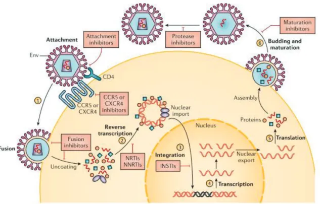
Figure 1.8: HIV-1 replication cycle with ART targets ... 15
Figure 1.9: Phases of natural HIV-1 infection ... 18
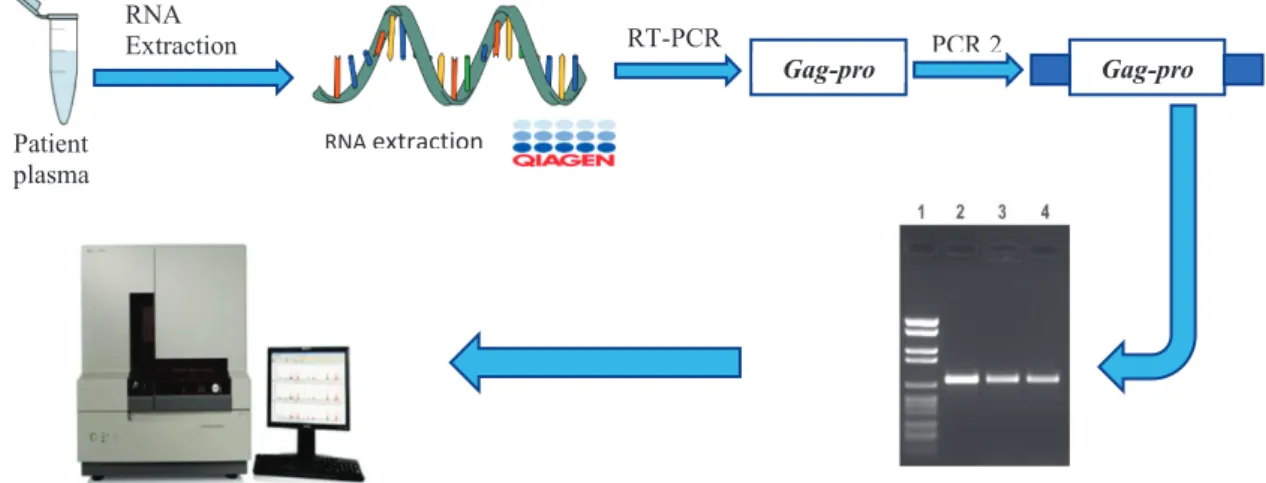
Figure 2.1: Amplification and sequencing summary ... 35
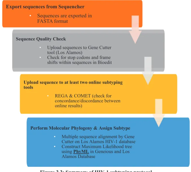
Figure 2.2: Summary of HIV-1 subtyping protocol... 37
Figure 2.3: Inter-subtype recombination analysis ... 38
Figure 2.4: Summary of co-transfection and viral harvest protocol ... 42
Figure 2.5: Summary of viral titration and replication capacity assay ... 45
Figure 3.1: Image of secondary amplicons resolved by 1% agarose gel electrophoresis. ... 49
Figure 3.2: Phylogenetic analysis of gag-protease sequences for West Africa ... 63
Figure 3.3:Phylogenetic analysis of gag-protease sequences for East Africa ... 65
Figure 3.4: Phylogenetic tree showing common subtypes in West and East Africa ... 66
Figure 3.5: Subtype distribution in West Africa ... 68
Figure 3.6: Subtype distribution in East Africa ... 70
Figure 3.7: Output of inter-subtype recombination analysis tools ... 73
Figure 3.8: Phylogenetic confirmation of sequence fragments ... 74
Figure 3.9: Inter-subtype recombination analysis ... 75
Figure 3.11: VRC and markers of disease progression... 78
Figure 3.12: Gag-protease VRC within West and East Africa ... 80
Figure 3.13: Gag-protease VRC within East and West Africa ... 81
Figure 3.14: VRC association with HLA class I expression ... 87
Figure 3.15: Amino Acid variation in Gag p6 region ... 89
ͳ͵
List of Tables
Table 1.1: Lentivirus Family... 1
Table 3.1: Demographic and clinical characteristics of participants ... 48
Table 3.2: Subtyping data for West Africa ... 50
Table 3.3: Subtyping data for East Africa ... 57
Table 3.4:Multivatiable model associating subtype and VRC for West Africa. ... 84
Table 3.5: Multivariable model associating subtype and VRC for East Africa. ... 85
Table 3.6: Codon-by-codon analysis for subtype CRF02_AG ... 91
Table 3.7: Codon-by-codon analysis for subtype A1 ... 92
ͳͶ
Abbreviations
ADDC Antibody dependent cellular cytotoxicity AIDS Acquired Immuno-deficiency syndrome APC Antigen presenting cells
APOBEC3G Apolipoprotein-B mRNA editing enzyme catalytic polypeptide-like 3G ART Anti-retroviral therapy
ARV AIDS Related Virus BSA Bovine serum albumin
CA Capsid
CCR5 Chemokine receptor 5
CD4+ Cluster of differentiation 4 positive CD8+ Cluster of differentiation 8 positive CDC Centres for disease control and prevention
cpx Complex
cpz Chimpanzee
CRF Circulating recombinant form CTL Cytotoxic T lymphocytes CXCR4 Chemokine receptor 4 DMSO Dimethyl sulfoxide DNA Deoxyribonucleic acid
DRC Democratic republic of Congo
dsDNA Double stranded Deoxyribonucleic acid EBF Early B cell factor
env Envelope
ER Endoplasmic reticulum FDC Follicular dendritic cells Gag Group specific antigen
GM-CSF Granulocyte-macrophage colony stimulating factor GWAS Genome wide association studies
gor Gorilla
gp Glycoprotein
HAART Highly active antiretroviral therapy HIV Human immunodeficiency virus HLA Human leucocyte antigen HMA Heteroduplex mobility assay HTLV Human T-lymphotropic virus IDU Intravenous drug user IFN-γ Interferon gamma
Ig Immunoglobulin
IL Interleukin
IN Integrase
LAS Lymphadenopathy syndrome LAV Lymphadenopathy associated virus
ͳͷ
LTR Long Terminal Repeat
MA Matrix
MACS Multicentre AIDS Cohort Study MHC Major histocompatibility complex MIP Macrophage inflammatory protein mRNA Messenger ribonucleic acid MSMs Men that have sex with men MTCT Mother to child transmission
mtDNA Mitochondrial deoxyribonucleic acid nef Negative factor
nt Nucleotide
Nκ Natural killer ORF Open reading frame
PAMPS Pathogen associated molecular patterns PAX Paired box gene
PBS Phosphate buffered saline PFA Paraformaldehyde
PIC Pre-integration complex
Pol Polymerase
PR Protease
Prep Pre exposure prophylaxis
PRRs Pathogen recognition receptors Ptt Pan troglodytes troglodytes rev Regulator of expression of virion RNA Ribonucleic Acid
RNAse H Ribonuclease H RT Reverse transcriptase
SIV Simian immunodeficiency virus smm Sooty mangabey monkey SU Surface protein
tat Transactivation protein TCR T cell receptor TLR Toll-like receptors TM Transmembrane protein
tMRCA Time to most recent common ancestor TNF-α Tumour necrosis factor alpha
TRIM5α Tripartite motif 5α URF Unique recombination form VCT Voluntary counselling and testing vif Viral infectivity factor
vpr Viral protein R vpu Viral protein U
VRC Viral replicative capacity WHO World Health Organisation.
ͳ
1 INTRODUCTION
1.1 Background
Since the identification of the human immunodeficiency virus (HIV) as the etiologic agent of the acquired immunodeficiency syndrome (AIDS) almost four decades ago (1-3), over 60 million people have been infected globally, with an excess of 35 million currently living with the virus (3-6). AIDS is a set of symptoms and illnesses that develop because of advanced HIV infection, whereby the host immune system becomes wholly compromised. In 1981, the United States Centers for Disease Control and Prevention (CDC) reported a characteristic set of symptoms which included Pneumocystis carinii pneumonia and Kaposi’s sarcoma that appeared to be prevalent within a community of homosexual men in the United States (7-9).
These would later turn out to be the first set of AIDS-related cases reported (7). Since then, the epidemic has grown widely, and extended beyond homosexual men into the general population.
Sub-Saharan Africa currently accounts for about 70% of the global burden of HIV infections (2-5). Over the years, the development of highly active antiretroviral therapy (HAART) and increased public awareness programs facilitated a significant reduction of the epidemic globally. However, the financial burden of life-long use of medication, patient non-adherence, social stigma among others are some of the challenges impeding the effectiveness of HAART globally. To complement the efficacy of HAART, the development of a safe, effective, and affordable vaccine against HIV infection remains a priority to reduce viral transmission.
Unfortunately, this has remained elusive due to the characteristic genetic diversity of HIV (2, 5, 10). I will give a brief history of HIV from its origins and discovery, its biology, pathogenesis, disease progression, host immune responses and conclude with the rationale for the current project in this chapter.
ͳ
1.2 Initial isolation of HIV
HIV is a lentivirus, a genus within the retroviridae family (1, 3). In 1983, Francoise Barre Sinuossi and colleagues reported the isolation of a virus from the lymph node of a homosexual man suffering from lymphadenopathy syndrome (LAS) which they named the Lymphadenopathy Associated Virus (LAV) (1, 9). Thereafter, the Gallo and Levy groups also independently reported the isolation of a human T-lymphotropic virus (HTLV III) (11), and an AIDS-related virus (ARV) (12) from patients within their respective studies. However, further characterization of these isolates showed that they shared similar biological properties with the LAV, indicating that they were the same (1, 13). To resolve the problem of having different names for the same isolate, a sub-committee was inaugurated and empowered by the International Committee on Taxonomy of Viruses to propose a unifying name. The proposed and adopted name for the isolates became the human immunodeficiency virus (HIV) (1, 14).
1.2.1 Origins of HIV
Following the discovery and isolation of HIV, it became imperative to understand the route of its introduction into the human population and factors that aided its efficient host-adaptation (3). HIV represents multiple distinct cross-species transmissions; lentiviruses were known to be more prevalent in non-human mammals prior to the discovery of HIV (3, 15-18) as summarised in Table 1.1. Retrospective sequence and phylogenetic analyses revealed distinct cross-species transmission events that led to the introduction of HIV into the human population (16). The search began for animal lentiviruses that could have been the source of HIV into the human population. Phylogenetic analysis showed that HIV was most closely related to lentiviruses found in non-human primates called simian immunodeficiency viruses (SIVs) (3, 18, 19).
ͳ
Table 1.1: Lentivirus Family *Table 1 adapted from (12) VirusHost Primary cell type infectedClinical disorder Equine infectious anemia virus (EIAV) Horse MacrophagesCyclical infection in the first year: hemolytic anemia and sometimes encephalopathy Visna virusSheepMacrophagesEncephalopathy Caprine arthritis-encephalitis virus (CAEV) GoatMacrophagesImmune deficiency, encephalopathy Bovine immune deficiency virus (BIV) CowMacrophagesLymphadenopathy, lymphocytosis, CNS disease Feline immunodeficiency virus (FIV) Cat T lymphocytes Immune deficiency Simian immunodeficiency virus (SIV) PrimateT lymphocytes Immune deficiency, encephalopathy Human immunodeficiency virus (HIV) Human Macrophages and T lymphocytes Immune deficiency, encephalopathy
ͳ
1.2.2 Primate reservoir theory
Forty different species of primates had been shown to harbour SIVs (6, 18, 20), indicating primates as reservoirs of the ancestral viruses that were transmitted into the human population (3, 15). Evidence to support this theory includes, similarities in the viral genome organization between SIVs and HIV, phylogenetic relatedness, the prevalence of SIVs in their natural hosts, a geographic coincidence of natural hosts of SIVs and the epicentre of the epidemic as well as plausible routes of transmission (15) as shown in Figures 1.1 and 1.2.
1.2.3 Primate sources of HIV-1 & HIV-2
The origin of HIV-1 was resolved in 1989 when a closely related virus was isolated from captive chimpanzees Pan troglodytes troglodytes (Ptt) in Gabon; the virus was designated SIVcpz (20). Corbet and colleagues were able to isolate additional samples of SIVcpz from wild chimpanzees (Ptt) (21, 22). Phylogenetic analyses showed that this virus gave rise to the Group M viruses responsible for the global pandemic and the N group that is largely restricted in West-Central Africa, while group O viruses were introduced into the human population by gorillas -Gorilla gorilla (SIVgor). The source of HIV-2 was also confirmed in the same year to be from sooty mangabey monkeys (SIVsmm) (3, 16). Phylogenetic analyses of mitochondrial DNA (mtDNA) obtained from African primates by Gagneux et al., (1999) showed four distinct sub-species of chimpanzees namely Pan troglodytes verus, Pan troglodytes ellioti, Pan troglodytes troglodytes and Pan troglodytes schweinfurthii and two main sub-species of gorilla- Gorilla gorilla and Gorilla beringei all spread across sub-Saharan Africa (10, 20, 23). My discussion hereafter will focus on HIV-1 since it is the subject of this study.
ʹ
Figure 1.1: Distribution of non-human primates in West-Central Africa
Figure 1.2: Phylogenetic analysis showing SIV and HIV relatedness
Figure 1.1: Map of West-Central Africa, showing the ranges of chimpanzee subspecies (colour coded). The gold circle denotes the region in southeast Cameroon where SIVcpz strains closely related to HIV-1 group M are found. (20). and Figure 1.2Evolutionary relationship of SIVcpz, SIVgor, and HIV-1 strains based on neighbour-joining phylogenetic analysis of partial env/nef sequences. Horizontal branch lengths are drawn to scale (3).
͵
1.2.4 HIV-1 host adaptation
Fitness is a parameter that measures the sum of all characteristics of a pathogen that enables it to successfully transmit its genetic material from one generation to another within its immediate environment and has been associated with the rate of transmission in HIV-1 infections (24, 25).
HIV-1 effectively adapted to its human hosts through several mechanisms including the transmission of mutation-prone viral particles that exist as a population of genetic variants or quasi-species within a single infection leading to positive selection of the fittest genetic variants (22, 26). HIV-1 has a short generation time, high mutation rate, high copy numbers and recombination of segments of its genome between circulating viruses of different clades (27- 29) facilitates its fitness. Variation in its envelope (env) sequence length and glycosylation patterns facilitate the utilization of receptors and co-receptors for viral entry into target cells.
High levels of intra-host amino acid variation within its hyper-variable (V1V2) region facilitates viral escape from immune effector functions such as antibodies and cytotoxic T- lymphocytes (CTLs) (26). Other examples of viral adaptation mechanisms include the inhibition of host antiviral immunity proteins such as the apolipoprotein-B mRNA editing enzyme catalytic polypeptide-like 3G (APOBEC3G) which inhibits the process of reverse transcription in HIV-1 (30), tripartite motif 5α (TRIM5α) which inhibits viral uncoating step (31) and tetherin which inhibits viral budding and the release of virions from infected cells (32). The entire sum of the viral characteristics plus the host cell environment, which includes the collection of CD4+, CD8+ cells and antibodies at the disposal of the host immune system ultimately determines the balance of viral adaptation whether positively or negatively (27).
Ͷ
1.2.5 Treatment and prevention strategies
Antiretroviral therapy (ART) lowers patient viral load to undetectable levels, thereby reducing the risks of further transmission (33); a concept called treatment as prevention (34). Following positive diagnosis of HIV-1 infected individuals, the World Health Organization (WHO) recommends the test and treat initiative, which simply encourages global public health systems to treat all infected individuals immediately, and not wait for depletion of CD4+ cells to 200/mm3 (35, 36). ART, however is unable to completely eradicate virus infected cells from the body (33), but successfully targets different steps in the viral replication cycle. There are currently, six classes of ARTs in clinical use namely: the nucleoside and non-nucleoside reverse transcription inhibitors, protease inhibitors, integrase inhibitors, the fusion inhibitor (enfuvirtide (T-20)), and the chemokine receptor antagonist (maraviroc) (37). ART regimens vary from country to country (33). Prevention strategies recommended by the WHO (35, 38) include advocacy for sexual health education, access to voluntary counselling and testing (VCT) services, advocating use of condoms between sexual partners and high-risk groups like sex workers and men who have sex with men (MSMs). Biomedical interventions have also not been left out in the prevention strategy against HIV-1 infection and transmission. Pre-exposure prophylaxis (PrEP) is given to high-risk groups like health workers, sex workers and MSMs (39-42). Also, the administration of short course of nevirapine during labour to prevent mother to child transmissions (MTCT) (43). Vaginal microbicides developed for women have also been shown to be moderately effective against HIV-1 acquisition, although not efficacious enough for licensure (39). Unfortunately, vaccine development efforts have failed to overcome the genetic diversity of HIV-1, although some concepts are showing promise and undergoing further testing (5, 44-46).
ͷ
1.3 HIV epidemiology
The HIV-1 epidemic remains a global public health concern with an estimated 37.9 million people currently infected (47). The pattern of transmission varies across regions based on adult prevalence and viral genetic composition. Some regions like Europe, Americas, and Australia experience a concentrated pattern of transmission where infection is high among key populations such as men that have sex with men (MSM) and intravenous drug users (IDUs), but generally low within the general population. Even in sub-Saharan Africa where the population prevalence rate of HIV-1 is high, key population groups are disproportionately affected (48-53). Global adult prevalence is reported to have stabilized at 0.8% between 2001 and 2007 (49), with a decline in new infections largely due to the successful expansion of ART coverage in poor developing countries resulting in the aversion of about 5.2 million deaths between 1995 and 2010 (54, 55). Adult HIV prevalence is highest within sub-Saharan Africa (5%), followed by the Caribbean (1%), Eastern Europe and Central Asia (0.8%), Central and South America (0.5%), South and Southeast Asia (0.3%), and East Asia (0.1%) (50).
Heterosexual transmission is largely responsible for the spread of the virus globally (48, 49), although other modes have been reported in key populations such as IDUs and MSMs, mother- to-child transmission (MTCT) and sharing of contaminated equipment in clinical settings (56).
Sub-Saharan Africa bears about 70% of the global burden of HIV-1 infection with an estimated 25.6 million currently living with the virus (47). However, prevalence rates in sub-Saharan Africa are heterogenous. Specifically, West Africa is reported to have an adult prevalence rate below 2%, except for Nigeria and Côte d'Ivoire with a prevalence of 5% and 11% respectively, while East Africa on the other hand has a higher prevalence rate above 5%, with Southern Africa having the highest prevalence rate of about 20% (48, 57-59).
1.3.1 HIV global subtype distribution
A major characteristic of HIV-1 is its genetic diversity. The virus evolves spontaneously in the absence of selection pressure and is found in infected persons in the form of a swarm of genetically diverse population termed quasi-species (60, 61). Viral evolution and thus diversity are driven by the multiple cross-species transmission events from non- human primates, the error prone RT (10-4/nt) enzyme, high viral turnover (1010 particles/day), high mutation rate (3
× 10-5 mutations per base pair per cycle) and host immune-selection (24, 61, 62). HIV-1 subtype distribution varies from region to region. The most recent estimates show subtype C as the most prevalent subtype globally, accounting for about 46.6% of all infections, while B (12.1%), A (10.3%), CRF02_AG (7.7%), CRF01_AE (5.3%), G (4.6%), and D (2.7%) are less common. Subtypes F, H, J and K combined account for 0.9%. Other circulating recombinant forms CRFs account for 3.7%, while unique recombinant forms (URFs) account for 6.1% of total infections (51). These subtypes are unevenly distributed globally with subtype A being predominant in Central and East Africa, and Eastern Europe, subtype B in Western and Central Europe, Americas, Australia, North Africa, and Middle East. Subtype C is predominant in Southern Africa, India, and Ethiopia. Subtype D on the other hand is locally predominant in East Africa especially Uganda, while G is predominantly found in West Africa, particularly Nigeria. CRF02_AG is the most predominant subtype in West Africa (6, 57, 60-64). The epidemic in sub-Saharan Africa is notably heterogeneous, with West-central Africa having the most genetically diverse epidemic globally. This genetic variation in HIV may have an impact on accurate diagnosis, disease progression, treatment, and development of a safe and effective vaccine (52, 63, 65-67).
1.3.2 HIV-1 classification system
Characteristics of viruses like transmissibility, pathogenesis and immunogenicity are determined by viral evolution and host genetic composition. Viral genetic factors play a critical role in virulence, accurate diagnosis, vaccine design and epidemiological outlook required to predict and possibly prevent future outbreaks (63, 68). Prior to 1992, HIV-1 strains were classified based on their geographical origin; namely North American and African variants.
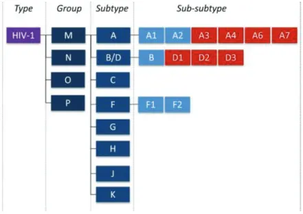
However, as more isolates began to emerge, a robust classification system based on phylogenetic analyses of env coding sequences was implemented (63, 68). HIV-1 is classified into 4 groups namely M (Major) (69), O (Outlier) (70), N (Non-major) (71), and a rare P (72) group. Group M variants are the most common and thus responsible for global infection, while other groups are restricted to West-Central Africa (3, 73, 74). Group M viruses are classified into 10 discrete clades or subtypes namely (A-D, F-H, J, K) as shown in Figure 1.3 and very recently, subtype ‘L’ was identified from archived samples collected from the Democratic Republic of Congo (DRC) (75). The first set of subtypes classified were A, B, C, D & E. These isolates varied in their env sequences by about 30% and by 14% in the gag sequences. Sub- subtypes are a category of viral isolates closely related to a specific subtype lineage, but not distant enough to be identified as a new subtype. For example, subtype A has sub-subtypes A1- A7 (76, 77) and recently subtype D has been shown to have sub-subtypes D1-D3 (77) (Figure 1.4). An alternative classification system is based on viral co-receptor usage (78), however, classification based on viral genetic variation is more commonly used. Observed increase in complexity of newly identified HIV-1 strains called for the re-evaluation of HIV-1 nomenclature and classification. A 1999 meeting to resolve this challenge was convened to properly articulate a new classification system that would maintain historical consistencies with already existing literature; details of the outcomes of this meeting are fully described in (68), which surmised that phylogenetic and distance analysis must be used to identify new subtypes
ͺ
and a branching index of 0.66 has been used as an acceptable cut-off to determine genetic distance (76, 78).
Figure 1.3: Updated classification of HIV-1 subtypes
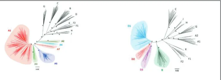
Figure 1.3 Updated classification of HIV-1 group M lineages. Figure 1.4: Maximum likelihood phylogenetic analysis of near full-length genome sequences of subtypes A and subtype D (77).
Figure 1.4: New sub-sub types for A and D
ͻ
1.3.2.1 Circulating recombinant forms
Circulating recombinant forms (CRFs) of HIV-1 group M represent a recombinant lineage that plays a critical role in regional pandemics. A CRF must share an identical genome structure that descends from the same recombination event (68). Generally, when people think of CRFs, the tendency is to presume that the recombination occurs because of fragment breakage and re- joining as is the case for double stranded deoxy-ribonucleic acid (dsDNA); however, this is inaccurate. CRFs are formed due to phenomenon known as template switching which occurs during the reverse transcription process in a dually infected cell. This is what leads to the recombination event (57, 60, 76). The naming system for CRFs include the acronym ‘CRF’, followed by a number to reflect the order in which it was identified in relation to previously identified CRFs and two letters to identify the subtype composition; for example, CRF02_AG is the second CRF identified and has components of subtype A and G in its genome. In the event that there are more than 2 subtype components within the genome it is referred to as a complex and designated ‘cpx’ for example CRF11_cpx (68). Currently, over a 100 CRFs have been identified according to the Los Alamos HIV database (https://www.hiv.lanl.gov/content/sequence/HIV/CRFs/CRFs.html). CRF01_AE and CRF02_AG are the most prominent. Subtype ‘E’ was initially identified in the env, but was not identified in gag, hence it did not ‘meet’ the criteria for becoming an independent subtype. In gag, it appeared to be more closely related to subtype A. At the 2000 HIV-1 classification meeting, it was proposed that subtype ‘E’ be designated CRF01_AE because it represented a putative recombinant (68), and to keep with historical consistencies of previous literature referring to it as subtype E. It is proposed that CRF01_AE formed early during the HIV-1 epidemic, and it is critical in regional epidemics in Asia and south east Asia, though it emerged from Central Africa (6, 68). CRF02_AG also plays a critical role in regional epidemic in Western and Central Africa. It is the most prevalent CRF, appearing to also have been formed
ͳͲ
by an early recombination event at the start of the epidemic. It was initially thought to be a
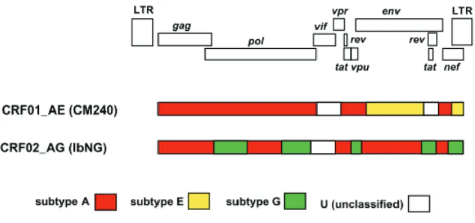
‘pure’ subtype, while subtype ‘G’ was thought to be a recombinant. However, time to most recent common ancestor (tMRCA) analysis has shown that the ancestral sequences of pure subtypes A and G predate that of CRF02_AG. This confirms that CRF02_AG is indeed a recombinant of pure subtype A and G (6, 71, 79). Figure 1.5 shows the genome map of CRF01_AE and CRF02_AG.
1.3.2.2 Unique recombinant forms
Unique recombinant forms (URFs) refer to unique mosaic sequences that currently lack evidence of onward transmission and thus do not meet the well-defined criteria to be classified as CRFs. They account for 6.1% of global HIV infections and are also critical to regional infections. For example, recombinants of AC, AD, CD and ACD are estimated to constitute about 30% of East Africa’s infections in countries like Kenya, Rwanda, Tanzania, and Uganda, while BF is predominant in South America, particularly Brazil and Argentina (51, 56, 80, 81).
Figure 1.5: Mosaic genome structures of CRFs
Figure 1.5: Mosaic genome structures of CRF01_AE and CRF02_AG. An alignment of representative near full-length strains was used and gap-stripped prior to all analysis. The HIV-1 genome map shows the position of each open reading frame in the gap-stripped multiple alignment. Below the genome map, each bar represents the mosaic pattern each CRF, adapted from (82)
ͳͳ
1.4 Morphology of HIV-1
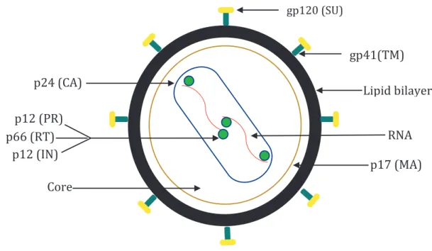
HIV-1 particles are circular in nature and comprise of 3 major components namely an envelope lipid bilayer, a viral matrix, and the core. The particle is surrounded by a lipoprotein membrane that is about 100-150 nm in diameter (83) containing 72 glycoprotein complexes integrated into this membrane. The lipoprotein membrane comprises of external glycoprotein trimers gp120 surface protein (SU) that are anchored by the transmembrane protein gp41 (TM) (84).
The matrix p17 (MA) is anchored inside of the viral lipoprotein membrane. The viral capsid p24 (CA) encases two copies of HIV-1 RNA genome (83-85). The HIV-1 RNA is part of a protein-nucleic acid complex composed of the nucleoprotein p7 and the reverse transcriptase p66 (RT) (85). The viral core contains the enzymatic machinery for replication, which are the reverse transcriptase (RT), integrase p32 (IN) and the protease p11 (PR) (86-88). A graphical representation of HIV-1 particle can be seen in Figure 1.6.
1.4.1 HIV-1 genome organisation
The HIV-1 genome is organized into nine open reading frames (ORF) just short of 10 kilo bases in length (84, 89). The composition of the genome can be broadly classified into structural and accessory genes. The group-specific antigen (gag) gene encodes the matrix, capsid, nucleocapsid and p6 proteins; while the envelope (env) gene encodes the gp120 and gp41 (84, 89). The polymerase gene (pol) codes the enzymatic machinery of the virus that facilitates effective replication which include the enzymes. All three genes make up the structural genes (89, 90). The virus also has 6 accessory/regulatory genes which include virion infectivity factor (vif), viral protein R (vpr), viral protein U (vpu), trans-activation of transcription (tat), regulator of expression of virion (rev) and the negative factor (nef). The proteins coded for by the genes all play important roles in the replication cycle of the virus (90, 91) and are well described in (86, 92). Figure 1.7
ͳʹ
Figure 1.6: Graphical representation of the morphology of HIV-1 particle and Figure 1.7:
HIV-1 genome map showing all three reading frames and nucleotide base positions.
ʹͶȋȌ
ͳʹȋȌ
ȋȌ
ͳʹȋȌ
ͳȋȌ
ͳʹͲȋȌ
ͶͳȋȌ
gag
pol vpr
tat
Reading frame
1 2 3
ͲʹͷͲͲͷͲͲͲͷͲͲͻͳ
env
vpu
rev
Figure 1.6: Structure of HIV-1 virion
Figure 1.7: HIV-1 genome map
ͳ͵
1.4.2 HIV-1 replication cycle
Host cell activation, which involves the induction of cellular division is a crucial requirement for effective replication of HIV-1 (92). The HIV-1 replication cycle can be broadly divided into the following stages: a) cell binding and entry, b) viral uncoating, c) reverse transcription, d) viral DNA integration e) virus protein processing and assembly and f) virion budding (89, 92, 93). Cell binding and entry is facilitated through the affinity of viral gp120 protein for CD4+ receptors found on a subset of T-lymphocytes and monocytes-macrophages. The binding between both molecules causes a conformational change allowing further interaction of gp120 with a co-receptor on target cells such as the chemokine receptor 4 (CXCR4) or chemokine receptor 5 (CCR5) resulting in the fusion of the viral envelope with the target cell membrane (92). Following successful entry, uncoating of viral core occurs releasing viral RNA into the host cell cytoplasm. This is immediately followed by the conversion of single stranded viral RNA into a double stranded DNA by the enzymatic action of RT. Through its ribonuclease H active site, RT degrades the viral RNA upon completion of the reverse transcription process.
The entire process occurs within a pre-integration complex (PIC) that comprises of MA (p17) protein, Vpr and IN. This PIC is then trafficked into the cell nucleus where the viral double stranded DNA is integrated into the host genome through the enzymatic action of IN (93). IN creates sticky ends at the 3’ ends of the host DNA allowing for the integration of the viral DNA into the host cell genome to form a pro-viral DNA. This process and the expression of the pro- viral DNA readily occurs when the host cell is in an activated state (93). This step is followed by the expression of the pro-viral DNA which encodes for the viral proteins and their pre- cursors. Expression of viral transcripts starts from the 5’ long terminal repeat (LTR) region of the pro-viral genome with Tat enhancing the initiation of the transcription process. Following successful transcription, full length and spliced viral RNAs are transported from the nucleus to the cytoplasm for translation and packaging of proteins. This process is facilitated by the Rev
ͳͶ
protein (89). Translation of viral messenger RNA (mRNA) occurs in the cytoplasm. Gag and Gag-Pol products are localized at the cell membrane, while translation of the env mRNA occurs in the endoplasmic reticulum (ER). Translation and post-translational processing of the mRNA result in the production of viral core proteins, MA, CA, NC, p6, PR, RT, IN and the accessory proteins such as Vif, Vpr, Nef, and genome RNA which leads to the gradual formation of immature virion and the initial step towards budding from the cell surface. The final stage of the process is the budding stage which also involves downregulation of surface CD4+ receptors by the Vpu and Nef proteins; this is to make way for the provision of SU and TM for the outer membrane coat during viral budding. Viral maturation occurs by proteolytic cleavage of Gag and Gag-Pol polyproteins by PR enzyme. At this stage, the mature virus is ready to begin infection of another target cell, continuing the cycle in the absence of ART (89, 92, 93). As mentioned in an earlier section, every step of the HIV-1 replication cycle as well as every viral gene product is a potential target for therapeutic interventions. Following the discovery of HIV/AIDS, several strategies to inhibit viral replication cycle are put into consideration during drug development; which include RNA based strategies such as antisense RNA, RNA decoys, ribozymes and protein-based strategies such as monoclonal antibodies, chimeric proteins, intracellular single-chain antibodies (94) and ARTs which target viral enzymes and block viral entry (37). Continuous investigation of HIV-1 replication cycle is critical for determining potential new targets for the development of novel ARTs (92) as shown in Figure 1.8.
ͳͷ
Figure 1.8: HIV-1 replication cycle with ART targets
Figure 1.8: HIV-1 replication cycle showing all known ART targets. Taken from (95).
ͳ
1.4.3 Group specific antigen (Gag)
The group specific antigen (gag) is one of the 3 structural genes in the HIV-1 genome; it codes for a Gag polyprotein precursor, which when acted upon by PR leads to smaller products namely MA, CA, NC and p6. HIV-1 particle assembly is primarily coordinated by products of the gag, a machinery that recruits building blocks required for formation of a fully infectious viral particle, a full description of gag products, their functions and intra-cellular trafficking can be found in (96, 97). Its examination is of import to this thesis, and in subsequent sections I will elaborate on its role in viral replication.
1.5 Pathogenesis of HIV-1
Viral pathogenesis is the series of steps that occur when a viral isolate infects a susceptible host resulting in disease induction (98). Pathogenesis of HIV-1 infection and subsequent progression to AIDS are a consequence of the characteristics of the infecting viral isolate and the host immune response; the balance of effectiveness of both components determines the outcome of the infection (93).
1.5.1 Primary (acute) infection
Acute infection occurs when HIV-1 infects a susceptible host cell (98). CD4+ and follicular dendritic cells (FDC) near the epithelial layer represent the initial target of HIV-1 infection following breakthrough into the mucosal barrier (93, 99, 100). Acute HIV-1 infection is generally characterized by three major events namely; an initial increase in plasma viral load, drastic depletion of naïve resting CD4+ cells, and establishment of latent viral reservoir (101).
The course of disease progression in an infected patient can be predicted within the first 6-12 months of infection. This finding is based on a 1996 Multicenter AIDS Cohort Study (MACS) data set, that showed rapid increase in plasma viral load following primary infection up to a
ͳ
median of 106 -107 RNA copies/mL, and depletion of CD4+ count can independently predict whether the patient will progress quickly or slowly to AIDS (101). Within the first 10-12 days post infection, viral RNA can be detected in the blood using RT-PCR amplification methods.
Following peak plasma viremia levels, HIV-1 antibodies can be detected in the patient blood due to humoral immune responses to the infection. This point is referred to as seroconversion and it is about 20-25 days on average post primary infection (93, 99). During this time the patient experiences flu-like symptoms such as fever, lymphadenopathy, mononucleosis, weight loss etc. which takes place for about 7-10 days post infection (93, 99).
1.5.2 Chronic infection and AIDS
Following the resolution of acute infection and the establishment of a virologic quasi-steady state, a prolonged period of asymptomatic chronic infection ensues (93, 101); though patients appear to be asymptomatic, ongoing viral replication and CD4+ depletion continues. During this phase, HIV-specific CTLs control viremia levels by recognizing viral antigens presented on the cell surface of infected cells (93). In addition, antibody dependent cellular cytotoxicity (ADCC) of infected cells is mediated by the natural killer (NK) cells. However, in the absence of therapeutic interventions, CD4+ T lymphocytes decline gradually and at a certain threshold (< 200 cells/μL), patients begin to suffer from certain symptoms, which constitute AIDS, such as prolonged fatigue, lymphadenopathy, oral and vaginal candidiasis, dermatological conditions, neurological conditions, atrophy, tuberculosis, and Pneumocystis carinii infection (93, 101).
ͳͺ
Figure 1.9: Phases of natural HIV-1 infection
Figure 1.9: (A) Showing phases of HIV-1 natural infection (Acute and Chronic), and (B) showing rate of CD4+ decline. Taken from (95).
A
B
ͳͻ
1.6 Host immunity
The human immune system comprises of a complex array of protective mechanisms including cells, chemicals, and processes to overcome unfortunate exposure to pathogens and their products that have gained entry through mucosal and epithelial surfaces of the body. An important feature of a functional immune system is its ability to properly discriminate between host cells (self) and antigens, allergens, or toxins of pathogenic origin (non-self) to avoid causing damage to physiological functions of the body (102). The immune system comprises of the innate and adaptive mechanisms which work in complement to one another to mount an appropriate immune response to pathogenic exposure (103-105).
1.6.1 Innate immunity
The innate immune system is organized into a network of cells (macrophages, dendritic cells, natural killer cells, neutrophils, mast cells, eosinophils, and basophils) and signals that act as a first line of defence against pathogens. The onset of HIV-1 acute infection occurs following breaching of the host epithelial and mucosal barriers - the host innate immune response is triggered by the presence of pathogen associated molecular patterns (PAMPs) which are recognized by pathogen recognition receptors (PRRs) found on the surface of cells of the innate immune system (106, 107). PRRs can be broadly divided into toll-like receptors (TLRs) and intracellular endosomes (108, 109). Dendritic cells are large cells encountered by HIV at mucosal surfaces. They transport the virus from the site of entry to the lymphoid tissue where follicular dendritic cells (FDCs) provide signals for activation of B-cells to attack the virus (109). Natural killer (NK) cells are also important for control of HIV replication (110). When activated they release cytokines such as interferon-γ (IFN-γ), tumour necrosis factor-α (TNF- α) and chemokines that facilitate T-cell proliferation and have been reported to show improved function in the presence of KIR3DS1 HLA subtypes (110, 111).
ʹͲ
Unfortunately, as the infection progresses it damages components of the innate immune system, especially macrophages which are linked to the adaptive immune system by inhibiting their antigen presenting function (112). As the first line of defence, the innate immune system is critical for sensing the presence of HIV particles and in turn setting in motion a series of immunological events to activate the adaptive immune response (108, 113).
1.6.2 Adaptive immunity
The adaptive immune system has evolved to provide a broad and fine-tuned collection of component assets for the recognition of both self and non-self thereby facilitating pathogen specific immune responses that result in the generation of long-term immune memory and regulation of immune homeostasis. The evolution of the adaptive immune system is driven by the vast variable antigenic structures of pathogens and their tendency to evade immune detection. Following initial pathogen encounters, cells expressing specific immune receptors for pathogen recognition persist in the host for life providing immunological memory and capacity for rapid response in the event of re-infection. The major components of adaptive immunity are the T-lymphocytes which are developed in the thymus and the antibody producing B-cells produced in the bone marrow (114). The adaptive immune system depends on customized receptors selected through somatic recombination events of specific germ-line segments which arose by gene duplication to form intact T cell receptor (TCR) and immunoglobulin (B cell antigen receptor; Ig) genes early in the evolution of vertebrates for the purpose of generating a highly flexible and specific immune response. The germline encoding genes elements facilitates the development of millions of different antigen receptors conferring each with the potential to be uniquely specific for the vast amount of antigen it may encounter during an infection (103).
ʹͳ
1.6.2.1 T-cell immunity and HIV infection
T-lymphocytes mature in the thymus following the migration of hematopoietic stem cells originating in bone marrow (104). They express a unique series of antigen-binding receptors on their cell membranes commonly referred to as T-cell receptors (TCR). Each T-cell expresses a single form of TCR and has the capacity to rapidly proliferate and differentiate based on reception of appropriate induction signals. Following HIV-1 infection, host experiences persistent immune activation, followed by decline in CD4+ cells and subsequent disease progression to AIDS (115) and have been shown to correlate directly with viral load and inversely with CD4+ count (116, 117). The major histocompatibility complex (MHC) class I on the cell surfaces present HIV-1 epitopes processed within the cells for recognition by TCRs on CD8+ T cells. CD8+ T cells then lyse HIV-1 infected cells and secrete cytokines such as IFN-γ, TNF-α, and chemokines such as macrophage inflammatory protein MIP-1 α and MIP β, that inhibit virus replication and block viral entry into CD4+ T cells. CD8+ T cells have been shown to be critical for control of HIV-1 replication (118-121). During acute infection, CD4+ T cells lose their ability to expand rapidly and therefore their contribution to viral control is minor, however, during chronic infection this is reversed with CD4+ T cells present and secreting interleukin-2 (IL-2) or cytokines, such as IFN-γ, to control viremia (117, 122). CTL escape is the variation in viral sequence that results in the loss of recognition by CTLs which is critical to the HIV pathogenesis (119). However, this in-turn impacts viral function positively or negatively on viral function and subsequent disease progression; and though it appears to be a mechanism of adaptation to its host, the range of variation of CTL epitopes is limited based on its impact on structure and function of the gene products (118), which further illustrates the effectiveness of CTL function on viral control albeit short-lived (123).
ʹʹ
1.6.2.2 B-cell immunity and HIV infection
The adaptive humoral immune system is driven by antibody production from B cells under the signalling influence of T cells and dendritic cells. B cells themselves, like T cell