REVIVAL AND CHARACTERIZATION OF AEROBIC ENDOSPORE- FORMING BACTERIA FROM AN ANCIENT SEDIMENT CORE OBTAINED FROM THE MFABENI PEATLAND, SOUTH AFRICA
by
SELISHA NAIDOO
Submitted in fulfilment of the academic requirements for the degree of Master of Science
in Microbiology
School of Life Sciences
College of Agriculture, Engineering and Science University of KwaZulu-Natal
Pietermaritzburg South Africa
June 2017
ii
PREFACE
The research contained in this dissertation was completed by the candidate while based in the Discipline of Microbiology, School of Life Sciences of the College of Agriculture, Engineering and Science, University of KwaZulu-Natal, Pietermaritzburg Campus, South Africa. The research was financially supported by the National Research Foundation (NRF).
The contents of this work have not been submitted in any form to another university and, except where the work of others is acknowledged in the text, the results reported are due to investigations by the candidate.
_________________________
Signed: Dr. C.H. Hunter (Supervisor) Date: June 2017
iii
DECLARATION
I, Selisha Naidoo, declare that:
i) the research reported in this dissertation, except where otherwise indicated or acknowledged, is my original work;
ii) this dissertation has not been submitted in full or in part for any degree or examination to any other university;
iii) this dissertation does not contain other persons’ data, pictures, graphs or other information, unless specifically acknowledged as being sourced from other persons;
iv) this dissertation does not contain other persons’ writing, unless specifically acknowledged as being sourced from other researchers. Where other written sources have been quoted, then:
a) their words have been re-written but the general information attributed to them has been referenced;
b) where their exact words have been used, their writing has been placed inside quotation marks, and referenced;
v) where I have used material for which publications followed, I have indicated in detail my role in the work;
vi) this dissertation does not contain text, graphics or tables copied and pasted from the Internet, unless specifically acknowledged, and the source being detailed in the dissertation and in the References sections.
Signed: Selisha Naidoo (Candidate) Student number: 211505643
Date: June 2017
iv
ABSTRACT
Peatlands contain an accumulation of organic material over extended periods of time which can provide biological and chemical proxies for determining palaeo-environments. The organic accumulation can be dated to provide a chronology of time that, integrated with various proxies, can help understand, interpret and infer past environmental conditions which prevailed at the time of deposition. One such proxy that has yet to be explored are aerobic endospore- forming bacteria (AEFB) which have the ability to form dormant endospores as a survival strategy to overcome unfavourable conditions. Dormant endospores may potentially serve as
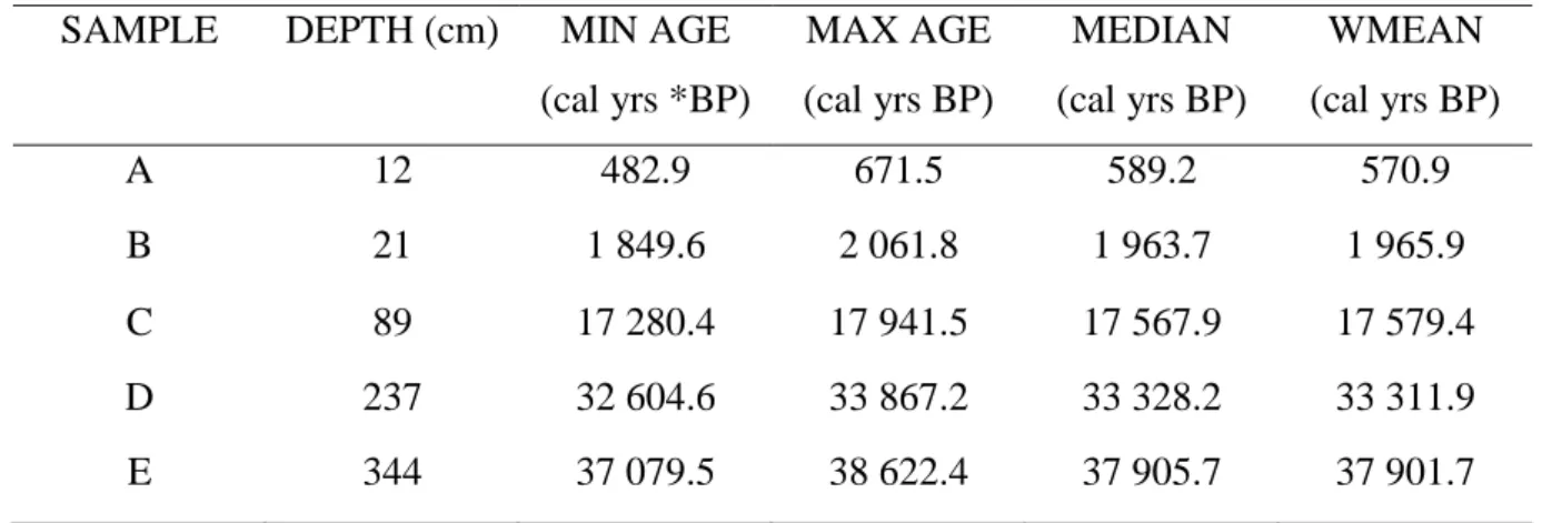
‘time capsules’ which could provide an insight into the changes in microbial diversity of AEFB, from ecologically-sensitive environments, in relation to past physico-chemical conditions. This study sought to revive and determine the genetic and physiological diversity amongst dormant AEFB sampled from an ancient sediment core obtained from the Mfabeni Peatland, KwaZulu- Natal, South Africa. Samples were taken from radiocarbon-dated sections of the core at ca.
589, 1 964, 17 568, 33 328 and 37 906 cal years BP and subjected to a sequential extraction protocol prior to dilution series plating onto selected media. A total of 270 isolates were selected for genetic fingerprinting and screened using Repetitive extragenic palindromic- Polymerase Chain Reaction (Rep-PCR), targeting the BOX mosaic repetitive element to evaluate genetic diversity, and High Resolution Melt Analysis (HRMA), which was evaluated as a means to distinguish between fingerprint groupings and to validate the genotyping results.
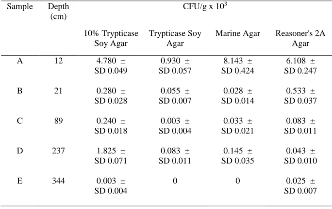
HRMA was carried out using primer sets targeting the spo0A gene and the V3 and V4 variable regions of the 16S rRNA gene. Taxonomic ranking and phylogeny of 83 representative genotypes were assessed through sequence analysis of amplified partial 16S rRNA gene fragments. As part of physiological testing, salt (0.5%–15% NaCl) and pH range (3–10) tolerances were determined using microtiter plate assays. Substrate utilization abilities of selected representative isolates were determined through physiological profiling using Biolog EcoPlates™ and statistically analyzed by means of Canonical Correspondence Analysis (CCA). Isolates were revived from all five depths and total colony-forming units (CFU) per gram were found to decrease with increasing depth and age of samples. The nutritionally-rich trypticase soy agar (TSA) medium produced lower counts of revived AEFB compared to the environmentally-based Marine Agar and nutritionally-limited Reasoner’s 2A agar and 10%
TSA media. Rep-PCR allowed for strain-level discrimination of the isolates. The V3 and V4 regions provided the best HRMA resolution and allowed for closely-related species to be
v
matched. Genotyping revealed that significant diversity was present amongst isolates from different sample depths. Ninety-four percent of operational taxonomic units (OTUs) distinguished were unique to their sampling depth. Phylogenetic analysis revealed the presence of several aerobic endospore-forming genera, namely, Bacillus, Brevibacillus, Paenibacillus, Domibacillus, Lysinibacillus, Solibacillus and Paenisporosarcina. Strains of Lysinibacillus and Brevibacillus were unique to sample depths corresponding to ca. 589 and 1 964 cal years BP, whereas Domibacillus sp. isolates were revived from samples dated at ca. 17 568 and 33 328 cal years BP. Eighteen percent of isolates had 16S rRNA gene sequences displaying
<97% similarity to reference species currently listed in the NCBI database, suggesting that some of these isolates may potentially represent novel species or strains. Substrate utilization profiling revealed that, collectively, isolates were able to utilize 27 of the 31 substrates tested;
the most commonly utilized substrates being Tween 40, Tween 80 and pyruvic acid methyl ester. Canonical correspondence analysis revealed, with significance, that the age of the samples from which the isolates were obtained did have an effect on the variations observed in the patterns of substrate utilization. Tolerance ranges to salt and pH depicted less significant variations between isolates from different depths. The highest percentages of isolates favoured salt concentrations up to 2.5%. Thirty-seven percent of selected isolates were capable of growth at a 15% NaCl concentration. The majority of isolates tested from each depth preferred a pH range of 7.5–8.5, whilst a smaller percentage, namely, 5.7% and 34%, were capable of growth at pH values of 3 and 10, respectively. The results suggest that peatlands do serve as a reservoir for dormant AEFB, some of which are potentially uncharacterized. This study highlights the changes in bacterial genetic and physiological diversity which occur along an environmentally- variable sediment profile. Dormant endospores may serve as a proxy which can be used to determine palaeo-environments.
vi
ACKNOWLEDGMENTS
The financial assistance of the National Research Foundation (NRF) towards this research is gratefully acknowledged.
I would also like to gratefully acknowledge the University of KwaZulu-Natal for the cum laude and GC-Weightman scholarships.
There are a number of people I would like to thank for their contributions, in various forms, throughout this research.
First and foremost I would like to thank my supervisor, Dr. Charles Hunter, for his invaluable assistance, expertise and advice throughout this research. All my gratitude goes to him for his continued guidance, support and time.
I am extremely indebted to Prof. Trevor Hill for letting me get my hands on the amazing sediment core which allowed this research to be possible. I am grateful for all the time taken for our meetings and for making me feel welcome into the Geography department.
Many thanks to Dr. Jemma Finch for always taking time out to provide me with all the knowledge I required regarding the Mfabeni Peatland.
Mr. Craig Morris, for all his assistance with data analysis. It was a great pleasure working with such a brilliant stats mind.
Heather Tredgold, for the constant advice and for being the best conference roommate a girl could ask for.
My appreciation also goes to the technical staff, Diane Fowlds and Celeste Clark, and the administrative staff, Tanya Karalic and Natalie Jones, for their assistance and help throughout the duration of this research.
Dr. Raymond Hewer, for all the support, encouragement and enthusiasm for my work.
Mariam Khan and Melanie Naidoo for the continuous assistance and reassurance throughout this research.
Finally, to my friends and family for their support.
vii
TABLE OF CONTENTS
PREFACE ... ii
DECLARATION... iii
ABSTRACT ... iv
ACKNOWLEDGMENTS ... vi
TABLE OF CONTENTS ... vii
LIST OF TABLES ... xi
LIST OF FIGURES ... xiii
LIST OF ABBREVIATIONS ... xvi
CHAPTER ONE: INTRODUCTION ... 1
CHAPTER TWO: LITERATURE REVIEW ... 4
2.1. INTRODUCTION ... 4
2.2. WETLAND MICROBIOLOGY ... 6
2.2.1. What are wetlands and why are they important? ... 6
2.2.2. Environmental characteristics of wetlands ... 6
2.2.3. The effects of climate changes on wetlands ... 7
2.2.4. Microbial activity in wetland environments ... 8
2.3 USING AEFB IN CLIMATE CHANGE STUDIES ... 9
2.3.1. The effects of climate changes on the ecology of bacteria ... 9
2.3.2. The use of endospore-forming bacteria in palaeo-ecological studies ... 9
2.4. AEROBIC ENDOSPORE-FORMING BACTERIA (AEFB) ... 10
2.4.1. Taxonomic diversity of AEFB... 10
2.4.2. Physiological diversity of AEFB ... 11
2.5. ENDOSPORES ... 12
2.5.1. Functions of endospores ... 12
2.5.2. Life cycle ... 12
2.5.2.1. Sporulation ... 12
2.5.2.2. Germination ... 14
2.5.3. Structure of endospores ... 16
2.5.4. Molecular mechanisms behind endospore resistance ... 19
2.5.5. Endospore dispersal ... 20
2.5.6. Mechanisms of destruction of endospores ... 21
2.5.7. Viability of endospore-forming bacteria ... 22 2.6. REVIVAL OF BACTERIAL ENDOSPORES TO A VEGETATIVE STATE . 23
viii
2.6.1. Isolation and revival of endospores from diverse locations ... 23
2.6.2. Applications of endospore revival ... 24
2.6.3. Challenges behind the analysis of ancient bacteria ... 25
2.7. TECHNIQUES FOR DETERMINING THE AGE OF ENVIRONMENTAL MATERIALS ... 25
2.7.1. Radiocarbon dating ... 25
2.7.2. Age-depth modelling ... 26
2.8. ANALYSIS OF BACTERIAL DIVERSITY ... 27
2.8.1. The study of bacterial diversity from the environment ... 27
2.8.2. Molecular fingerprinting techniques for the analysis of bacterial genetic diversity ... 29
2.8.3. Use of High Resolution Melt Analysis (HRMA) for genotyping... 31
2.8.4. Characterization of microbial metabolic diversity ... 32
2.8.4.1. Community-level physiological profiling (CLPP)... 32
2.8.4.2. Statistical analysis of CLPP data ... 33
2.9. PHYLOGENETIC EVALUATION OF ENDOSPORE-FORMING BACTERIA ... 34
2.9.1. 16S rRNA gene sequencing and applications ... 34
2.9.2. Gene mapping for the comparison of ancient and modern bacterial genes 35 2.10. CONCLUSION ... 36
CHAPTER THREE: ISOLATION AND ASSESSMENT OF THE PHYLOGENETIC DIVERSITY OF AEROBIC ENDOSPORE-FORMING BACTERIA REVIVED FROM AN ANCIENT PEATLAND SEDIMENT CORE ... 37
3.1. INTRODUCTION ... 37
3.2. MATERIALS AND METHODS ... 38
3.2.1 Site description ... 38
3.2.1.1. Introduction into the Maputaland region ... 38
3.2.1.2. Location, climate and hydrology of the Mfabeni Peatland ... 39
3.2.1.3. Age of the Mfabeni Peatland ... 39
3.2.2. Sample collection ... 40
3.2.3. Radiocarbon Dating ... 40
3.2.4. Subsampling from the sediment core ... 43
3.2.5. Evaluation of extraction methods to recover endospores from peat sediment ... 43
3.2.5.1. Mechanical treatment via agitation ... 44
3.2.5.2. Calcium chloride treatment ... 44
ix
3.2.5.3. Buffer-mediated extraction accompanied by bead-beating ... 45
3.2.6. Genomic fingerprinting and phylogenetic analysis of revived AEFB ... 46
3.2.6.1. DNA extraction using a kit protocol ... 47
3.2.6.2. DNA extraction using a colony ‘pick-off’ approach ... 47
3.2.6.3. Repetitive extragenic palindromic-Polymerase Chain Reaction (Rep-PCR) ... 48
3.2.6.4. 16S rRNA gene amplification ... 49
3.2.6.5. 16S rRNA gene sequencing and phylogenetic analysis ... 49
3.3. RESULTS ... 50
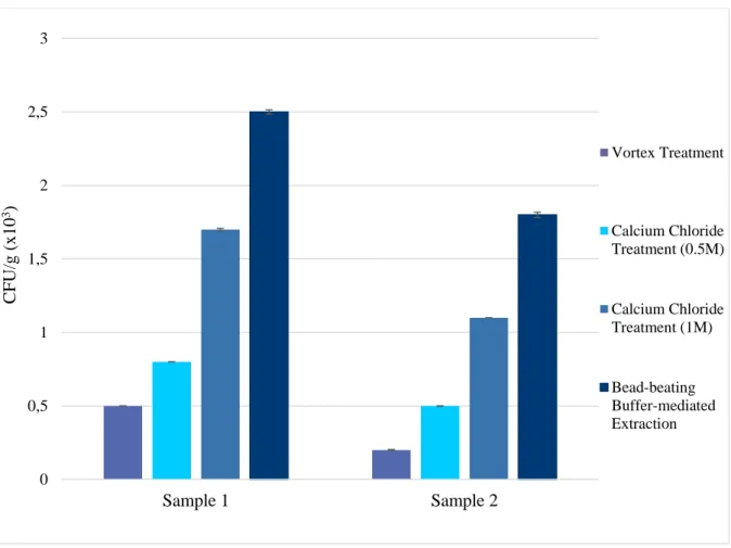
3.3.1. Evaluation of extraction methods to recover endospores from peat sediment ... 50
3.3.2. Endospore revival from sediment core samples ... 52
3.3.3. Rep-PCR fingerprinting ... 54
3.3.4. 16S rRNA gene sequencing and phylogenetic analysis ... 63
3.3.5. Strain comparison of taxonomically-related isolates ... 69
3.4. DISCUSSION ... 72
CHAPTER FOUR: ASSESSMENT OF HIGH RESOLUTION MELT ANALYSIS (HRMA) AS A GENOMIC FINGERPRINTING METHOD FOR THE SCREENING AND GROUPING OF AEROBIC ENDOSPORE-FORMING BACTERIA ... 80
4.1. INTRODUCTION ... 80
4.2 METHODS AND MATERIALS ... 82
4.2.1. High Resolution Melt Analysis (HRMA) ... 82
4.2.1.1. HRMA of the V3 variable region of 16S rRNA ... 83
4.2.1.2. HRMA of the V3 and V4 variable regions of 16S rRNA ... 83
4.2.1.3. HRMA of the spo0A gene ... 83
4.2.2. Generation of sequence matrixes for AEFB isolates based on partial amplified 16S rRNA gene fragments... 84
4.3. RESULTS ... 85
4.3.1. PCR amplification using the three primer sets evaluated ... 85
4.3.2. HRMA of PCR amplicons ... 87
4.3.2.1. V3 variable region of 16S rRNA ... 88
4.3.2.2. V3 and V4 variable regions of 16S rRNA ... 89
4.3.2.3. Spo0A gene ... 90
4.3.3. The ability of HRMA to identify similar ‘unknown’ isolates ... 91
4.3.4. Determination of the ability of HRMA to distinguish between isolates ... 93
x
4.3.5. Comparison of V3 and V3+V4 variable regions as gene targets for HRMA
... 94
4.3.6. Grouping of AEFB isolates based on HRMA……….……….97
4.4. DISCUSSION ... 99
CHAPTER FIVE: DETERMINATION OF PHYSIOLOGICAL DIVERSITY OF SELECTED REVIVED AEROBIC ENDOSPORE-FORMING BACTERIAL ISOLATES... 104
5.1. INTRODUCTION ... 104
5.2. METHODS AND MATERIALS ... 106
5.2.1. Determination of salinity and pH tolerance of selected AEFB isolates ... 106
5.2.1.1. Salinity testing using microtiter plate assays ... 106
5.2.1.2. pH testing using microtiter plate assays ... 107
5.2.1.3. Multivariate analysis of salinity and pH results ... 107
5.2.2. Physiological profiling for the determination of metabolic diversity of isolates ... 108
5.2.2.1. Determination of substrate utilization abilities of selected AEFB isolates using Biolog EcoPlates™ ... 108
5.2.2.2. Analysis of Biolog EcoPlate™ data ... 108
5.2.2.3. Multivariate analysis of physiological profiling data ... 109
5.3. RESULTS ... 110
5.3.1. Screening of AEFB isolates for salinity and pH tolerance ... 110
5.3.1.1. Salinity tolerance testing ... 110
5.3.1.2. pH tolerance testing ... 116
5.3.2. Physiological profiling of AEFB isolates to determine metabolic diversity ... 121
5.4 DISCUSSION ... 136
CHAPTER SIX: GENERAL DISCUSSION AND CONCLUSIONS ... 144
6.1. RESEARCH OVERVIEW ... 144
6.2. SUMMARY OF FINDINGS ... 145
6.3. DO DORMANT AEFB MEET THE REQUIREMENTS OF A PALAEO- ECOLOGICAL PROXY? ... 148
6.4 ADDITIONAL APPLICATIONS OF ANCIENT AEFB ... 149
6.5. CONCLUSION ... 149
REFERENCES ... 151
APPENDIX A ... 175
APPENDIX B ... 187
xi
APPENDIX C ... 189 CONFERENCES...……….. 194
LIST OF TABLES
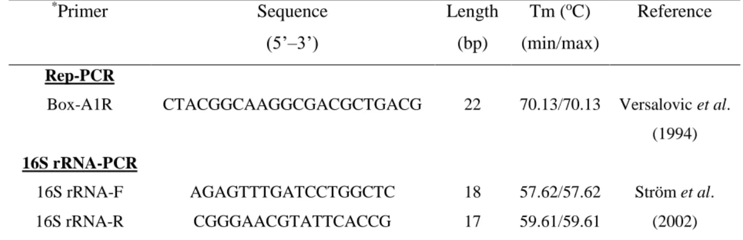
Table 3.1. Interpolated ages for each of the five sub-samples taken from an Mfabeni Peatland sediment core ... 42 Table 3.2. Nucleotide sequences for primers used for Rep-PCR and 16S rRNA gene sequence amplification ... 48 Table 3.3. Colony-forming units per gram of sediment CFU/g (x103) showing revival efficiency across depths on four media types ... 52 Table 3.4. Assignment of Operational Taxonomic Units (OTUs) to AEFB isolates from sections of a sediment core based on Rep-PCR fingerprinting profiles and their distribution across sample depths and cultivation media ... 59 Table 3.5. OTU richness and relative abundance for the four media at each depth ... 60 Table 4.1. Nucleotide sequences for primers used for HRMA ... 84 Table 4.2. Groupings of isolates based on the target variable regions for HRMA, Rep-PCR fingerprinting data and partial 16S rRNA gene sequence similarities ... 97 Table 5.1. Tolerance of AEFB isolates to varying salt (NaCl) concentrations using microtiter plate assays (OD595) ... 111 Table 5.2. Summary of statistical parameters calculated for AEFB isolate responses at varying concentrations of NaCl ... 113 Table 5.3. Tolerance of AEFB isolates to pH variation tested using microtiter plate assays (OD595) ... 116 Table 5.4. Summary of statistical values calculated for AEFB isolate responses to varying pH values ... 118 Table 5.5. Summary of Biolog EcoPlate™ substrate utilization frequency parameters of isolate responses to each of the 31 substrates ... 123 Table 5.6. Variation in substrate utilization as a function of age for Biolog EcoPlate™ substrates ... 130 Table A1. BLAST similarity matches and percentage identity scores of isolates from sample depth A (12 cm) ... 180
xii
Table A2 . BLAST similarity matches and percentage identity scores of isolates from sample depth B (21 cm) ... 182 Table A3. BLAST similarity matches and percentage identity scores of isolates from sample depth C (89 cm) ... 184 Table A4. BLAST similarity matches and percentage identity scores of isolates from sample depth D (237 cm) ... 185 Table A5. BLAST similarity matches and percentage identity scores of isolates from sample depth E (344 cm) ... 186 Table B1. Sequence similarity matrix based on partial 16S rRNA gene sequences for selected isolates generated through BioEdit software (v 7.0.9.0) ... 187 Table C1. The layout of the Biolog EcoPlate™ containing 31 carbon sources in triplicate sets ... 189 Table C2. The layout of the microtiter plate used for salinity testing displaying the varying concentrations of NaCl (%) in duplicate allowing for the inoculation of six isolates per plate ... 190 Table C3. The layout of the microtiter plate used for pH testing displaying the varying pH values in duplicate allowing for the inoculation of six isolates per plate ... 191
xiii
LIST OF FIGURES
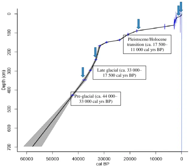
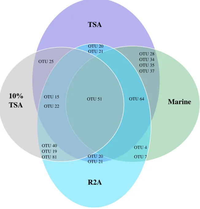
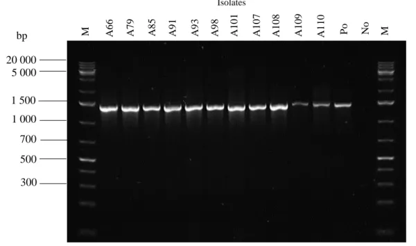
Figure 2.1. Cycle of sporulation and germination in AEFB………....16 Figure 3.1. Location of the Mfabeni Peatland, the site used in the current study, on the eastern shore of Lake St. Lucia, northern KwaZulu-Natal, South Africa ... 41 Figure 3.2. Core extraction using a vibracorer, Mfabeni Peatland ... 41 Figure 3.3. Age depth model based on linear interpolation depicting the calibrated 14C dates for the Mfabeni Peatland region generated using the classical age-modelling software, CLAM . 42 Figure 3.4. The recovery of viable endospore-forming bacteria from a peatland sediment core using three different extraction procedures ... 51 Figure 3.5. Total AEFB colony-forming units (CFU) per gram of sediment for each media type across five sampling depths examined along a sediment core from the Mfabeni Peatland. …53 Figure 3.6. Agarose gel electrophoresis comparing Rep-PCR fingerprints of selected isolates using template DNA from GeneJET Genomic DNA extraction kits (K) and colony pick-offs (C). ... 54 Figure 3.7. Examples of Rep-PCR fingerprint profiles of selected AEFB isolates from each sediment core sample visualized using 1.5% agarose gel electrophoresis ... 57 Figure 3.8. Venn diagram illustrating the distribution of Operational Taxonomic Units representing AEFB isolates revived on different agar media ... 61 Figure 3.9. Venn diagram illustrating the distribution of Operational Taxonomic Units which were present at more than one sample depth examined ... 62 Figure 3.10. Agarose gel (1.5%) electrophoresis image displaying 16S PCR amplification products of selected AEFB isolates using 16S rRNA forward and reverse primers ... 63 Figure 3.11. Maximum-likelihood phylogenetic tree based on partial 16S rRNA gene sequences inferring the evolutionary relationships between selected AEFB isolates and reference sequences ... 65 Figure 3.12. Sub-clades of the Maximum-likelihood phylogenetic tree (Figure 3.11) illustrating the evolutionary relationships of revived AEFB isolates matched to reference strains of Domibacillus spp. (A) Brevibacillus spp. (B) Lysinibacillus spp. (C) and Paenibacillus spp.
(D) ... 68 Figure 3.13. Rep-PCR comparison of sediment core AEFB isolates exhibiting 16S rRNA gene sequence similarity (98–99%) to strains of Bacillus acidicola and B. shackletonii. ... 69 Figure 3.14. Rep-PCR comparison of fingerprint banding profiles of isolates C2, E1 and E2 which displayed 16S rRNA gene sequence similarity (97%) to strains of Paenibacillus elgii and P. ehimensis... 70
xiv
Figure 3.15. Rep-PCR comparison of fingerprint banding profiles of isolates A1 and C26 which displayed 16S rRNA gene sequence similarity (99%) to strains of Bacillus aquimaris and B.
vietnamensis ... 70 Figure 3.16. Rep-PCR comparison of fingerprint banding profiles of isolates C24, D23, D10 and D21 which displayed 16S rRNA gene sequence similarity (94–99%) to strains of Domibacillus robiginosus ... 71 Figure 3.17. Rep-PCR comparison of fingerprint banding profiles of isolates B53 and D18 which displayed 16S rRNA gene sequence similarity (99%) to strains of Paenisporosarcina quisquiliarum and P. indica ... 72 Figure 4.1. Amplification plots displaying RT-PCR amplification represented by increases in fluorescence for 42 AEFB isolates (A) the V3 region of 16S rRNA (B) the V3 and V4 regions of 16S rRNA and (C) spo0A ... 86 Figure 4.2. Melt curves displaying the HRMA of the V3 region amplicons for selected AEFB isolates... 88 Figure 4.3. Melt curves displaying the HRMA of V3+V4 region amplicons for selected AEFB isolates... 89 Figure 4.4. Difference plots displaying the melt curves of Isolates B11 (blue curve) and B63 (dark red curve) of amplified variable regions V3 and V4 against the selected baseline isolate A10 (red line) ... 91 Figure 4.5. Difference plots displaying the melt curves of Isolates B11 (blue curve) and B63 (dark red curve) of the amplified variable V3 region against the selected baseline isolate A10 (red line) ... 92 Figure 4.6. Difference plots displaying the melt curves of Isolates A98 (blue curve) and B45 (purple curve) of amplified variable regions V3 and V4 against the selected baseline isolate A10 (red line) ... 93 Figure 4.7. Difference plots displaying the melt curves of Isolates A25 (blue curve) and B30 (purple curve) of amplified variable regions V3 and V4 against the selected baseline isolate A10 (red line) ... 94 Figure 4.8. Difference plots displaying HRMA melt curves for Isolates A93 (pink curve) and B51 (green curve) using the V3 (A) and V3+V4 (B) amplicons compared to the baseline isolate A10 (red line) ... 95 Figure 4.9. Difference plots displaying the melt curves of Isolates A39 (purple curve) and D45 (green curve) of amplified variable regions V3 and V4 against the selected baseline isolate A10 (red line) ... 98 Figure 5.1. Distribution of relative growth intensity of revived AEFB isolates cultured at NaCl concentraions ranging from 0.5–15%. ... 114 Figure 5.2. The effect of increasing salt concentration on AEFB isolate growth ... 115
xv
Figure 5.3. Distribution of relative growth intensity of revived AEFB isolates cultured at six
pH values ranging from 3–10… ... 119
Figure 5.4. The influence of pH on AEFB isolate growth ... 120
Figure 5.5. Substrate utilization profile determined for isolate B11 using a Biolog EcoPlate™ after 7 days incubation at 30oC ... 121
Figure 5.6. Substrate utilization profile determined for isolate A37 using a Biolog EcoPlate™ after 7 days incubation at 30oC ... 122
Figure 5.7. The distribution of Biolog EcoPlate™ substrate utilization by AEFB isolates from each depth across a LnAge scale ... 126
Figure 5.8. A CA plot depicting the level of variation amongst AEFB isolates (log (X+1) transformed values) based on their substrate utilization profiles ... 127
Figure 5.9. CA plot depicting the centroid locations of the utilized substrates to determine the pattern of utilization amongst the AEFB isolates ... 128
Figure 5.10. CCA plot of AEFB isolates along a LnAge (natural logarithm of age) gradient..129
Figure 5.11. The centroid positions for Biolog EcoPlate™ substrates along an age gradient according to the CCA plot (Figure 5.10) ... 130
Figure 5.12. CCA plots of substrates which were utilized by AEFB isolates exclusively from sample depth A (A) Glycyl-L-Glutamic acid and (B) L-arginine ... 132
Figure 5.13. CCA plots of substrates utilized by AEFB isolates exclusively from sample depths A and B for (A) α-D-lactose, (B) Glucose-1-phosphate and (C) L-threonine. ... 133
Figure 5.14. CCA plot displaying utilization of Tween 80 across sampling age ... 134
Figure 5.15. CCA plot displaying utilization of Tween 40 across sampling age ... 135
Figure A1. Volume weighted average particle size at each depth along the Mfabeni Peatland sediment core………..175
Figure A2. Percentage size fraction of sand, silt and clay along the sediment core ... 176
Figure A3. Percentage of total nitrogen at each depth along the sediment core ... 177
Figure A4. Percentage of total organic carbon at each depth along the sediment core ... 178
Figure A5. pH values of each depth selected for examination in the current study ... 179
xvi
LIST OF ABBREVIATIONS
AEFB - Aerobic endospore-forming bacteria AMS - Accelerator mass spectrometry ANOVA - Analysis of variance
ARDRA - Amplified ribosomal DNA restriction analysis ATP - Adenosine triphosphate
CA - Correspondence analysis
CaCl2 - Calcium chloride
Cal years BP - Calibrated years before present (before 1950 AD) CCA - Canonical correspondence analysis
CFU - Colony-forming units
CLPP - Community-level physiological profiling
DGGE/TGGE - Denaturing/temperate gradient gel electrophoresis DNA - Deoxyribose nucleic acid
DPA - Dipicolinic acid
EDTA - Ethylenediaminetetraacetic acid G+C - Guanine and cytosine
HRMA - High resolution melt analysis LnAge - Natural logarithm of age MST - Multilocus sequence typing NaCl - Sodium chloride
OTU - Operational taxonomic unit PCA - Principal component analysis PCR - Polymerase chain reaction
xvii R2A - Reasoner’s 2A agar
RAPD - Random amplified polymorphic DNA RDA - Redundancy analysis
Rep-PCR - Repetitive extragenic palindromic-Polymerase chain reaction RFU - Relative fluorescence units
RISA - Ribosomal intergenic spacer analysis RNA - Ribonucleic acid
rRNA - Ribosomal ribonucleic acid SASPs - Small acid soluble proteins
SSCP - Single strand conformation polymorphism
T-RFLP - Terminal restriction fragment length polymorphism TSA - Trypticase soy agar
TSB - Trypticase soy broth
UV - Ultraviolet
1
CHAPTER ONE INTRODUCTION
Wetlands are biologically diverse ecosystems that occupy approximately 6% of the total land surface on earth (Batzer and Sharitz, 2014). These systems can be sub-divided into areas of fresh-water, estuarine, fen, marsh or peatland. Of these, peatlands make up approximately 50%
of wetlands in the world (Grundling et al., 2013a).
Within peatlands, the rate of biomass production exceeds that of decomposition (Whittle and Gallego-Sala, 2016). This causes an accumulation of organic material to occur in layers, over potentially thousands of years, in which biological organisms can become entrapped. Both anoxic and oxic conditions occur within these ecosystems (Bodelier and Dedysh, 2013).
Aerobic endospore-forming bacteria (AEFB) are routinely found in the oxic zone of wetland environments and contribute towards the productivity and turnover within such environments (Ding et al., 2005; Nieder and Benbi, 2008; Kumar et al., 2012). When anoxic conditions dominate, AEFB have the ability to form dormant endospores for survival.
Endospore formation is a survival strategy which is triggered during adverse conditions and is employed by certain genera of Gram positive bacteria (Schlegel, 1993). The endospores are dormant, highly differentiated and exceedingly resilient structures which are formed within the bacterial cell and serve the function of providing resistance to the bacteria (Schlegel, 1993;
Hilbert and Piggot, 2004). The structural composition of the endospore ensures DNA protection from the harmful effects of environmental conditions which may include exposure to chemicals, ultraviolet (UV) radiation or fluctuations in temperature, pH, salinity, moisture or oxygen levels (Hilbert and Piggot, 2004). This enables endospores to survive for extended time periods under unfavourable and potentially diverse conditions.
Dormant endospores may become deposited and preserved during the accumulation of peat material over time. As a result, they may remain trapped within layers over a significant time period in an unaltered form. The organic material forming the peatland can be dated to provide a chronology of time and age. In this way, dormant endospores may serve as ‘time capsules’, or environmental proxies, having the potential to provide information regarding changes in bacterial diversity and physiological activity arising from physico-chemical changes occurring within a given environment. By examining shifts in the presence, abundance and environmental tolerances of proxies trapped within the organic material, inferences can be made regarding
2
past environmental conditions, which is useful in the field of climate change (Meadows, 2014).
By developing new proxies, a greater insight into palaeo-environments can be gained and could contribute to the list of existing proxies used, which include pollen, diatoms and macro-fossils, to allow for a more accurate and precise reconstruction of past environments (Meadows, 2014).
Whilst dormant endospores have the potential to serve as palaeo-ecological indicators, their application in the examination of past environmental conditions has not been examined in great detail (Renberg and Nilsson, 1992). Therefore, by examining palaeo-environments, the physiological and taxonomic diversity, as well as the metabolic capabilities of AEFB, may be studied. Furthermore, the potential of AEFB to serve as proxies in palaeo-ecological research can be examined.
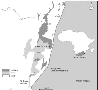
Within the iSimangaliso World Heritage Site, a site which houses approximately 3 280 km2 of diverse ecosystems, lies St. Lucia, the largest estuarine-linked lake system in Africa (Grundling et al., 2013b). Approximately 66% of recorded peatlands in South Africa are contained within the greater St. Lucia Wetland Park, thereby making it one of the most significant peatland ecoregions in the country (Grundling et al., 2013a). Located within St. Lucia is the study site, the Mfabeni Peatland.
The Mfabeni Peatland is a system that is receiving research attention due to its age, dating back beyond the Holocene, which is exceptional in the region (Meadows, 2015). However, to date, this region has not been explored from a microbiological viewpoint and hence, there is a lack of information regarding the functional and genomic diversity and microbial communities which resided in this area. This is a region which is ideal for palaeo-environmental research (Finch and Hill, 2008) and due to the age of this peatland, would be suitable for examining the feasibility of reviving AEFB present in a sediment core extracted from the site.
The study was undertaken with the aim of reviving dormant AEFB from sections of an ancient sediment core obtained from the Mfabeni Peatland, KwaZulu-Natal and determining their genetic and physiological diversity. As such, the objectives of this study are as follows:
i) Isolation and revival of dormant AEFB from sections of an ancient sediment core that had been radiocarbon dated to ages ranging from ca. 589 to 37 906 cal years BP.
ii) Assessment of the genetic diversity of revived AEFB through utilization of two techniques, namely Repetitive extragenic palindromic-Polymerase Chain Reaction (Rep-PCR) and High Resolution Melt Analysis (HRMA).
3
iii) Determination of taxonomic ranking and phylogeny of representative genotypes through 16S rRNA gene amplification and sequence analysis.
iv) Assessment of the salt and pH tolerance ranges of selected representative isolates.
v) Determination of the metabolic diversity amongst isolates by examining substrate utilization capabilities through the use of Biolog EcoPlates™.
This study focussed on characterizing the diversity of AEFB at varying radiocarbon-dated ages down a sediment core, namely 589, 1 964, 17 568, 33 328 and 37 906 cal years BP. This will be the first report providing information regarding the bacterial diversity in an ecosystem within the Mfabeni Peatland. In addition, the changing climatic conditions of this peatland through time make it the ideal study site for examining the changes in bacterial genetic and physiological diversity which occur along an environmentally-variable sediment profile. This study fits into a multidisciplinary approach based on palaeo-environmental research for a fragile and important ecosystem in southern Africa and provides the potential to examine a new proxy which could contribute towards future studies of palaeo-environments.
4
CHAPTER TWO LITERATURE REVIEW
2.1. INTRODUCTION
Wetlands are areas that are saturated with water on a temporary or permanent basis and are considered to be distinct ecosystems (Ramsar Convention Secretariat, 2013). These regions contain water which is either flowing or in a static state and support a diverse range of plant and animal life (Batzer and Sharitz, 2014). Wetlands play important roles in a number of ecological processes such as biogeochemical cycling, water purification, greenhouse gas emissions, shoreline stabilization and flood mitigation (Bodelier and Dedysh, 2013). These regions are extremely susceptible to climatic changes. Coastal wetlands may become flooded through sea level rise and are prone to erosion, runoff and depreciation in water quality (Jin et al., 2009). These changes have an impact on the biota and microorganisms which reside there, consequently affecting the diversity, abundance and distribution of species in these ecosystems (Jin et al., 2009). Microbial communities play a significant role in biogeochemical cycling and the overall maintenance of wetland processes; however, the study of microbial diversity in wetland environments is often overlooked (Bodelier and Dedysh, 2013).
Peatlands make up approximately half of all wetlands in the world (Grundling et al., 2013a).
They are characterized by a build-up of organic material, consisting of partially-decomposed plant matter, which occurs over extended periods of time as a result of anaerobic conditions which predominate (Kroetsch et al., 2011). These regions are suitable for palaeo-environmental research, which is centred on the reconstruction of past environments (Gorham et al., 2001).
Biological and chemical proxies contained within the organic material can be used for determining palaeo-environments (Gorham et al., 2001). These proxies generally exist in unaltered forms over long time periods and can be used to interpret and infer past environmental conditions which prevailed at the time of deposition. Proxies currently used include pollen, ichnofossils, diatoms and macro-fossils (Meadows, 2014). One such proxy that has yet to be explored are aerobic endospore-forming bacteria (AEFB) which have the ability to form dormant endospores, which are highly differentiated structures formed within the bacterial cell to provide protection against unfavourable conditions during metabolic dormancy (Hilbert and Piggot, 2004).
5
The occurrence of anoxic and oxic zones within peatlands support the presence of anaerobic and aerobic microbial communities (Bodelier and Dedysh, 2013). AEFB are heterotrophs that would be expected to be metabolically active within the aerobic zones. When anaerobic conditions prevail, AEFB form dormant endospores for survival. As peat layers are deposited over time, endospores would consequently remain trapped and preserved over long periods.
Endospores are able to remain in sediment once deposited (Wunderlin et al., 2014) and viable endospores have been successfully isolated from ancient sediments dated to 13 000 years (Rothfuss et al., 1997), 9 000 years (Renberg and Nilsson, 1992) and 5 800 years (Bartholomew and Paik, 1966). Sediments may, therefore, serve as a suitable archival material which could provide stable conditions that allow for endospore survival. Since endospores are able to remain deposited and inactive, they could provide reflections of AEFB species diversity and abundance present at the time of deposition within ecologically-sensitive environments. In this way, dormant ‘ancient bacteria’, i.e. bacteria revived from ancient, radiocarbon-dated environmental samples, may serve as bioindicators of prevailing conditions for a given palaeo- ecological era.
The study of bacterial diversity across environmentally-variable profiles has been made possible through a wide range of molecular fingerprinting techniques which allow for microbial diversity to be differentiated at the species or strain-level (Mandic-Mulec and Prosser, 2011).
Through the combination of sequencing technologies and phylogenetics, the taxonomic ranking and evolutionary relationships amongst isolated bacteria can be determined. In addition to genetic diversity, physiological dynamics may also provide insight into the changes in bacterial diversity occurring in palaeo-environments. By examining the physiological diversity of endospore-forming bacteria at different time scales, tolerance ranges to various environmental parameters can be ascertained (Jones et al., 1993). In addition, metabolic profiles can be established, thereby providing information on the potential functions specific bacteria may have played or the niches which they may have occupied within these ecosystems.
This review shall, therefore, address the following topics:
i) Wetland microbiology.
ii) Examination of AEFB as suitable candidates for palaeo-ecological studies.
iii) The diversity of AEFB.
iv) An evaluation of endospores, their structure, the mechanisms behind their resistance and their viability.
v) The revival of ‘ancient’ endospores and the associated challenges.
6
vi) Techniques used for the evaluation of bacterial genetic and physiological diversity.
2.2. WETLAND MICROBIOLOGY
2.2.1. What are wetlands and why are they important?
Several definitions exist which explain what wetlands are; however, from an ecological point of view, wetlands can be described as ecosystems which are characterized by land saturation with water (Bergh et al., 2004). The Ramsar Convention on Wetlands describes wetlands as areas of water, fen, marsh or peatland, which may be temporary or permanent, and contain water which is either flowing or in a static state (Ramsar Convention Secretariat, 2013). The water may either be saltwater, brackish or freshwater. In these areas, shallow water may cover the land or the water table may be present at the land surface. Due to the significant animal and plant life supported by wetlands, they are considered to be one of the most biologically diverse types of ecosystems.
Approximately 6% of the land surface on earth is occupied by wetlands (Batzer and Sharitz, 2014). Due to the significant effect that wetlands have on nutrient cycles, greenhouse gas emissions, hydrological processes, biogeochemical transformations and vegetation; wetland systems are considered to be of high ecological and economical value (Bodelier and Dedysh, 2013). Wetlands serve as buffers to run-off from terrestrial regions, thereby protecting inland and coastal waters from eutrophication. They also contribute to erosion control, stabilization of shorelines, water storage and the mitigation of floods (Ramsar Convention Secretariat, 2013). In addition, they play a pivotal role in water purification, nutrient retention, sequestration of carbon dioxide and groundwater control. Microbial communities are able to thrive in these systems due to the presence of oxic as well as anoxic conditions. The presence of these microbial communities boosts the productivity of wetland systems as they contribute significantly towards the biogeochemical cycling of nutrients and a global cycling of elements (Richardson, 1994; Bodelier and Dedysh, 2013). Ecologically, wetlands support a great range of aquatic and terrestrial biota and economically, they contribute towards fisheries, agriculture, food production, tourism and the assimilation of wastewater (Richardson, 1994).
2.2.2. Environmental characteristics of wetlands
Topography, geology and climate have an influence on the distribution and type of wetland.
Wetlands are present in temperate lowlands, deserts, tropical, subtropical, alpine regions and
7
even the Arctic (Scott et al., 2014). The driving factor which dictates the characteristics, composition, productivity and biology of wetlands is hydrology (Mitsch and Gosselink, 1993).
Wetlands experience saturation of water at the surface of the soil for extended periods of time annually. The hydrology includes many factors such as the level of groundwater, drainage characteristics, the degree of saturation of the soil and surface water outflow. The second major feature is the occurrence of hydric soil i.e. soil which is saturated with water (Mitsch and Gosselink, 1993). These soils generally have low oxygen contents. The third defining characteristic is the aquatic and plant life which is present (Mitsch and Gosselink, 1993). This generally includes plants such as hydrophytes which have adaptations to wet conditions.
2.2.3. The effects of climate changes on wetlands
The greatest effect that climate changes have on wetlands is the alteration of hydrological systems, since wetlands rely on the availability, supply and flow of water (Erwin, 2009). Inland wetlands may experience droughts when water inputs through groundwater recharge and surface runoff are outweighed by outputs through seepage and evaporation. Coastal wetlands can experience an increase in seawater due to the rise in sea levels (Jin et al., 2009). Extreme climate events can result in soil erosion, flooding, runoff and mudslides. These effects have a direct impact upon the recharging of floodplain aquifers, organic sediment oxidation and damage to local vegetation (Erwin, 2009).
Water quality is affected as a consequence since water temperatures in wetlands rise as a result of higher atmospheric temperatures. Certain plant species thrive with higher temperatures, resulting in algal blooms and a decrease of dissolved oxygen, which then promotes anaerobic decomposition (Jin et al., 2009). Additionally, the pH and salinity levels of wetlands may become significantly altered, thereby impacting upon the biota and microorganisms which reside there, consequently affecting the diversity and distribution of species in these regions (Jin et al., 2009). With the continued impact of climate changes, the functional capacity of wetlands can decline and the number of wetlands with high productivity may decrease (Erwin, 2009).
8 2.2.4. Microbial activity in wetland environments
Plant roots are largely responsible for the presence of anoxic and oxic conditions in wetlands, which generally occur in close proximity to each other (Bodelier and Dedysh, 2013). Due to the presence of these conditions, both anaerobic and aerobic microbial communities are active in these ecosystems. Nutrient recycling by these microbial communities contributes towards the productivity in wetlands (Bodelier and Dedysh, 2013). This occurs through the biogeochemical conversion of nutrients including carbon, nitrogen and sulfur, which are primarily derived from plant biomass (Bodelier and Dedysh, 2013).
Wetland microbiota comprise of microbial communities which belong to various functional guilds (Bodelier and Dedysh, 2013). Heterotrophic rhizosphere and phyllosphere microbial communities in wetland ecosystems may be involved in interactions with living plants and may contribute towards plant growth and productivity (Kumar et al., 2012). When plants die, heterotrophic aerobic and anaerobic microbial communities are responsible for the degradation of organic carbon through the production of extracellular enzymes. Carbon is one of the most predominant nutrients in wetlands and arises from plant breakdown into organic matter (Bodelier and Dedysh, 2013). The organic matter then gets broken down to release carbon dioxide (CO2) and methane (CH4). Methanogenic Archaea are responsible for methane production in these ecosystems, whereas aerobic methanotrophs play an important role in the oxidation of methane (Kolb and Horn, 2012). These communities thus contribute largely towards the atmospheric methane flux. Within the anoxic zones, denitrifiers are responsible for the conversion of nitrates to nitrous oxide (N2O) and nitrogen gas (N2) (Kolb and Horn, 2012).
Nitrifiers are also present in the oxic zones and play a role in the conversion of ammonia (NH3) to nitrate (NO3-), whilst diazotrophs carry out nitrogen fixation (Lovell and Davis, 2012).
Peatlands are generally characterized by a low availability of nitrogen, thereby resulting in high C: N ratios, which contributes to the slow degradation process encountered under anaerobic conditions (Sheppard et al., 2013). Sulfate-reducing microorganisms also make up a part of wetland microbiota and are found to closely interact with microbes involved in carbon and nitrogen cycling (Pester et al., 2012).
Various species of AEFB have been isolated from wetland and peatland environments (Albert et al., 2005; Bae et al., 2010; Baik et al., 2011a; Baik et al., 2011b; Liu et al., 2015), thereby providing evidence for the occurrence of AEFB in these ecosystems. In addition to their role in degrading and metabolizing various forms of organic carbon as primary sources of carbon and energy, AEFB also contribute towards the solubilisation of phosphates, nitrogen fixation
9
and ammonification in sediment regions (Oren, 2002; Kumar et al., 2012). Endospore-formers are commonly associated with the plant rhizosphere and some species are responsible for the promotion of plant growth (Kumar et al., 2012). This generally occurs through the production of plant hormones and the solubilisation of elements such as phosphate and phosphorous (Han et al., 2006; Kumar et al., 2012). These elements are then released in forms which are available to surrounding plants. Endospore-forming bacteria may therefore also potentially contribute towards these functions within wetland environments.
2.3 USING AEFB IN CLIMATE CHANGE STUDIES
2.3.1. The effects of climate changes on the ecology of bacteria
Due to the roles that microbial communities play in biogeochemical processes and in maintaining ecosystem stability, it is important to examine the effects that climate changes may have on bacterial community structure and functionality. While many of the climate change effects might be indirect, such as impacts on sediment quality and on the abundance and types of plants present, the most common direct effects include fluctuations in moisture content and temperature (Singh et al., 2010). These factors can result in changes in microbial species richness and abundance.
The moisture content of peatlands impacts on the availability of oxygen and the rates of gas diffusion, thereby influencing the physiological activity of the extant microbial community.
For instance, periodic reductions in sediment moisture may increase the availability of oxygen, thereby enhancing nutrient cycling, whereas an opposite effect is observed when moisture levels are high (Singh et al., 2010). The carbon: nitrogen ratio of a peatland environment can also be affected by climate changes, which can directly influence the rates of decomposition and microbial biomass in soils.
Temperature has an effect on the rate of growth, which has an impact on the rate and occurrence of microbial processes such as respiration (Singh et al., 2010). Temperature fluctuations within peatlands may thus potentially impact certain activities for some microbial species.
2.3.2. The use of endospore-forming bacteria in palaeo-ecological studies
Palaeo-ecology involves the reconstruction of previous environmental conditions, including the nature of past communities, using proxies (Gorham et al., 2001). The reconstruction of past
10
environments provides an insight into past climates as well as the effects of climate changes in specific regions and can be used to assess the impacts of future environmental changes (Meadows, 2014). A primary requirement for a biological indicator to serve as a proxy is that the organism, or associated structures, should exist in the preserving material over a significant period of time in an unaltered form (Gorham et al., 2001). Indicators which fit this requirement, and are commonly used, include siliceous microfossils and pollen grains. Past environmental conditions which prevailed at the time of deposition can be inferred using these proxies. It is necessary to explore and establish new proxies to add to the current list of proxies to aid in improving resolution and detail when reconstructing past environments (Meadows, 2014).
Reconstructing past histories can enable various aspects to be studied, such as the effect of disturbances on ecosystem structure or the effect of climatic changes and anthropogenic activities on microbial diversity (Wunderlin et al., 2014). One such proxy which has not been examined in great detail is dormant bacterial endospores.
Wunderlin et al. (2014) used endospore-forming bacteria from a sedimentary record as an indicator to assess the impact of eutrophication on bacterial diversity and composition. Renberg and Nilsson (1992) concluded that whilst endospore-forming bacteria could serve as potentially useful contributors towards palaeo-ecological studies, their application is relatively unexplored. The isolation and identification of endospores, combined with molecular genetics and biochemical techniques could allow for past environmental conditions to be deduced (Renberg and Nilsson, 1992).
2.4. AEROBIC ENDOSPORE-FORMING BACTERIA (AEFB) 2.4.1. Taxonomic diversity of AEFB
The first genus of AEFB described, Bacillus, was proposed in 1872 by Ferdinand Cohn, a German biologist (Wipat and Hardwood, 1999). Bacillus is classified under the phylum Firmicutes and the family Bacillaceae (Wipat and Hardwood, 1999). This genus comprises of members which are facultatively anaerobic or aerobic, rod-shaped, Gram positive, capable of endospore-formation, with low guanine and cytosine (G+C) contents. Initially, variation in G+C content amongst different species ranged from 33–67 mol%; demonstrating higher levels of genomic heterogeneity within the Bacillus genus than expected (Priest, 1993). Subsequently, this genus underwent further taxonomic revision following 16S rRNA gene sequence analysis and phylogenetic evaluation (Ash et al., 1991). Since 1990, certain species which were
11
previously assigned to Bacillus were reclassified into 16 additional genera. Over 220 species are incorporated into these genera, with greater than 150 assigned to Bacillus (Logan and Halket, 2011). In addition to this, 38 new genera of AEFB have also been proposed since 1990.
Collectively, there are currently 54 listed genera of AEFB (Logan and Halket, 2011). Together, these genera contain greater than 460 new species which have been proposed since the 1986 edition of Bergey’s Manual of Determinative Bacteriology (Logan and Halket, 2011).
Paenibacillus is the second largest genus of AEFB after Bacillus, containing 110 species (Logan and Halket, 2011).
2.4.2. Physiological diversity of AEFB
Physiological diversity amongst AEFB is extensive. Examples include: aerobic chemoheterotrophs such as Bacillus, Sporolactobacillus and Paenibacillus; sulfate reducers, such as Desulfotomaculum spp.; acidophiles, such as Alicyclobacillus; alkaliphiles, such as Halalkalibacillus; microaerophilic lactate fermenting bacteria, such as Sporolactobacillus; as well as coccoid representatives with morphological characteristics similar to actinomycetes e.g.
Thermoactinomycetes and Sporosarcina (Nicholson et al., 2000; Slepecky and Hemphill, 2006; Ammann et al., 2011; Logan and Halket, 2011).
Endospore-formers have adapted to a wide range of environmental conditions and are capable of inhabiting diverse habitats (Nicholson et al., 2000). Endospore formation also provides bacteria with the ability to successfully colonize specific regions and out-compete other bacteria (Wipat and Hardwood, 1999). Representatives of the Halobacillus, Thermobacillus and Psychrobacillus genera have been isolated from halophilic, thermophilic and psychrophilic regions respectively (Ammann et al., 2011). AEFB have also been isolated from diverse environments including marine sediment, salt marshes, thermal acid waters, desert soils, volcanic soil, glaciers, subantarctic soil, marine sponges, wetlands, peat bogs and geothermal vents (Logan et al., 2000; Logan et al., 2004; Albert et al., 2005; Slepecky and Hemphill, 2006;
Margesin and Miteva, 2011; Phelan et al., 2011; Cappa et al., 2014; Sonalkar et al., 2014;
Aanniz et al., 2015; Liu et al., 2015). Many of these strains possess useful abilities, such as proteolytic, amylolytic and antimicrobial activities (Phelan et al., 2011; Aanniz et al., 2015).
Members of the Bacillus genus are capable of the production of various compounds including antibiotics, enzymes such as cellulases, proteases and amylases, and lipopeptides, which have
12
anti-fungal and anti-bacterial activities (Wipat and Hardwood, 1999). They may therefore be useful candidates in the biotechnological and medical sectors.
2.5. ENDOSPORES
2.5.1. Functions of endospores
Endospores are dormant, exceedingly resistant, highly differentiated structures which are formed within the bacterial cell (Hilbert and Piggot, 2004). These specialized structures serve the function of providing resistance to the cell during periods of unfavourable environmental conditions (Schlegel, 1993). The sporulation process has been described as a ‘specialization of morphological characteristics’ which is initiated in direct response to adverse conditions (Hilbert and Piggot, 2004). Unfavourable environmental conditions may include exposure to high temperatures, lack of moisture or nutrients and high concentrations of disinfectants.
Protection needs to be present against factors which may result in direct damage to DNA, which could include UV or gamma radiation (Setlow, 1995). The resistance of endospores to heat is 105 times greater than that of vegetative cells and is a hundred-fold more resistant towards radiation (Roberts and Hitchins, 1969). Once favourable conditions arise, a vegetative state may once again resume. This may also occur through the relocation of the endospores to new environments through various agents of dispersal (section 2.5.5). Environmental samples are often abundant with bacterial endospores, which demonstrates the ability of endospore formation to serve as a successful means of dispersal and survival (Nicholson et al., 2000).
Endospore formation is one of the primary reasons behind the effective establishment and colonization of endospore-forming bacteria in various niches, thereby allowing for vital roles in various ecosystem processes to be fulfilled.
2.5.2. Life cycle 2.5.2.1. Sporulation
Sporulation is described as the transitioning stage between a cell in the vegetative state and the completed endospore (Roszak and Colwell, 1987). Approximately 4% of the genome of Bacillus subtilis (ca. 4.2 Mbp) is linked to processes involved in endospore formation (Kunst et al., 1997). In the presence of abundant nutrients, there is a repression of the sporulation process (Trach et al., 1985). However, when conditions change, the sensing proteins SfrA and
13
NtrC are able to transmit a signal regarding the environmental conditions, in turn directly impacting upon the guanosine-5'-triphosphate levels within the endospore (Trach et al., 1985).
Due to numerous similarities between sporulation and division in vegetative cells, it is believed that the process of endospore formation is a modified form of cell division found within prokaryotes (Slepecky and Hemphill, 2006).
A range of environmental and internal signals act as triggers for systems which are involved in regulation. The most important transcriptional regulator involved in these systems is known as Spo0A, which is responsible for the control of greater than 10% of the genes present in the Bacillus genome (Errington, 2003). Approximately 200 genes are involved in the entire sporulation process and the first stage in the development of endospores is referred to as endosporulation (Slepecky and Hemphill, 2006). This process takes approximately eight hours to reach completion. This step commences when Spo0A undergoes phosphorylation through the action of numerous kinases, each of which responds to a range of stimuli from the environment (Errington, 2003; Piggot and Hilbert, 2004). Once Spo0A has been phosphorylated, it serves to activate the transcription of genes such as spoIIE and spoIIA, which have key roles in the sporulation process. Remaining genes which are vital in the sporulation process are transcribed by the RNA polymerase enzyme, which is regulated by a sigma factor known as σH (Errington, 2003). Initially, the cell undergoes DNA replication. A protein known as FtsZ forms two distinct rings, one at each pole. The cell then divides through the formation of a membrane referred to as a septum, which occurs as a result of one of the Z-rings (Errington, 2003). This results in the production of a distinct pre-spore and a mother cell and is termed septation, also known as an asymmetric sporulation division (Piggot and Hilbert, 2004). The pre-spore and the mother cell follow independent developmental pathways (Figure 2.1).
Approximately one-third of the chromatin material is captured in the pre-spore during septum formation. The remainder of the chromatin material is translocated by means of a protein known as SpoIIIE (Errington, 2003). Cell-specific gene expression is co-ordinated by means of intercellular communication systems through the activation of specialized sigma factors (Piggot and Hilbert, 2004). These sigma factors are responsible for the initiation of gene expression by directing RNA polymerase to the required DNA sites. σE is the sigma factor present in the mother cell, whereas σF is active in the pre-spore. The mother cell then produces proteins which result in the degradation of the septum, with one of these proteins being SpoIIB, which is responsible for catalysis of the process. Once the developing spore obtains a double membrane, it is also sometimes referred to as a forespore (Piggot and Hilbert, 2004). The
14
double membrane occurs as a result of the cell plasma membrane surrounding the septum and pinching off. This process is termed engulfment and follows the degradation of peptidoglycan in the septum (Roszak and Colwell, 1987). At the completion of engulfment, different sigma factors become activated, namely σG in the forespore and σK in the mother cell, respectively.
These sigma factors are responsible for the activation of gene transcription, which function towards building the endospore structural components (Piggot and Hilbert, 2004). The forespore is protected by the presence of a combination of calcium and dipicolinic acid (DPA), also referred to as calcium dipicolinate (Piggot and Hilbert, 2004). At this stage, the forespore is still present in the cytoplasm of the mother cell as a protoplast. The forespore consequently becomes the endospore core and the outer endospore layers are produced by the mother cell.
The cortex, which consists primarily of peptidoglycan, is then added between the two membranes (Roszak and Colwell, 1987). An endospore coat then develops around the entire structure, after which an optional exosporium may be laid down in certain bacteria. One of the concluding stages involves the dehydration of the endospore, a stage which is vital for its resistance. The endospore now also contains pyrimidine nucleotides and nucleoside triphosphates in low concentrations (Roszak and Colwell, 1987). Upon maturation of the endospore, lysis of the mother cell occurs (Errington, 2003). This transpires through a process known as apoptosis, after which the release of the endospore into the surrounding environment takes place. The differences in resistance between species may be attributed to the variation in the structure of the polypeptides incorporated into the coat. The average time for sporulation to occur in Bacillus subtilis is 6–8 h (Errington, 2003).
Endospores may be produced in different regions of the cell, which may also aid in distinguishing bacterial species. The three locations include the end of the cells, referred to as terminal endospores, the middle of the cell, known as central endospores and lastly, the region between these two locations which is referred to as the sub-terminal endospores (Errington, 2003).
2.5.2.2. Germination
Once favourable environmental conditions arise, the endospore then undergoes germination.
This arises through the activation of specific receptors present within the inner membrane of the endospore (Hudson et al., 2001). These are known as germinant or nutrient receptors and are protein in nature. They act as sensors and are therefore able to detect the occurrence of
15
changes in the surrounding environment, for instance, the presence of nutrients or germinants such as amino acids (Hudson et al., 2001). Germinant receptors such as gerA only identify one type of germinant, whereas other receptors such as gerK and gerB are able to distinguish multiple types of germinants (Moir et al., 2002). Other studies have found evidence for the formation of complexes through the interaction of various germinant receptors (Igarashi and Setlow, 2005). The conditions under which activation occurs determines the rate at which the germination process takes place. The transmission of a signal occurs when the germinant binds to the specific receptor (Setlow, 2003). Following this, a partial loss of resistance occurs as the core becomes hydrated. The fluidity of the endospore membrane increases due to conformational changes in receptor proteins located within the inner membrane (Foster and Johnstone, 1989). The amounts of zinc ions and DPA within the endospore core decrease due to the release of the DPA and cations. It is also believed that a proton gradient may be established due to the formation of a proton motive force (Setlow, 2003). The removal of the endospore coat also occurs. The subsequent stage entails the hydrolysis of the endospore cortex region which allows for a greater degree of hydration and expansion to take place within the core (Setlow, 2003). The endospore now loses a greater degree of dormancy and becomes less resistant to factors such as radiation, chemicals and heat. The succeeding stage in the germination process involves the commencement of metabolic activities, through utilization of high-energy reserves present in the endospore. Since amino acids are not synthesized in the early stages of germination, the degradation of small acid soluble proteins (SASPs) takes place through utilization of specific enzymes which break down these proteins (Setlow, 2003). These are then used for the biosynthesis of macromolecular compounds and new proteins. GPR, an endoprotease which is sequence-specific, is responsible for the cleavage which initiates the degradation of the SASPs (Setlow, 2003). The two major genes involved in this process are the ger and spo genes, for germination and sporulation respectively (Moir et al., 2002).
DNA replication and division occurs during the outgrowth stage, which is controlled by the out genes. Outgrowth is described as the phase in which the endospore transitions into the characteristics of a vegetative cell. If conditions remain favourable, biochemical and morphological transformations occur, resulting in symmetric cell division (Slepecky and Hemphill, 2006). A variety of proteases is also present, such as esterase A and B (Moir et al., 2002). These enzymes perform numerous vital functions such as the degradation of intracellular proteins and the inactivation of sporulation-specific enzymes (Slepecky and Hemphill, 2006). ATP synthesis also commences during the primary period of germination,
16
prior to the production of macromolecules (Roszak and Colwell, 1987). The primary molecules involved in ATP synthesis are basic proteins and phosphoglycerate (Roszak and Colwell, 1987).
Figure 2.1. Cycle of sporulation and germination in AEFB (Adapted from McKenney et al., 2013)
2.5.3. Structure of endospores
The endospore is a multi-layered structure which comprises of six basic components. The innermost region of an endospore is referred to as the core (Setlow, 1988). The core consists of the cytoplasm as well as various components including RNA, DNA, ribosomes, enzymes and proteins. The cytoplasm of the core contains proteins in an immobile form and has reduced quantities of high-energy yielding compounds which are present in the vegetative cell cytoplasm. One of the significant differences between the composition of the core and that of the vegetative cell is the total water content (Potts, 1994). The cytoplasm of the vegetative cell is largely hydrated, with a total water composition of approximately 70–88% (Potts, 1994).
However, the water content of the endospore core is a much reduced 28–57% (Leggett et al., 2012). This change plays a substantial role in the survival of the endospore. Durability, as well