• Tidak ada hasil yang ditemukan

Fe III mineral formation and cell encrus (1)

N/A
N/A
Protected

Academic year: 2018

Membagikan "Fe III mineral formation and cell encrus (1)"

Copied!
11
0
0

Teks penuh

Loading

Gambar

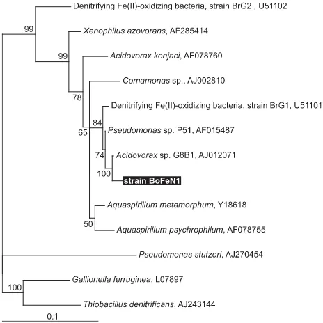
Fig. 1 16SrRNA sequence comparison of strain BoFeN1 with other strains10 000 puzzling steps were employed
Fig. 4 Confocal microscopy pictures of 4-day-old dead-live stained cells ofstrain BoFeN1 (green = alive; red = dead) grown with dissolved Fe(II) (4 mM),nitrate (5 mM) and (A) 2 mM acetate and (B) 1 mM acetate showing the viabilityof BoFeN1 cells and different levels of cell-mineral encrustation.
Fig. 6 X-ray diffractograms of precipitates collected from BoFeN1 culturesoxidizing dissolved Fe(II) when inoculated from a culture grown either withFe(II), nitrate plus acetate (‘Fe-/acetate-grown’) or grown with nitrate plusacetate (‘acetate-grown’)
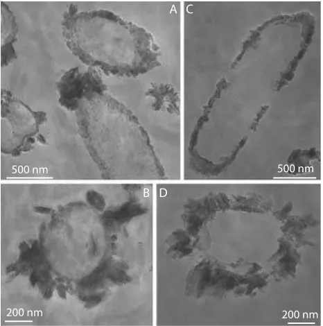
Fig. 8 Transmission electron micrographs of stained (A,B) and nonstained(C,D) thin sections of cells of strain BoFeN1 grown with dissolved Fe(II) (4 mM),nitrate (5 mM) and acetate (2 mM), and inoculated with a culture pregrown onthe same substrates

Referensi

Dokumen terkait