Horizontal Gene Transfer: An Insight into Antimicrobial Resistance
By:
Mahbubul Hoque Tanveer ID-18136008
Atihar Arzu Rafa ID-18336027 Arnob Sharma
ID-18136054 Tashfia Islam
ID-19136059
A Thesis Submitted to the Department of Mathematics and Natural Sciences in Partial fulfillment of the requirements for the degree of
Bachelors of Science in Biotechnology
Biotechnology Program
Department of Mathematics and Natural Sciences BRAC University
October, 2022
© 2022. BRAC University All rights reserved.
Declaration
It is hereby declared that;
1. The thesis was created by us as part of the Bachelor of Science in Biotechnology program at BRAC University;
2. The thesis does not include any content that has been published before or that was written by a third party unless it is properly cited with complete and accurate referencing;
3. It has been made explicit where any part of this thesis has already been submitted for a degree or other qualification at this University or another institution;
4. The thesis does not include any content that has been approved or submitted for consideration for any other degree or certificate at a university or other institution;
5. All significant sources of help have been acknowledged.
Student’s Full Name & Signature:
Mahbubul Hoque Tanveer 18136008
Atihar Arzu Rafa 18336027
Arnob Sharma 18136054
Tashfia Islam 19136059
Approval
The thesis titled "Horizontal Gene Transfer: An Insight into Antimicrobial Resistance" submitted by Atihar Arzu Rafa (ID: 18336027) of Summer 2018, Mahbubul Hoque Tanveer (ID:
18136008), Arnob Sharma (ID: 18136054), and Tashfia Islam (ID: 19136059) of Spring 2018 has been accepted as satisfactory in partial fulfillment of the requirement for the degree of Bachelors in Biotechnology.
Examining Committee:
Supervisor:
(Member)
Iftekhar Bin Naser Assistant Professor
Department of Mathematics and Natural Sciences BRAC University
Biotechnology Program Director:
(Member)
Dr. Munima Haque Associate Professor
Department of Mathematics and Natural Sciences BRAC University
Departmental Head:
(Head)
Dr. A F M Yusuf Haider Professor and Chairperson
Department of Mathematics and Natural Sciences BRAC University
Acknowledgment
We would like to proceed by expressing our sincere appreciation to the Almighty for endowing us the opportunity to pursue this research course and for then bestowing us the courage we required along the way to conclude it successfully. We want to convey our sincere gratitude and admiration to our renowned supervisorDr. Iftekhar Bin Naser, Assistant Professor, BRAC University, without whom it would have been impossible for us to work on several crucial topics during the COVID-19 epidemic. During this time, we were able to develop as researchers thanks to our supervisor's ongoing support and encouragement of this work, for which we are really appreciative. We were able to tackle a variety of unpredictable situations because of his extraordinary research abilities. This allowed us to quickly accumulate a substantial amount of information and various study materials, which allowed us to conduct novel research projects. We would like to express our gratitude to our Biotechnology Program DirectorDr. Munima Haque, Associate Professor, BRAC University, for providing us the support we needed to complete our research with diligence. We would also like to convey our gratitude toProfessor A F M Yusuf Haider, Ph.D., Chairperson of the Department of
Mathematics and Natural Sciences at BRAC University, for endorsing our thesis proposal. Last but not least, we would like to express our profound gratitude to our family for their unwavering support and prayers, which have inspired us to aim higher and pursue ambitions that can only be achieved after overcoming adversity.
Mahbubul Hoque Tanveer Atihar Arzu Rafa Arnob Sharma Tashfia Islam
Table of Contents
Declaration 1
Approval 2
Acknowledgment 3
Abstract 7
Keywords 7
Introduction 8
Antibiotic Resistance Transmission via Conjugation 11
Kinetics of the conjugation reaction 16
Quantification of antibiotics' effects on conjugation 17
HGT quantification in dynamic conditions 21
The measurement platform for conjugation in experiments 22 Antibiotic Resistance Transmission via Transformation 23
Kinetics of Transformation Reaction 24
In-vitro Process of Transformation 27
Transfer Rate End-point Equation 29
Transformation Method of Gram-positive and Gram-negative Bacteria 30 Five Models Explaining Bacterial Uptake of DNA from the Environment 31
Effects of Antimicrobial Resistance in Transformation 33 Antibiotic Resistance Transmission via Transduction 36
Classification of Transduction 37
Transduction Related to Lysogenization 40
Transduction in Clinical Settings 41
Kinetics of Transduction Reaction 41
Potential for transduction of soil coliform bacteria by biosolids-derived
phage 44
Roles of transduction in antibiotic resistance 45
Spread of Antibiotic Resistance 47
Distribution of prospective host bacteria with ARG in the environment 49
Transmission of Antibiotic Resistant in the Environment 52
The Antibiotic Resistome 53
The Clinical Antibiotic Resistome 55
The Intrinsic Antibiotic Resistome 58
Adept Outlook into Antibiotic Resistome 59
Conclusion 61
References 62
List of Tables
Number Name Page
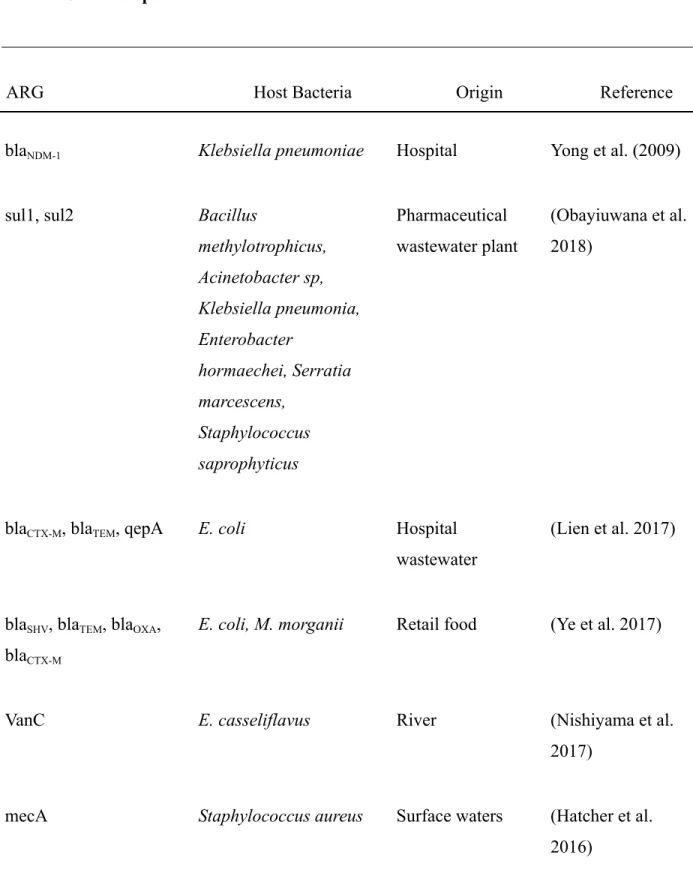
1 Genes for partial resistance and their host bacterium
51-52
List of Figures
Number Name Page
1 Horizontal gene transfer mechanisms
14 2 Antibiotics' potential impact
on conjugation
19 3 Prospective effects of
antibiotics on conjugation dynamics
20
4 Flowchart of the overall transformation process
25-26
5 The in-vitro transformation
process 28
6 Generalized transduction mechanism
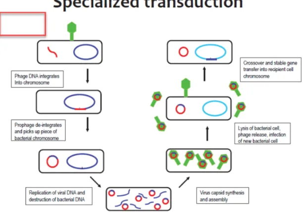
42 7 Specialized transduction
mechanism
43
8 Cumulative Resistance 55
9 The development of
antibiotic resistance.
57
Abstract
Antimicrobial resistance (AMR), which affects a variety of infectious pathogens, is a pivotal public health issue for many countries and corporations. Governments across the globe are starting to take heed of an issue that is so grave that it jeopardizes the advancements of advanced medication such as antibiotics. A key factor in the spread of antibiotic resistance is horizontal gene transfer (HGT). Contrarily, it is periodically presumed that antibiotics stimulate HGT. A comprehensive review of the research, however, indicates that there is insufficient credible data available to support such a hypothesis in principle. This is primarily due to the insufficiency of concise quantitative studies to answer this query. We assess how significantly HGT correlates to the antibiotic resistance spread in this study, as well as what is documented about how antibiotics regulate the mechanics of HGT. Our attention is on conjugation, the predominant HGT method that is primarily responsible for the global spread of antibiotic resistance. According to our research results, trials to quantify HGT must be planned in a systematic way in order to critically evaluate the outcomes. Such experiments are crucial for constructing cutting-edge approaches to inhibit the propagation of resistance by HGT. In this study, we explore about how much HGT contributes to the spread of antibiotic resistance and explore what is known about how antibiotics affect the dynamics of HGT including a brief discussion on antibiotic resistome.
Keywords: antibiotics, antibiotic resistance, horizontal gene transfer
Introduction
Antimicrobial substances that are effective against bacteria are antibiotics. Since antibiotics are quite frequently utilized in both the treatment and prevention of bacterial infections, they are the most crucial form of antibacterial agent for countering such infections since bacterial growth may be inhibited or even eliminated by them. Antibiotics uphold modern medicine; they are essential for invasive surgery and therapies like chemotherapy and have decreased pediatric mortality and improved life expectancy.
However, all kinds of antibiotics now used in clinical settings are now facing resistance.
Antibiotic resistance has been shown to be inevitable, and it frequently develops promptly after an antibiotic is introduced to a clinic. Determining the causes, extent, and development of antibiotic resistance is consequently of significant importance (Wright, 2010). Anti-bacterial, anti-parasitic, anti-viral, and anti-fungal medication efficacy is reduced considerably due to AMR, making patient treatment challenging, expensive, or perhaps unattainable. The effects on highly susceptible individuals are most evident, leading to chronic illness and a greater mortality rate (Mazaheri Nezhad Fard et al., 2011).
Antibiotic resistance in bacteria, particularly multidrug resistance (MDR), has emerged as a major problem that threatens the safety of the environment, food, and both human and animal health. By 2050, it is predicted that MDR will be responsible for 10 million human fatalities, outnumbering those caused by cancer (World Health Organization, 2014). MDR bacteria are becoming more common in diseases across the world, and the threat of untreatable infections is beginning to develop. A major hazard to human health, according to the most recent World Economic Forum Global Risks assessments, is antibiotic resistance (Blair et al., 2014). In addition, to be degraded resistance to antibiotics through chromosomal gene changes and horizontal gene transfer, bacteria can also be innately resistant to certain antibiotics. A bacterial species' capacity to withstand the effects of an antibiotic due to innate structural or functional traits is known as intrinsic resistance.
In contrast to the vertical inheritance of genes from parents to offspring, lateral or horizontal gene transfer (HGT) describes the exchange of genetic material across or within species. Bacteria
can acquire new genetic material either naturally through internal genetic mutation or indirectly through HGT. After being discovered in 1928 (Griffith, 1928), HGT is becoming more well recognized as a major driver in microbial evolution as it allows bacteria to acquire complex new traits (Boto, 2009; Ragan & Beiko, 2009). According to estimates, previous HGT phenomena caused the derivation of 17% of the Escherichia coli genome and up to 25% of genome of other bacterial species (Ochman et al., 2000).
There are three primary mechanisms by which HGT occurs in bacteria: conjugation, transformation, and transduction (Stewart, 2013). For conjugation to occur, the donor and recipient cells must come into direct contact. The donor cell creates a multiprotein bridge that connects the mating pair of cells, allowing DNA to be transferred between them. Most of the time, the DNA that has to be transferred is single-stranded in the donor cell before being converted to double-stranded DNA through replication process of the recipient cell. The bulk of newly found conjugative systems are plasmid-encoded, and DNA from plasmids serves as their conventional substrate (Wozniak & Waldor, 2010). But it has recently become clear that conjugation mechanisms are common in chromosome-borne mobile genetic components (MGEs) as well. The terms integrative and conjugative elements (ICEs) are frequently used to describe such components (Wozniak & Waldor, 2010).
In transformation, extracellular naked DNA is absorbed by cells that have acquired genetic competence, horizontal gene transfer (HGT) is one method used by bacteria. Transformation may be distinguished from DNase-resistant HGT processes by their sensitivity to DNase, which breaks down bare DNA (Hasegawa et al., 2018). Meanwhile, bacteriophages facilitate gene transfer during transduction (Ozeki & Ikeda, 1968). A broad variety of sequences, including antibiotic resistance cassettes, may be transferred by transduction and transformation (Lopatkin et al., 2016). Following precise identification between the phage and its corresponding receptor on the bacterial surface, transduction takes place. Since many phages have a limited host range due to their specialization, conjugation is thought to have had a greater impact on the spread of antibiotic resistance (Smillie et al., 2010). Natural transformation frequently takes place in a brief physiologically competent condition brought on by environmental signals like nutrient availability or cell density (Thomas & Nielsen, 2005). Even though bioinformatic investigations show that most bacteria have genes that are similar to known competence genes (Johnsborg et
al., 2007), it is uncertain if these bacteria go through transformation (Mell & Redfield, 2014;
Johnston et al., 2014). As a result, studies that claim there are not many naturally transformable species tend to believe that transformation does not significantly contribute to the dissemination of antibiotic resistance genes, albeit more research is necessary (Lopatkin et al., 2016).
Genes that have been horizontally transmitted can encode many different properties, including as metabolic characteristics, virulence components, and antibiotic resistance (Lopatkin et al., 2016).
Resistance-carrying MGEs are part of the shared gene pool used by a variety of microorganisms (Norman et al., 2009). The transmission of resistance genes can be significantly sped up by HGT since it can traverse phylogenetic barriers (Hawkey & Jones, 2009). In fact, it has been hypothesized that HGT has made it possible for resistance to most commercially available antibiotics to spread (Bennett, 2008; Davies & Davies, 2010; de la Cruz & Davies, 2000). On the other hand, the usage of antibiotics may have an impact on HGT dynamics and, as a result, the emergence of antibiotic resistance. An antibiotic, however, has the potential to both selectively modify the general dynamics of HGT and the efficiency of HGT at the single-cell level.
Experiments must be carefully designed in order to separate these various impacts in order to fully understand these effects (Lopatkin et al., 2016).
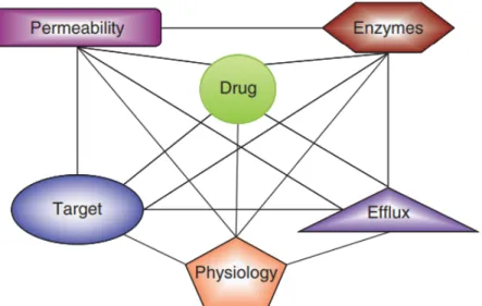
The resistome is a complex collection of genes whose actions either directly or indirectly work to inhibit antibiotic activity. Combinatorial resistance is oftentimes caused by the presence of many processes in resistant strains, which further complicates the issue and makes it complex to understand how each process affects the entire phenotype in regards to clinical treatment (Institute of Medicine et al., 2011; Wright, 2010).
Antibiotic Resistance transmission via Conjugation
Conjugation is the process by which DNA is transferred from one cell to another via cell surface pili or adhesins. The conjugative mechanism, which is either encoded by genes on plasmids capable of independent replication or through integrative conjugative components in the chromosome, facilitates it (Wozniak and Waldor, 2010; Smillie et al., 2010). Furthermore, as seen for the very broad host range IncQ plasmids, this conjugative mechanism might permit the mobilization of non-conjugative plasmids (Meyer, 2009). Conjugation is unquestionably the best researched of the several processes that may enhance HGT (Figure 1) (Guglielmini et al., 2013;
Norman et al., 2009). In many instances, conjugative elements like plasmids or transposons are linked to ARGs. Although these components can also be transferred by transformation and perhaps even transduction, conjugation is frequently regarded as the most plausible causal agent.
This is because it offers superior environmental defense, a more effective method of accessing the host cell over transformation, and frequently a wider host spectrum than bacteriophage transduction. Additionally, whereas the process of conjugation is intended to transmit bacterial genes, the transfer of bacterial DNA through transduction is a byproduct of incorrect bacteriophage replication (Norman et al., 2009).
Given the extensive research in this field on human infections, it is evident that after resistance genes successfully establish themselves onto viable plasmids, they may swiftly spread through various strains as well as species (von Wintersdorff et al., 2016). TheblaCTX-MESBL genes, that have spread across several Enterobacteriaceae plasmids with both restricted and wide host ranges in addition to various opportunistic human infections, serve as an excellent example of this (von Wintersdorff et al., 2016; Canton et al., 2012). Since these genes have become incredibly common in people, animals, and the ecosystem (Woerther et al., 2013; von Wintersdorff et al., 2016; Hartmann et al., 2012). Additionally, the transfer of plasmids in infections has caused the dissemination of multiple ARGs that code for resistance to aminoglycosides, β-lactams, sulfonamides, tetracyclines, quinolones, and many other drug classes around the world (von Wintersdorff et al., 2016; Huddleston, 2014). A growing number of reports of the spread of plasmids containing carbapenem resistance (Carattoli, 2013) and the recent identification of plasmid-encoded colistin resistance in China (Liu et al., 2015) are of particular concern. These developments may lead to Enterobacteriaceae becoming truly pan-drug resistant (Arcilla et al.,
2015). Additionally, numerous ARGs are frequently co-localized on the same plasmid, thus makes it possible for multidrug resistance to propagate quite quickly (von Wintersdorff et al., 2016).
Among the three types, the far more complex type of horizontal gene transfer (HGT) that occurs in bacteria is conjugation, and it offers a platform for the dissemination and maintenance of virulence and antibiotic resistance genes (von Wintersdorff et al., 2016; Norman et al., 2009).
Integrative conjugative elements (ICEs) and conjugative plasmids (CPs) serve as the transfer carriers (ICEs). Different forms of CPs can be distinguished using phylogenetic analysis. a group of conjugative plasmids classified as F-like because, in terms of their DNA transfer genes, they are related to the well-established F plasmid or F, or F-factor. Since Lederberg and Tatum's (LEDERBERG & TATUM, 1946) first depiction of bacterial conjugation, F and F-like plasmids plays a major role in elucidating the molecular processes and architectures enabling DNA transfer between bacterial cells. While F-like plasmids appear to be confined to closely related enterobacterial genera like Citrobacter, Enterobacter, Escherichia, Klebsiella, Salmonella, and Shigella conjugation is pervasive throughout the bacterial and archaebacterial world (as are ICEs and CPs) (Koraimann, 2018). These facultatively anaerobic, Gram-negative bacteria may thrive in a variety of environments, including the gastrointestinal tracts of people and animals, where they can live commensally or cause moderate to severe illnesses.
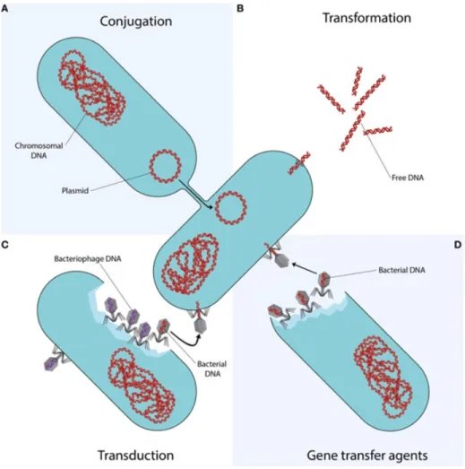
Figure 1: Horizontal gene transfer mechanisms. The technique of gene transfer represented by each quadrant is distinct. (I) The process of conjugation, in which DNA is transmitted from the donor cell to the recipient cell, requires cell to cell contact via cell surface pili or adhesins. In (II), transformation, bare extracellular DNA fragments are taken up, incorporated, and functionally expressed. (III) Bacteriophages can transmit bacterial DNA from an infected donor cell to an uninfected target cell by specialized or generic transduction. Bacterial DNA may inadvertently be injected into the phage head during generalized transduction (represented as a phage with a red DNA strand.). It is co-extracted and loaded into a new phage during specialized transduction of genomic DNA that is close to the prophage DNA (not represented). (IV) Bacteriophage-like particles called gene transfer agents (GTAs) transport random genome fragments from the generating cell. The transfer of GTA particles to a recipient cell might occur as a result of cell lysis (von Wintersdorff et al., 2016).
The worldwide resistance landscape is shaped in part by HGT and in part by antibiotic selection (Martínez, 2008; Aminov, 2009). In a limited setting, such as a patient's intestines, HGT facilitates the local dissemination of resistance genes across an origin species to different strains or species (Karami et al., 2007; Egervärn et al., 2009). People, animals, and objects that contain newly resistant germs are continually shifting and interacting. Due to this enhanced diffusion, the newly acquired resistant bacteria can spread globally. Through HGT, resistant bacteria spread the resistance determinants farther in new situations. By enhancing for the resistant offspring, the use of antibiotics might enhance the overall impact of HGT. Selection may increase the number of cells competent of HGT, increasing the likelihood of more HGT events (Stecher et al., 2012).
Collectively, these factors have undoubtedly assisted resistance expand across the globe (Stokes
& Gillings, 2011; Berendonk et al., 2015).
The proliferation of β-lactamase (Bla) varieties serves as the finest illustration of this. Among the earliest and most frequently given antibiotic classes, β-lactams, are resistant to bla enzymes (Davies & Davies, 2010). Numerous bla genes presumably propagate by conjugation (Jacoby &
Sutton, 1991; Johnson & Woodford, 2013), and bla variations, particularly extended-spectrum β-lactamases (ESBLs) (Meini et al., 2014), are frequently encoded on plasmids (Shaikh et al., 2015; Vaidya, 2011). For instance, lateral acquisition is suggested by MGE-associated CTX-M (a frequent bla variation) in Enterobacteriaecae species (Olson et al., 2005), which shares adjacent gene sequences with Kluyvera species. Similar to this, the variety of species bearing conjugative plasmids with the sequence-identical metallo-β-lactamase blaNDM-1 (Table 1) which confers resistance to carbapenem medicines, implicates the significance of conjugation. In fact, one investigation from New Delhi's sewage and water faucet discovered the prevalence ofblaNDM-1in upwards of 20 different strains, many of which previously had not been connected to ESBL resistance; each isolate is capable of transmitting their plasmid through conjugation under specific experimental circumstances (Walsh et al., 2011). The occurrence of such cases has probably been aided by the strong β-lactam selection pressure during the past few decades.
Numerous resistance genes are found in human gut microbiota (Bailey et al., 2010; Marshall et al., 2009), and this microbiota can serve as an accumulation for other dependent or pathogenic bacteria to pick up resistance through horizontal gene transfer (Liu et al., 2012; Huddleston, 2014; SALYERS et al., 2004). By enhancing for low-level resistance already existent, antibiotics
can create an environment that is conducive to transmission (Karami et al., 2007; Stecher et al., 2012). If two (or more) of the selected genes are physically connected to the same MGE, this selection may also affect how frequently organisms develop resistance to other drugs. In the gut flora of children who received non-FQ antibiotic treatment, for instance, one research found elevated frequencies of qnr-mediated FQ resistance (Vien et al., 2012). According to the environment, however, different elements could be what drives resistant landscapes. According to one investigation, the main factor influencing the resistance landscape in unmanicured soil conditions is not HGT but rather the makeup of the bacterial population (Forsberg et al., 2014).
Considering how frequently resistance is passed along via conjugation, it becomes plausible to anticipate that using antibiotics helps increase HGT rates. It is undeniable that the use of antibiotics in both human and animal medicine has altered the ecology of resistance (Martínez, 2008), and that this shift in the ecology of resistance has presumably had an impact on HGT by altering the number of species that carry resistance on MGEs (Baquero et al., 2013). Antibiotics may potentially modify the rate of HGT by favoring cells with higher transfer efficiencies (Stokes & Gillings, 2011). Antibiotic usage does not necessarily lead to the emergence of antibiotic resistance, though. For instance, ESBL genes have developed and propagated through conjugation in the course of millions of years prior to the current use of antibiotics (Barlow &
Hall, 2002; von Wintersdorff et al., 2016). A complex issue that depends on a number of variables is the degree to which antibiotics modify the HGT rate. The concepts of HGT rate must be made clear, and trials must be well planned to yield definitive findings, in order to shed light on this subject (Lopatkin et al., 2016).
Kinetics of the conjugation reaction
Conjugation may be modeled at the population level as a biomolecular reaction between the donor (D) and the receiver (R), culminating in the transconjugant (T), where D, R, and T each signify the corresponding cell densities (Lopatkin et al., 2016; Levin et al., 1979). T's rate of generation through the two parents can be expressed as:
𝑑𝑇 (1)
𝑑𝑡 = η
Here the conjugation efficiency, denoted by the symbol η, is the interaction's rate constant.
If R and D are generally constant and T is largely produced by the two parents (i.e., secondary conjugation by T is minor) within a brief window of conjugation (Δt), the conjugation efficiency may be estimated as follows (Lopatkin et al., 2016):
η≈ 𝐷𝑅Δ𝑡𝑇
The conjugation readout has previously been described as the ratio of T between two experimental conditions (for instance, with and without antibiotics) or as the relative frequency, defined asT per either parent(T/D or T/R), relative increase, defined asTper either initial parent density (T/Di or T/Ri) (Sørensen et al., 2003). Regardless of the nomenclature, it is possible to ascertain if certain elements contribute to the reported rise in T by comparing the conjugation readouts under various settings (Lopatkin et al., 2016).
However, as is conclusive from (1), these alternative measurements may confuse various influences on T. T in particular is produced either by cell division or conjugation. Therefore, various experimental settings (i.e., shifting D or R starting values, Δt) or antibiotic-mediated selection (growth/death of D, R, or T) can likewise cause a change in T. When evaluating if a factor such as an antibiotic does, in fact, alter the conjugation efficiency η, these effects should be avoided or reduced (Lopatkin et al., 2016).
Quantification of antibiotics' effects on conjugation
Two factors—the pace of conjugation or the conjugation efficiency and the subsequent expansion of transconjugants—affect the overall dynamics of conjugation (Figure 2 a) (Lopatkin et al., 2016). An antibiotic may influence these dynamics by altering the efficiency of conjugation, acting as a selection factor that influences the dynamics of the population after conjugation, or by doing both (Figure 2 b, c). Limitations of decoupling among these two aspects in earlier research made it difficult to quantify the impact of antibiotics on conjugation efficiency (Rensing et al., 2002; Johnsen & Kroer, 2007; Sørensen et al., 2005). Consequently, it is rather unclear whether or whether antibiotics facilitate conjugation (Blázquez et al., 2012). Generally, transconjugants have been quantified after a growth phase has been allowed to pass during in vitro conjugation studies in the presence of an antibiotic (Schuurmans et al., 2014; Smet et al., 2010). Based on the observed rise in transconjugants in the presence of an antibiotic compared to the untreated control (Barr et al., 1986), it has been concluded that antibiotics stimulate conjugation. Unfortunately, this experimental setup cannot discriminate between the antibiotic's effects on conjugation efficiency as well as its impacts on selection dynamics, and it also does not demonstrate how antibiotic concentration could affect these effects. Results from in vivo and case studies have also suggested a connection between antibiotic use and conjugation-mediated transfer of resistance, presumably because antibiotic choice creates a conducive environment for transfer (Goren et al., 2010; Karami et al., 2007; Cavaco et al., 2008). Due to the complexity of the research, much like with in vitro studies, this theory has yet to be proven beyond a reasonable doubt.
Figure 2: Antibiotics' potential impact on conjugation. a, Overall, there are two stages to conjugation dynamics: conjugation and the subsequent changes in the populations of the donor (green), recipient (red), and transconjugant (yellow). b,c, Antibiotics can change the population dynamics by modifying the growth rates of single or multiple populations (c), or they can modify the conjugation efficiency (b). In this example, we presume that, generally, antibiotics improve conjugation (b,c), relative to the untreated state (a) (Lopatkin, Huang, et al., 2016).
It's important to tell the difference between an alteration in and one in R, D, and T when evaluating antibiotic effects on conjugation. The latter may be brought on by a confluence of the possible impact of antibiotics on and the ensuing processes of selection (Rensing et al., 2002;
Johnsen & Kroer, 2007; Lopatkin et al., 2016; Smets & Barkay, 2005). Antibiotics may indirectly or directly encourage (Figure 3). Antibiotics are thought to cause a general cellular response that indirectly promotes or inhibits conjugation when transfer mechanism expression is unrelated to antibiotic treatment. Antibiotics have the ability to directly induce a series of molecular activities that lead to the development of transfer machinery (Andersson & Hughes, 2014; HASTINGS et al., 2004).
Figure 3: Prospective effects of antibiotics on conjugation dynamics. a. Conjugation happens at a certain pace in the lack of antibiotics, and the resistant progeny develop normally but are not chosen for. b. There are two hypothesized possibilities for how antibiotics alter conjugation efficiency when present. Antibiotics are believed to indirectly modify the conjugation efficiency in the first case. In this instance, antibiotics cause the cell to react universally. In these situations, it is feasible that the efficiency has either (i) increased or (ii) decreased in comparison to (a), but following a time of development and selection, all three possibilities lead to the same conclusion.
Antibiotics directly induce the development of the conjugation mechanism in this scenario (iii), which is demonstrated by the excision of the resistance gene from the chromosome into a circularized plasmid. This is a typical method for integrating conjugated elements and transposables with a partner (ICE). When this occurs, the conjugation efficiency rises even before selection takes place (Lopatkin, Sysoeva, et al., 2016).
Previous research on these topics has produced what appear to be inconsistent findings (Blázquez et al., 2012), such as the fact that the same or comparable antibiotics might have different effects depending on the experimental setting. Transconjugants are often tested after a period of time Δt to assess the effects of the antibiotic. Typically, donors and recipients are combined in the presence of an antibiotic (or other stimuli) (Lopatkin, et al., 2016). The combination of sub-inhibitory kanamycin (Kan) and streptomycin (Strep) doses increased conjugation for three separate conjugative plasmids in E. coli, according to one study's T/Ri calculations (Lopatkin, et al., 2016; Zhang et al., 2013).
However, a rigorous analysis of the data suggests that the measureT/Ri, which revealed on the time-dependent expansion of the transconjugant population, may have been more accurate in reporting the conjugation efficiency improvement. Antibiotics in particular may have had a considerable influence on growth rates. As time passes and cells multiply, these variations become more pronounced. In fact, there were hardly any variations between the readout with and without treatment during the first four hours after conjugation, once these influences of antibiotic-mediated selection were probably least noticeable. Similar findings have been reported in other investigations (Zhang et al., 2013; Al-Masaudi et al., 1991; Barr et al., 1986), which link an increase in frequency to the antibiotic's indirect induction of conjugation. It is less obvious if the conjugation efficiency also increased.
It is generally known that conjugation contributes significantly to the spread of antibiotic resistance on a global scale. Contrarily, it's not always correct to say that using antibiotics encourages conjugation. In general, mechanical, physiological, and selective hurdles must be surmounted for conjugation to succeed (Smets & Barkay, 2005; Thomas & Nielsen, 2005). In fact, the findings by Lopatkin et al. suggest that there may be two reasons why the role of antibiotics to the development of conjugation is overstated (2016). Initially, when the conjugation mechanism is expressed constitutively in a system, antibiotics do not appreciably improve conjugation efficiency. Second, only a tiny group of characteristics encourage a rise in the percentage of transconjugants, even when selection dynamics are taken into account. This unexpected result results from the conflicting functions an antibiotic performs during conjugation. If an antibiotic has no impact on either of the parent strains, there is no selective advantage for the transconjugant. If the antibiotic inhibits one or both of the parent strains, the
transconjugant does get a selection advantage. To counteract the effects of positive selection on the transconjugant, the antibiotic may still reduce the frequency of conjugation by decreasing the ratios of one or both parental populations. The results of the study by Lopatkin et al. suggest that other factors, particularly the physiological state of cells prior to conjugation as well as energy accessibility throughout conjugation, have a significant impact on conjugation efficiency even when the presence of an antibiotic need not significantly increase conjugation efficacy intrinsically (Figure 2 c) (2016). Population structure predominates when selection processes are taken into account to forecast the appearance of transconjugants. The findings highlight the significance of assessing microbial population growth patterns (either with or without antibiotic treatment), in order to assess cell physiology and determine the impacts of antibiotic-mediated selection (Lopatkin et al., 2016). The evaluation of the risk posed by the spread of resistance can also benefit from this knowledge. For instance, a recent study (Korem et al., 2015) showed that it is possible to use sequencing to estimate the abundances and growth rates of various microbial communities. Quantitative assessments of the level of HGT in various natural habitats may be made using these data along within vitroestimations of conjugation efficiency.
HGT quantification in dynamic conditions
Quantifying HGT and the impact of antibiotics on HGT becomes more difficult when settings are more complicated, like those of natural soils, toxic water, or biofilms (Bellanger et al., 2014;
Aminov, 2011). However, it is difficult to accurately evaluate the impact of a medication on growth rates, as well as the process of gene transfer is affected by geographic dispersion and motility. These challenges make it difficult to quantify conjugation in these conditions. To differentiate between various antibiotic effects, appropriate control studies must be created.
Studies demonstrating that antibiotics may increase conjugation efficiency in purified and activated sludge cultures (Kim et al., 2014) and decrease it in sewage water (Ohlsen et al., 2003) may be deceptive without these data. These findings collectively imply that HGT rates might vary based on the environment. When exposed to sub-inhibitory quantities of Kan (instead of imipenem, a -lactam), another study found increased conjugation in biofilms of plasmids expressing Kan resistance (Ma & Bryers, 2012). The authors, however, fail to distinguish between an impact on conjugation efficiency and antibiotic-induced modifications to the dynamics of biofilm growth. Growth rate predictions were ignored while quantifying
conjugation, which was additionally complicated by the interchangeability of several conjugation measures. Therefore, it would be premature to draw the inference that bacteria may "detect the antibiotics to which they are resistant" and, in turn, enhance the horizontal transmission of resistance genes (Lopatkin et al., 2016). The outcomes of these investigations may more indicate how antimicrobials affect biofilm development or community makeup than how they affect gene swapping. In order to understand how antibiotics, alter conjugation dynamics in such settings while accounting for compounding variables, new experimental methodologies and quantitative metrics need be put into place; several research have in fact started to go in that direction (Jutkina et al., 2016).
The measurement platform for conjugation in experiments
By minimizing the bias present in some experimental methodologies, selecting a suitable experimental platform may assist quantification. Colony-forming units (CFU) are used to quantify conjugation by selecting markers (such antibiotic or heavy metal resistance, which often reside on plasmids) that may identify transconjugants from parental strains (Johnsen & Kroer, 2007; Neilson et al., 1994; Andrup & Andersen, 1999; Singh et al., 2013). By maintaining total separation amongst conjugation and selection, as previously mentioned, their effects on the conjugation process should be easily discernible if the antibiotic being tested is comparable to the one used for plating. Phenotypic readouts, in contrast, provide a benefit since higher throughput technologies, including plate readers as well as flow cytometry for fluorescence, may be employed (Sørensen et al., 2003). With the right reporters, single-cell and microfluidics analyses are made possible by quantification employing microscopy (Babić et al., 2008). In fact, utilizing fluorescently tagged donors, recipients, and transconjugants, a microfluidics platform was recently employed to measure conjugation dynamics (Lopatkin et al., 2016). However, the detector must be capable of picking up events that happen at low frequencies. In some systems, the conjugation efficiency may alternatively be determined via nucleic acid measures, such as qPCR (Wan et al., 2011); this method of quantification does not need transconjugant enrichment (for example, through selection), and it may be able to avoid biases brought on by growth.
Experiments should be planned to provide comparable outcomes regardless of the platform. As an illustration, the impact of antibiotics on various systems might introduce additional
compounding variables, such as variations in the antibiotic sensitivity of donors and receivers.
The minimal inhibitory concentration (MIC) or IC50 value of a medication, as well as whether the antibiotic examined correlates to the resistance conferred, must be understood in relation to the concentrations employed in each specific investigation. When comparing efficiency, it is important to take into account the fact that various systems may need varying timescales.
Antibiotic Resistance Transmission via Transformation
HGT is the transfer of genetic information from one bacteria to another bacteria within the same generation. The active uptake of free DNA by bacterial cells and the heritable inclusion of its genetic material constitute natural genetic change. The cellular processes involved in transformation have been extensively studied through in vitro experimentation with a few transformable species since Griffith's famous discovery of transformation in Streptococcus pneumoniae in 1928 and the demonstration of DNA as the transforming principle by Avery and coworkers in 1944. Transformation maybe a potent horizontal gene transfer method in naturally occurring bacterial populations, although this idea has only recently come to light (LORENZ &
WACKERNAGEL, 1994). DNA internalization and chromosomal integration are required for natural bacterial transformation, which has been observed in about 80 species so far, requires DNA internalization and chromosomal integration. Recent research has shown that phylogenetically distinct animals differ in the inciting cues and regulatory processes involved, whereas they share conserved uptake and processing proteins.
There are three types of gene transfer systems, i.e., conjugation, transformation, and transduction. In the previous conjugation method, we saw that in order to transfer genetic information, bacteria must be physically attached to each other. In contrast, the transformation process is a genetic recombination method in which lysed and degraded bacteria are enter into the competent recipient bacterium and incorporate the piece of DNA into the recipient bacterium.
That means, the transformation process directly uptakes the foreign DNA from the surrounding environment. Again, normal bacteria are not able to uptake the foreign DNA particles directly via the membrane. For that reason, the competent cell can easily take the foreign DNA particle.
During the growth of bacteria, they pass certain phase like the exponential phase, which is a
competent phase. In this phase, the cell membrane is a permeable surface that allows the liquid-type materials in or out. Some examples of naturally competent cells are Neisseria gonorrhoeae, Neisseria meningitides, Hemophilus influenzae, Legionella pneomophila, Streptococcus pneumonia, and Helicobacter pyloria(Libretexts Biology & Kaiser, 2022).
Kinetics of Transformation Reaction
During transformation, the competent cell directly uptakes the foreign DNA from the surrounding environment. In the surrounding environment presents billions of bacteria which are created by manmade activities, animals, industries and so on (Prussin & Marr, 2015). Most of them are pathogenic or nonpathogenic. Pathogenic are those bacteria that are disease-causing, whereas nonpathogenic bacteria are not the cause of the disease. Moreover, these pathogenic bacteria may be AMR, meaning these bacteria can fight against medicine (World Health Organisation, 2020). So, these existent bacteria are broken down every day, naturally or unnaturally, via different factors like lack of oxygen, enzymes, detergents, and so on. After being lysed, the genetic materials are released into the environment. After that, the lysed DNA will bind with the cell wall of the recipient bacterium. Here, one DNA fragment of foreign DNA will be hydrolyzed by the envelope exonulease of the recipient cell. Therefore, only one DNA strand will enter the recipient cell. That means a linear DNA fragment entered. Once the fragment is entered, it will find the homologous region with the recipient cell after determining that it will be integrated into the host chromosome. Thus, if that happens, it will be a successful transformation.
If the entered fragment does not find a homologous region in the recipient cell, it will degrade inside the cell and is referred to as an unsuccessful transformation. For example, a plasmid uptakes a green fluorescent protein gene from the surrounding environment and it turns into a green fluorescent bacterium.
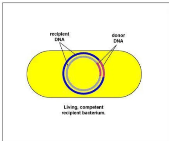
Figure 4: Flowchart of the overall transformation process. In the very first stage donor bacteria must lysed and released the genetic material in the environment. In the second step, the lysed DNA around 10 genes long band bind with the living recipient competent bacterium cell wall to transformation. In the third step, one DNA fragment of foreign DNA will degrade by envelope exonuclease by the recipient cell and another DNA fragment will entered into the recipient. Here, to prevent from being degrade in the cytoplasm the foreign DNA bind with the Competence-specific single-stranded DNA-binding proteins. In the fourth step, exchange a fragment of genetic information to promote RecA proteins between a donor foreign DNA and recipient bacterium. And the foreign DNA is incorporated into the recipient chromosome as can be seen in the picture.
The fifth step shows picture of successfully complete transformation and the flowchart pictures are taken from (Libretexts Biology & Kaiser, 2022).
In-vitro Process of Transformation
Till now, it has been described how the transformation occurs in the environment naturally.
Additionally, the transformation process can also be performed in vitro, meaning in the lab setup.
This experiment is done in the laboratory to produce multiple copies of our targeted gene via plasmid. Therefore, first insert the targeted gene into the circular DNA, which is called a plasmid. In this process, restriction enzymes and DNA ligase are used, which help in ligation.To perform this experiment first, our targeted DNA will be designed. Here, for cutting, the designed foreign DNA fragment will be cut by using a restriction enzyme. After that, the recipient bacteria must be competent phage. As all bacteria are not naturally competent to uptake foreign DNA, they must be converted to competence via chemical manipulation in the laboratory. Different ways to convert competent cells, e.g., treating cells with solutions of CaCl2 or chlorides of other elements such as Mg, Ba, Rb, Sr, and mixtures of them; (ii) treating cells with chelating agents (e.g., EDTA); (iii) treating cells with enzymes (muraminidases or peptidases), resulting in the formation of spheroplasts or protoplasts; (iv) fusing cells or protoplasts with DNA (bio listic transformation). Here, we commonly use calcium chloride, which helps to permeabilize the cell membrane so that the bacteria can uptake our targeted foreign DNA (LORENZ &
WACKERNAGEL, 1994, P565).Now, use the same restriction enzyme to open the plasmid gene and it will be ready to accept the piece of new DNA (Cecchetelli, 2019). Finally, the incorporated foreign DNA is ligated with the recipient plasmid with the help of a ligase enzyme.
Once the experiment is successfully done, it will be cultured in the lab and make a multiple number of copies (Science Learning Hub, 2014). Furthermore, because the foreign DNA is circular, the degradation of double-stranded DNA is less than that of single-stranded DNA, as previously mentioned.Also, plasmids can replicate independently, and they can replicate multiple numbers of genes inside the cell and produce a good amount of desired product.
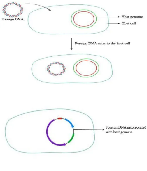
Figure 5: The in-vitro transformation process. Pictures are made by biorender.
Transfer Rate End-point Equation
The end-point equation is used to determine the transfer rate from experimental data.
γ=1Δt (V0−R0) ( ln (V0−R0+R2R1) – ln (V0R0))
Here, Δt = time interval of transfer; V0 & R0 = initial concentrations of vector and recipient cells; R1 = final concentration of recipient cells and γ = transfer rate per recipient cell concentration and vector concentration. (von Wintersdorff et al., 2016; Frontiers in Microbiology et al., 2018)
For example, we used the end-point equation to calculate the value of γ transformation with data taken from the published transformation study (Lu et al., 2015). ). Here, we used average DNA and mass DNA fragments of about 30 kb and converted between count and DNA concentration.
The published paper reported a DNA concentration of 2.5 μg/mL at one data point, which translates to a V0 of 8.33 × 10¹¹ fragments/mL and an artificially induced recipient cells' competence rate is about 0.2. As the same data point, the reported recipient concentration of 107 cells/mL translates to an R0 of 2 × 10⁶ cells/mL. So, at that point, the value of R1 is calculated as 1.9998 × 10⁶ from the stated transformation frequency. According to these three values Δt of 30 min, so, γ = 4.00 × 10⁻¹⁷ mL DNA fragment-1 min-1. Again, its repeat for each amount reported in the paper generates a mean of γ = 4.35 × 10⁻¹⁷ mL DNA fragment-1 min-1. Now, to get the final γ transformation, the receiver competence rate and the ratio of free DNA to donor cells must be scaled into this rate constant. Although different bacterial species have different competence rates in nature, this model uses a suitable competence rate of 0.01(von Wintersdorff et al., 2016; Lorenz & Wackernagel, 1994).So, this competence rate is directly proportional to the γ transformation. Based on estimations of 106 cells/mL and 0.01 g/mL of DNA in ocean water, the ratio of vector concentration to donor cell concentration employed translates to a ratio of 333 DNA pieces per donor cell assuming an average fragment size of 30 kb (Jiang & Paul, 1998; Lorenz & Wackernagel, 1994; Lu et al., 2015; von Wintersdorff et al., 2016). From these two factors, the final γ transformation is approximately 10-16 mL cell-1 min-1. Only 1% of bacterial species can naturally change. Since these species are widely scattered among the taxa, it is appropriate to choose each species' transformability at random with a probability of 0.01 to mimic natural transformation (Jhontson et al., 2014, Thomas & Nielsen, 2005).
Transformation Method of Gram-positive and Gram-negative Bacteria
Gram positives transformation method is based on the agitation of bacterial protoplasts with glass beads in the presence of DNA and polyethylene glycol (Rattanachaikunsopon &
Phumkhachorn, 2009). One of the main obstacles to genetic studies of Gram-positive bacteria is the challenge of delivering DNA into the cells of Gram-positive bacteria. Their cell walls' substantial peptidoglycan coating is seen as a potential impediment to DNA uptake. These organisms cannot be transformed spontaneously, but they can be altered using specific transformation techniques like protoplast transformation and electroporation. To produce protoplasts by enzymatically removing the cell wall, a process known as protoplast transformation was created. Polyethylene glycol makes it easier for protoplasts to absorb DNA.
For several Gram-positive bacteria, it was created (Morelli et al., 1987). This approach is infrequently utilized due to its low and extremely variable efficiency and time-consuming methodology. For Gram-positive bacteria, electroporation is now the most popular transformation technique. To stimulate the creation of transitory holes in cell walls and membranes, a high voltage electric pulse with a brief duration is applied. The pores may allow DNA from the surrounding environment to enter under the right circumstances. DNA was successfully inserted into numerous types of Gram-positive bacteria using this technique.
Although electroporation can be used to change bacteria with high effectiveness, due to the demand for expensive and specialized equipment, it cannot be carried out by small and underequipped laboratories. Gram-positive bacteria were used as the recipients for pGK12, an erythromycin resistance gene-carrying 4.4 kbE. coli/Lactococcusshuttle vector, to achieve glass bead transformation. Enterococcus faecalis TISTR 927, Lactobacillus casei ATCC 393, Lactococcus lactis DSM 20481, Leuconostoc dextranicum ATCC 19255, Listeria innocua DSM 20649, Staphylococcus aureus ATCC 25923, and Streptococcus pneumoniae ATCC 10015 were the bacteria employed in the transformation studies (Rattanachaikunsopon & Phumkhachorn, 2009).
Additionally, in Gram-negative by using FT-IR analysis of the frequency shifts of the acyl chain methylene symmetric stretching band as a monitor, temperature-induced order/disorder transition profiles were discovered in the membranes of intact Gram-negative bacterial cells. Different
transition profiles were produced by cells grown at various temperatures. But at each growth temperature, the virtually same frequency readings showed that the bacterial membranes were in a very similar "state of order." A GC analysis of the fatty acid composition of entire cells was used to supplement the FT-IR findings. The in vivo FT-IR data provided clear proof that bacterial membranes can change their "state of order" and "fluidity" to a range of growth temperatures (Schultz & Naumann, 1991).
Five Models Explaining Bacterial Uptake of DNA from the Environment
According to the first hypothesis, DNA is ingested by the bacterial cell and used as a source of nutrients (Finkel & Kolter, 2001). Studies demonstrating that in nutrient-limited circumstances, some bacterial strains are capable of assimilating DNA corroborate this concept (Redfield, 1993). By switching the cultures from a rich medium to a starving media, many bacterial species, including Gallibacterium anatis, Haemophilus influenza, and Aeromonas salmonicida26, are brought to competence in vitro (Huddleston, 2014). More particular, purine deprivation causes H. influenzae to become competent.
The second hypothesis postulates that natural transformation arose so that altered DNA molecules might function as templates for DNA repair, explaining the evolutionary roots of competence development and natural transformation (Huddleston, 2014). When DNA is applied to Bacillus subtilis cells that have been exposed to ultraviolet light after damage has occurred, the cells' survival rate is higher than when DNA is applied before damage. It is likely that the additional DNA acts as a template for re-combinational repair (Michod et al., 1998). When exposed to the DNA-damaging chemical mitomycin C, Streptococcus pneumoniaecan develop competence, more specifically the com regulon.
A third hypothesis holds that natural processes evolved to enable cells to acquire novel genetic information on purpose in order to broaden their genetic variety and better withstand natural selection. But according to research on the transformability ofAcinetobacter baylyi, competent and incompetent strains both acclimated to laboratory circumstances at the same rate (Bacher et al., 2006). Competence did not bring about any observable benefits, and the competent lineages
even evolved a lower level of transformability in lab settings. In their recently published complementary model, Engelstädter and Moradigaravand hypothesized that competence evolved as a result of bacterial cells' ability to take up DNA from their environment that has endured for an infinite amount of time and incorporate genes into their genomes in order to return to a previous genetic state (Huddleston, 2014).
The fourth hypothesis proposes that periodic stressful situations, which may unintentionally favor non-growing competent cells, are responsible for the evolution and maintenance of natural transformation. Competent cell populations, like Bacillus subtilis, need many hours to grow. This state is regarded as "per-sistering." The per-sister state was initially identified in 1944 in penicillin-treated Staphylococcus cells that survived. These cells survived not because they were mutants with different antibiotic targets, but rather because they were not growing and unaffected by the antibiotic (Huddleston, 2014). Instead of being resistant to antibiotics, these cells are now thought to be tolerant of them. When the cell population is exposed to stressful situations that kill growing cells more quickly than per-sister cells that are not expanding, this process of episodic selection of per-sister cells takes place. If these per-sister cells are also competent, as some of them are, then the stress also indirectly selects for natural transformation ability. The use of antibiotics (penicillin-G) as episodic selection in populations of B. subtilis, where capable cells outlived non-competent mutants, has been used to support this idea (Huddleston, 2014; Johnsen et al., 2009).
The final model that Natural transformation is a byproduct of the twitching motility and cell adhesion activities of type IV pili (Huddleston, 2014). It is well known that Type IV pili absorb free DNA during natural transformation. The tip of the type IV pilus, PilC, may unintentionally bind DNA as the cells move over surfaces using their twitching motility. The PilQ pore in the outer membrane is blocked by bound DNA as the pilus retracts, and it eventually passes through after one strand is broken down by nucleases. According to this hypothesis, environmental stress activates the SOS response, a DNA repair system that is prone to errors, which causes the development of type IV pili, allowing the cell to move by twitching motility to a more advantageous environment. The active type IV pili result in the binding and uptake of free DNA by the cell (Huddleston, 2014; Bakkali, 2013). The capacity to naturally convert is accidental in this scenario, but it can be useful if the cell can use the new DNA as a source of nutrients or as a
model for genome repair. The recipient cell's genotype changes as a result of transformation, regardless of the biological cause for the uptake. Microorganisms frequently undergo transformation, which has a long-lasting effect on bacterial genomes that is challenging, if not impossible, to measure (Huddleston, 2014; Beiko et al., 2005).
Effects of Antimicrobial Resistance in Transformation
Antibiotic-resistant illnesses are seen as a serious worldwide health concern. It is thought that the era of antibiotic therapy's greatest success is coming to an end and that we are on the verge of returning to a time before antibiotics, when there were few effective therapies for infectious diseases brought on by bacteria. Both in terms of the economic costs to society and the morbidity and mortality of those affected, these resistant illnesses are expensive. Infections with third-generation cephalosporin-resistant Escherichia coli and methicillin-resistant Staphylococcus aureus alone cause hundreds of fatalities, countless additional infections, and millions of dollars' worth of medical expenses each year (de Kraker et al., 2011). Pharmaceutical companies have drastically reduced the amount of time they spend developing antibiotics, now making up only approximately 0.2% of all new medication development, even though infectious diseases are still the largest cause of death globally. Treatment choices are becoming more difficult as antibiotic resistance particularly that of bacteria resistant to multiple drugs, continues to evolve (Huddleston, 2014). Moreover, a favorable environment for the emergence and dissemination of antibiotic resistance genes in bacterial populations is provided by the human gastrointestinal tract. The presence of many cells is one of these factors.
For example, a kind of bacteria that often picks up additional DNA Neisseria is a spontaneous transformation involving horizontal gene exchange. They're among the most prevalent and resilient Neisseria gonorrhoeae is a species ofNeisseria. This species is capable at all phases of development and ingests DNA from contains a 10 kb DNA uptake sequence peculiar to the genus (GCCGTCTGAA) (Hamilton & Dillard, 2006). In N. gonorrhoeae, this sequence arises around once per 1000 bp. Neisseria autolysis, Type IV secretion, and other less well-known secretion techniques can all release donor DNA into the extracellular environment. Neisseria transformation is obviously significant and may be conserved due to its involvement in producing variable surface antigens that help evade the host immune response. Multiple
resistance determinants have been successfully transmitted among N. gonorrhoeae by frequent transformation. The significant prevalence of drug resistance to fluoroquinolones and β-lactams has seriously hampered their ability to effectively treatN. gonorrhoeae infections. Amino acid changes in PBPs are the mechanism by which N. gonorrhoeae is resistant to β-lactam antibiotics.
One study discovered a mosaic PBP-2 protein in β-lactamresistant N. gonorrhoeaethat differed from PBP-2 in susceptible bacteria by 60 amino acid changes and was thought to be a contributing factor to β-lactam resistance. By transforming the DNA of resistant, non-pathogenic, commensal Neisseria, Mosaic PBP-2was able to acquire the mutations required for the conferral of resistance (Spratt et al., 1992). Although they are not pathogenic, the species N. flavescens and N. cinereaare naturally more resistant toβ -lactamsthanN. gonorrhoeae. As a result, they have spread the infection and made it more difficult to treat N. gonorrhoeae infections. For the purpose of researching interspecies transformational events, the evolution of resistance genes in theStreptococcus and Neisseriaspecies has made for a useful model. Without the strong selection that antimicrobials subject bacteria to and the careful attention paid to emerging resistance phenotypes, it is doubtful that transformation in S. pyogenes would have been detectable (Ojha et al., 2021). Without the transfer of antimicrobial-resistance determinants, it is also doubtful that the transfer of genes from commensal to pathogenic species ofNeisseria would be as easily discovered. These instances of the transformational transfer of resistance determinants highlight the value of utilizing antimicrobial resistance as a model to investigate fundamental microbial mechanisms and population genetics (Ojha et al., 2021; Barlow, 2009).
Another example, a crucial mechanism for the propagation of resistance among thestreptococci is transformation, or the uptake and integration of DNA from the environment. The pathogen Streptococcus pneumoniae is significant and widespread. It also causes meningitis, severe sinusitis, otitis media, septic arthritis, and other diseases in addition to respiratory illnesses (López, 2006). S. pneumoniae frequently gains fresh DNA through natural evolution. The competence factors underlying S. pneumoniae's inherent competence have been thoroughly investigated and discussed. Penicillin resistance is common in S. pneumoniae, and it appears to be connected to how frequentlystreptococcichange (Ojha et al., 2021; Desai & Morrison, 2006).
The frequent occurrence of transformation within S. pneumoniaepopulations may be a factor in the pathogen's high level of penicillin resistance. Another extremely common infection found around the world is Streptococcus pyogenes. Although it can cause more serious infections as
well, it most usually colonizes in the nasopharynx and produces superficial infections of the epithelium (Ojha et al., 2021; Passàli et al., 2007). S. pneumoniae is frequently resistant to penicillin, although S. pyogenesis generally thought to be penicillin-susceptible despite frequent exposure. It doesn't happen often for S. pyogenes to transform. One theory for S. pyogenes' continued vulnerability to penicillin is because the organism rarely transforms. Strong selection for fluoroquinolone resistance, however, has uncovered elements of transformation in S.
pyogenes that were not previously known. Fluoroquinolones have become widely utilized in recent years to treat infections caused by streptococci (Pletz et al., 2006). Fluoroquinolone resistance is caused by mutations in the parC component of topoisomerase IV and the gyrA gene, which codes for gyrase. Fluoroquinolone resistance-causing substitutions tend to happen in specific "hotspots." The "quinolone resistance determining region" (QRDR) is what is known as this region and it is conserved in both Gram-positive and Gram-negative bacteria.
Fluoroquinolone resistance in S. pyogenes is uncommon, but it has been observed (Ojha et al., 2021; Yan et al., 2000). Interestingly, HGT of the QRDR of parC was the best explanation for resistance in some strains, according to the analysis of the resistant strains, which revealed that point mutations in parC were the cause of resistance in some strains. Even more surprisingly, the sequence of the recombined DNA segments revealed that the segment causing fluoroquinolone resistance had originated from the absorption of DNA from a different species, S. dysgalactiae (Ojha et al., 2021; Pletz et al., 2006). As a result, transformation among S. pyogenes might happen more frequently than previously thought. The data also suggest that S. pyogenes is capable of consuming DNA from many species. These findings show that using antimicrobial resistance as a model for researching HGT and evolution in bacteria allows for the detection of unusual events (Ojha et al., 2021; Barlow, 2009).
Antibiotic Resistance Transmission via Transduction
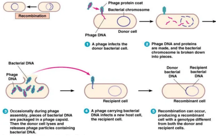
Transduction is a method through which organisms acquire DNA.whereby non-viral DNA can be transmitted by infectious or non-infectious virus particles from an infected host bacteria to a new host. When the phage particle forms, host DNA is inadvertently packaged into the empty phage head. Infectious phage particles that are liberated from lysed host cells have the ability to bind to fresh host cells and deliver the DNA contained in the capsid. The recipient's genome may incorporate the injected bacterial DNA. Although the majority of bacteriophages only infect a small number of hosts, this method of gene transfer has the benefit that transducing phages may be rather tenacious in their environment, DNA in the transducing phage particles is protected, and there is cell-cell interaction. Recent research on the number of bacteriophages in various environmental settings and data from bacterial genome sequences are the major sources of evidence for the significance of transduction as an HGT mechanism under environmental circumstances. By using electron or epifluorescence microscopy to count bacteria, bacteriophages were shown to be around ten times more numerous than bacteria in fresh or marine water samples. Directly counting bacteriophages in soil is clearly more difficult, and only lately have studies found that soil viruses are also abundant. (HEUER, 2007).(Fard, 2011).
Transduction is one instance where bacteria have been hypothesized to benefit from phages.
Transduction is regarded as the primary factor causing the dissemination of antibiotic resistance genes and the pathogen's success.As a result of an abnormal prophage excision, DNA bordering the prophage attachment site (attB) is transferred during specialized transduction. Recently found lateral transduction results in extremely effective packaging of bacterial DNA several hundred kilobases downstream of the integration site by late excision and in situ replication of an integrated prophage (Institute of Medicine et al., 2011; Salom, 2019). Transduction has historically been undervalued as a key method of horizontal gene transfer in naturally occurring ecosystems. Up to 50% to 60% of bacteriophages may include functional bacterial genes of all sorts, according to a metagenomic investigation of viromes. These particles may potentially operate as a conduit for the transfer of genes between bacteria.When a bacteriophage replicates in one bacterial cell before moving on to another, the process known as transduction takes place.
The donor bacterial cell's genome is packed into the phage head and transferred to the recipient bacterial cell. It is believed that transduction is an unevolved process that results from the errors
made during the excision of bacteriophage DNA from the donor genome. (Huddleston, 2014).Microbial organisms can acquire additional genetic material from sources other than their clonal ancestry thanks to (HGT). Microbes may sample and share a sizable gene pool through HGT, which may contain features that are advantageous in their immediate context. For instance, horizontal acquisition of antibiotic resistance genes (ARG) permits diversity of genomes and generates a possibility for fast fitness gains when bacteria are subjected to strong selective pressures, such as the presence of antimicrobials. In fact, HGT can produce the genes needed for survival more quickly than spontaneous mutations.It is recognised that transduction, particularly between individuals of the same species, may play a role in the spread of ARGs. When viral particles transfer bacterial genes, transduction takes place. Bacteriological DNA may inadvertently get wrapped in a bacteriophage capsid during bacteriophage infection. A recipient cell may connect to a capsid harboring bacterial DNA and receive the foreign DNA with no problems. Transduction has taken place if the bacterial DNA has been recombined into the receiving cell's genome. (Lerminiaux, 2018).
Classification of Transduction
Transduction can be divided into broad categories. Specialized transduction takes place when only bacterial DNA close to a temperate bacteriophage's attachment site, such as that found in E.
coli, is unintentionally packed into the bacteriophage and then transported to a recipient cell.When any gene from a host gets arbitrarily packed with viral DNA into a bacteriophage head, as happens in Salmonella P22, and then transmitted to a new recipient, this is known as generalized transduction. As bacteriophages can remain in the environment for variable length of time, the donor strain and the receiver strain do not necessarily need to be near to one another in terms of time or space. Almost every DNA sequence that can be identified in the bacterial genome may be transferred, including antibiotic resistance. Chromosome sequences and mobile genetic elements like plasmids, transposons, and insertion elements are included in this.
(Huddleston, 2014).
Generalized transduction was a potent genetic tool when it was first discovered, revolutionizing microbial genetics at the time. It was also discovered early on that the transductants, or cells that