This thesis titled “Multi Categorical Detection of Common Eye Diseases Using Convolutional Neural Network: A Transfer Learning Approach” submitted by Abu Kowshir Bitto, ID at Department of Software Engineering, Daffodil International University has been accepted as satisfactory for the partial fulfillment of the requirements. for the degree of B.Sc. I also acknowledge that neither this thesis nor any part of it has been submitted elsewhere for the award of any degree previously by others. This thesis that I represent was only possible to complete with guidance from a few conscientious people.
Specially obliged to Daffodil International University for the guidance and constant supervision by my esteemed teacher Dr. Because the human eye is the most important of the four sense organs, it is necessary to detect external eye diseases early. In this paper, we use different architecture of Convolutional Neural Network to detect normal eyes, conjunctivitis eyes and cataract eyes where ResNet50, VGG 16 and Inception v3 are applied.
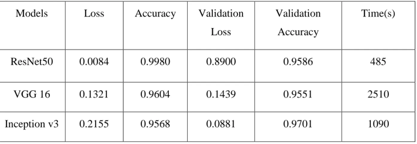
Among them, ResNet50 performs with 99 percent accuracy to detect eye diseases, with a detection time of 485 seconds.
- INTRODUCTION
- RESEACRH OBJECTIVES
- RESEARCH GAPS
- ORGANIZATIONS OF THESIS
The human eye is protected from dust particles by a thin layer known as the conjunctiva. It acts as a lubricant in the eye and prevents friction during opening and closing of the eye. The white part of the eye, known as the sclera, turns red due to the dilation of the blood vessels [11].
The human eye is the most essential of the four senses; Early identification of external eye disease is critical. The conjunctiva is a thin, transparent tissue that borders the inside of the eyelid and covers the white part of the eye. Cataract is a clouding of the lens of the eye that is the leading cause of blindness and vision loss worldwide.
The slip caused by a cataract can damage only a small part of the eye's lens, leaving the patient to forget about any loss of vision.
LITERATURE REVIEW
According to the network configuration used, the maximum test accuracy of the input image with the lowest resolution of 31*35 pixels is 80.93 percent. 5] provides a systematic review of studies published in the medical literature before May 2019 that used CNNs for medical image analysis. Samples were taken from both eyes of 104 infants with conjunctivitis and from the left or right eye of 104 control children, and the lateral sample was matched to that of the child affected by conjunctivitis.
Neonatal conjunctivitis was observed to occur 8.2% of the time, and no neomycin-resistant disease was detected during the study. The technique was able to eradicate about 95% of the unwanted red-eye artifacts in the test bed. The technology correctly identified about 98 percent of the red-eye anomalies in the test samples.
The algorithm missed the majority of the artifacts as the red-eyes were not visible to the naked eye. The primary goal of this study was to develop a CNN-based classifier that could distinguish between a healthy and a red human eye in the shortest possible time. The results of the classification using tree-based algorithms revealed that, given enough data, the proposed framework works well.
Due to the organized organization of data, the prediction rate of random forest and decision tree algorithms is over 90% when compared to more advanced approaches such as neural networks and naive Bayes algorithm. The suggested technique shows how to use HOG to extract the best components from the pupil and parts of the sclera of the eye in order to diagnose cataract and conjunctivitis. The difficulty caused by brightness and clarity is handled by the distance of the location of interest.
The input images were taken from the Structured Analysis of the Retina (STARE), Digital Retinal Images for Vessel Extraction (DRIVE), High Resolution Fundus (HRF), and Digital Retinal Images for Optic Nerve Segmentation (DRIONS). The proposed method aims to help medical practitioners make more accurate diagnoses, reduce infant mortality due to pneumonia and detect the severity of eye disease early. In the system's evaluation testing, the ORIGA "Online Retinal Fundus Image Database for Glaucoma Analysis" dataset was used.
When compared with current approaches, it showed outstanding performance in terms of efficiency and effectiveness in the face of blur, noise, and light variations.
- METHODOLOGY MODEL
- DATASET
- PRE-PROCESSING
- DATA VISUALIZATION
- MODEL DESCRIPTION
For a quick image, tag or categorize images using a search engine. search by keyword at this time all the transcribed images had been saved. There are several models; However, how many layers must be used and how many retraining must be carried out depends on the situation. For example, Keras has nine pre-trained models for computer vision tasks, transfer learning, forecasting, feature extraction and fine-tuning.
For all these models, we used three predefined models: VGG16, ResNet50 and Inception V3. Since fine-tuning a CNN model that has already been trained is usually faster and significantly less time-consuming than training a CNN model with random data, TL was recently updated to include weights that were initialized from scratch. In reality, the implementations are the softmax layer, the fully connected layer, and the output categorization layer of the CNN model.
The padding on the first two levels is the same and the first two layers have 64 channels with 33 filter sizes. Then there are two layers with filter size 256 and convolution layers with filter size, followed by a step (2, 2) maximum pooling layer (3, 3). There are 256 filters in total, as well as two convolution layers with three and three filter sizes.
Then there are two sets of three folding layers, as well as a max pool layer. In some of the layers, 11 pixels are also used to control the number of input channels. After each convolution layer, 1-pixel padding (same padding) is added to avoid losing the spatial information in the image.
There are three convolution layers in each convolution block and three convolution layers in each identity block. It is the third version of Google's Inception Convolutional Neural Network, which was first showcased at the ImageNet Recognition Challenge.
RESULT ANALYSIS
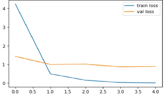
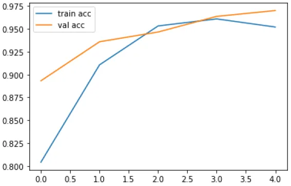
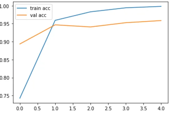
In ResNet50 we used the adam optimizer and a batch size of 64 and for the loss function we used categorical cross entropy. We were able to increase the amount of shots using this method, whereas most previous attempts had fewer shots. We are trying to train our data using 5 epochs for ResNet50, with the goal that these learning algorithms traverse the entire training dataset 20 times respectively.
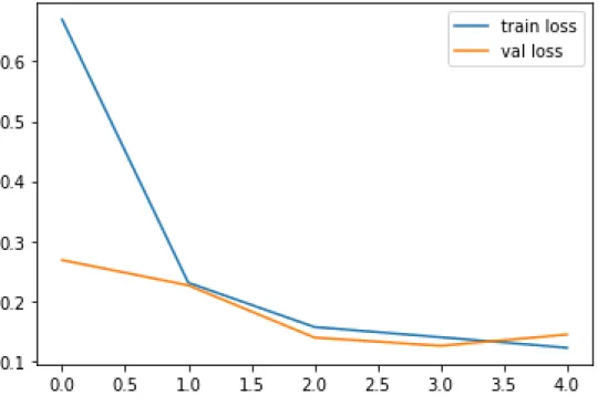
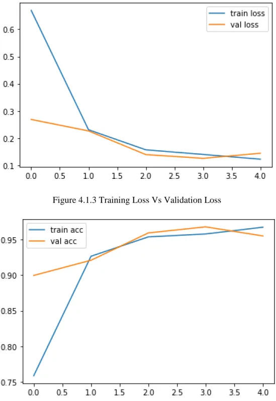
In VGG 16 the adam optimizer was used and a batch size of 32 and for loss function we used categorical cross entropy. Using this strategy, we were able to increase the number of photographs taken when most previous experiments had fewer photographs taken. We attempt to train our data with 5 epochs for VGG 16 with the goal of having these learning algorithms run through the complete training dataset 20 times.
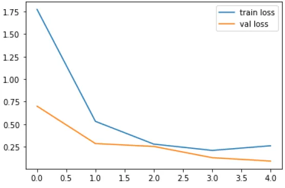
After the instruction, the testing period begins to ensure that the instructions have been followed correctly. In Inception v3 the adam optimizer and a cluster size of 32 were used and for the loss function we used categorical cross entropy. For Inception v3, we are trying to train our data using 5 epochs, with the goal that these learning algorithms will explore the full training data set 20 times each.
In this proposed work, we try to evaluate the newly trained model using the test data set. The training dataset, which included both the original and enhanced images, was provided to our model as an option to continue training. The performance of the model was evaluated using test photographs after it was trained on the eye disease dataset using ResNet50, VGG16 and the Inception v3 architecture.
This was done to see how our model compares to other well-known networks that have been previously trained for transfer learning. From Table 4.1.1, we can see that ResNet50 performs with a maximum accuracy of 99 percent with less time of 485 s to detect eye disease.
FINDINGS AND CONTRIBUTIONS
This research describes the detection efforts of transfer learning and deep feature extraction for the identification of eye diseases using images collected from around the world. Deep feature extraction and transfer learning are performed using three popular deep CNN architectures: ResNet50, Vgg16 and Inception v3.
FUTURE WORK
Among all models ResNet50 performs the highest accuracy with 99 percent and less time 485s to detect eye disease. Detection of bacterial and viral organisms from the conjunctiva of cats with conjunctivitis and upper respiratory tract disease. 8] Prentice MJ, Hutchinson GR, Taylor-Robinsin DA microbiological study of neonatal conjunctivae and conjunctivitis. British Journal of Ophthalmology.
Early detection and classification of living bacteria using time-lapse coherent imaging and deep learning. D., "Detection of Conjunctivitis with Deep Learning Algorithm in Medical Image Processing Third International Conference on I-SMAC (IoT in Social, Mobile, Analytics and Cloud) (I-SMAC), 2019, pp. Bansal, "Retinal Eye Disease Detection Using Deep Learning Fourteenth International Conference on Information Processing (ICINPRO), 2018, pp.
Yang, "Klassifikasie van katarak fundus beeld gebaseer op diep leer IEEE Internasionale Konferensie oor Imaging Systems and Techniques (IST), 2017, pp. Danisman, "Outomatiese opsporing van Adenovirale Konjunktivitis Disease from Facial Images using Machine Learning IEEE 14th International Conference on Machine Learning and Applications (ICMLA), 2015, pp. 27] Tahira Nazir, Aun Irtaza, Valery Starovoitov, "Optic Disc and Optic Cup Segmentation for Glaucoma Detection from Blur Retinal Images Using Improved Mask-RCNN", International Journal of Optics, vol.
Anand, "Eye Spy Face Detection and Identification Using YOLO International Conference on Intelligent Systems and Inventive Technology (ICSSIT), 2019, pp. Eye Detection," in IEEE Access, vol. Data and its (un)content: A survey of the development and use of data in machine learning research.
34;Transfer learning." I Handbook of research on machine learning applications and trends: algorithms, methods and techniques, s. 34;Deep residual learning for image recognition." I Proceedings of the IEEE-konference om computersyn og mønstergenkendelse, s.