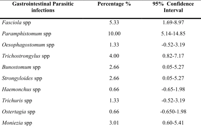
The ideal temperature for the development of larvae of many species in the microclimate of the tuft of grass or vegetation is between 22° and 26°C. The rate at which acquired resistance develops in the host, which is influenced by the species of the parasite and host, genetic factors, nutrition and physiological stress (e.g. parturition). After slaughtering the tracer animals, examination of the gastrointestinal tract revealed six species of nematodes and one cestode. The nematode herbs were Haemonchus contorsus, Trichostrongylus axei, Mecistocirus digitatus, Oesophagostomum spp, Trichuris spp and Bunostomum spp. The cestode belonged to the genus Moniezia. .
1994) conducted a pathological examination study of 173 slaughtered buffaloes which revealed some cases of the disease. Identified diseases. The rate of hydatid cyst infection was reported to be 26.4% in spring, summer, autumn and winter. They investigated the level and location of cysts in different organs, which were approximately 78% in the left lung and approximately 10% in the right lung. and examined the gastrointestinal tracts of 218 crossbred goats representing the dry zone of Sri Lanka during a one-year study period.217 nematodes. Faecal samples collected from 102 sheep and 147 goats and examined using a modified McMaster technique showed that and of the samples contained at least one nematode egg.
The other egg types encountered during the study did not show much variation with the season of the year. Liver condemnation rates differed significantly between cattle, goats and pigs (p≤ 0.05) for F. The total loss by condemnation of both F. The ratio of loss in cattle, goats and sheep was 76%, 17% and 7% respectively. . 2006) conducted a study to determine the prevalence and risk factors associated with gastrointestinal parasitism in western Oromia, Ethiopia during 2003-2004. The highest conservation infections in sheep and goats were recorded in June (33.33%) and August (41.16%), respectively. 2006) examined the gastrointestinal (GI) organs of 50 goats in Burdur region, Turkey for the prevalence of GI nematodes and the seasonal activity of the parasites.
Amphistome FECs followed a seasonal pattern, with an increase in counts during the warmer months of the year (September–April).
CHAPTER-III
MATERIALS & METHODS
- Description of the study area and duration
- Selection of animals and Survey Design .1 Target age groups and sample collection
- Sample collection and preservation
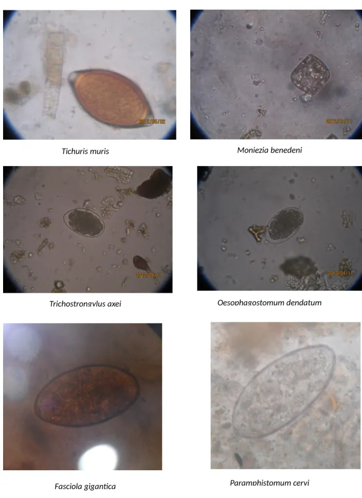
- Examination of samples .1 Fecal Samples Examination
- Statistical Analysis
In addition to the gross examination of fecal samples (color, consistency, blood or mucus, etc.), three different types of qualitative tests, namely direct smear, flotation and sedimentation techniques, were used to examine the fecal samples (Hendrix, 2006).
CHAPTER-IV RESULTS
Prevalence of gastrointestinal parasitic infections in Black Bengal goat .2 Overall prevalence of gastrointestinal parasitic infections
- Age specific prevalence of gastrointestinal parasitic infections
- Sex-specific prevalence of gastrointestinal parasitic infections
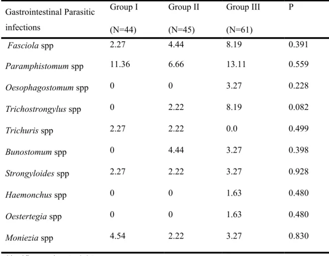
During this investigation, it was observed that older goats (category III) were more affected by different gastrointestinal parasitic infections. Among different parasitism, Paramphistomum spp infection (13.11%) and Fasciola spp infections (8.19%) were highest in age group III. On the other hand, Bunostomum spp (4.44%) infection was more in age group II, while Oesophagostomum spp, Ostertagia spp and Haemonchus spp were recorded only in adult buck of this study (Table 2).
In the current study, it was revealed that female goats were more susceptible to several gastrointestinal parasites than males, but this was not statistically significant. However, the prevalence of Paramphistomum spp infections (14.28%) was highest in male black Bengal goats, while Fasciola spp infections (4.76%) were more common in female goats. However, Trichuris spp and Moniezia spp were more commonly found in male goats, while Bunostomum spp. and Strongyloides spp infections were more common in female goats.
CHAPTER-V DISCUSSION
Prevalence of gastrointestinal parasitic infections in Black Bengal goat .1 Overall prevalence of gastrointestinal parasitic infections
- Sex-specific prevalence of gastrointestinal parasitic infections
Prevalence of Oesophagostomum spp infection from this investigation was partially similar to the report of Lima et al (2006) who recorded 0.54% in Brazil and the report of Pathak et al. In the present study, Oesophagostomum spp infection was observed to be low, which may be due to the relatively long life cycle and low resistance to desiccation of the pre-infection stages of this genus (Pfukeny et al., 2007 and Rivera et al., 1983). . Prevalence of Trichostrongylus spp infection of this study showed consistent with the findings of Gadahi (2008) who recorded 4.51% in Pakistan.
The lower prevalence of Trichostrongyltus spp in the current study may be due to its high sensitivity to preparasitic phages (Soulsby.1982) and improved husbandry practices (Alim et al., 2011). The prevalence of infection with Bunostomum spp in this study showed variation from Uddin et al. 1998) who recorded a prevalence of bunostomiasia of 55.83% in Bandarban district of Chittagong. The prevalence of Strongyloides spp infection in this study was found to be consistent with the report of Gadahi et al.
Differences in the occurrence of such infection in goats may be due to the free-living nature of the parasite and the different bionomics of the parasites (Urquhar et al., 1996 and Soulsby, 1982). The occurrence of Trichuris spp infection in this study was consistent with the findings of Lima et al. Variations in the incidence of Trichuris spp infection in this study may be due to the geoclimatic conditions of the study areas and animal husbandry practices.
Prevalence of Moniezia spp infection was found in accordance with the reports of Mohanta et al. 2005) observed 2.5% in Kenya which also supported the findings of this study. Lower incidence of Moniezia spp may be due to less distribution of eggs in the faces of the gravid segments (Radostits et al., 1994). Higher prevalence of gastrointestinal parasitic infections in older goats of this study showed consistency with the observation of Biu et al. 2009), Uddin et al., (2006), Soulsby (1982) who reported that goats older than 2 years are more susceptible to gastrointestinal parasitism. 2006) also noted that paramphistomiasis had a significant influence on the age of the goat.
The results of this study were found consistent with the reports of Shahiduzzaman et al., (2003), Uddin et al., (2006), Biu et al., (2009) who also reported higher prevalence of helminths in male goats. On the other hand Pramphistomum spp, Trichostrongylus spp, Moniezia spp, Ostertegia spp, Trichuris spp infection was more in hanged. In this study, variation in the prevalence of such helminths in male and female animals may be due to the variation in sample size (Bachal et al., 2002), reduced resistance in females, temporary loss of acquired immunity near birth (Garcia et al., 2007 and Barger , 1993), stress, genetic resistance in the host and insufficient/unbalanced feed against higher requirements (Raza et al., 2010 and Hansen and Perry, 1993).
CHAPTER-VI CONCLUSION
CHAPTER -VII REFERENCE
Gastrointestinal helminthiasis: prevalence and associated determinants in domestic ruminants in district Toba Tek Singh, Punjab, Pak. Pernambuco State, Brazil, through faecal egg counting and larval culture. Evaluation of gastrointestinal helminth infection in goats from the metropolitan area of Recife. Prevalence and population dynamics and pathological effects of intestinal helminthes in black Bengal goats Bangl.
The distribution and economic importance of Fasciola gigantica and Stilesia hepatica in slaughtered animals in semi-arid coastal Kenya. Mollah, MMR., Islam, A., WMS and Islam, MK., (1996). Epidemiology of abomasal helminthes of Black Bengal goats in Bangladesh, Indian Journal of Veterinary Medicine 16:29-31. Epidemiology of gastro-intestinal helminthiasis in small ruminants in the tree crop grazing zone in Senegal Veterinary Research.
Occurrence of parasitic worms among small ruminants reared under traditional husbandry system in Owerri, south-eastern Nigeria. Epidemiological studies of parasiticastrointestinal nematodes, cestodes and coccidia infections in cattle in the communal grazing areas of the high and lowveld of Zimbabwe. To investigate the incidence of gastrointestinal parasites in goats in East Pakistan Agricultural University campus.
Final Report of the Pilot Project for the Control of Animal Parasitic Diseases in Bangladesh. An abattoir study on the prevalence of gastrointestinal nematodes of goats in the dry zone of Sri Lanka. Monthly prevalence of gastrointestinal trematodes, cestodes and nematodes infecting Damani sheep and goats in D.I district.
Seasonal influence on the incidence of Haemonchus contortus infection in slaughtered Black Bengal goats in Bangladesh. Coproscopic and hematological approaches to determine the prevalence of helminthiasis and protozoan diseases of red chittagong. Gastrointestinal parasitic infections of sheep and goats in semi-arid areas of Machakos District, Kenya.