Helena Khatoon, Associate Professor, Department of Aquaculture, Chattogram Veterinary and Animal Sciences University for her valuable suggestions, intellectual guidance, constructive and constant inspiration throughout the study period and in the preparation of this manuscript. 34-35 4.3 Isolated microalgae from different sampling sites 35 4.4 Characterization of isolated microalgae 36–40 4.5 Growth phases of isolated microalgae 40-46 4.6 Specific growth rate (SGR), cell duplication time, cell. 06 4.5 Protein content (% dry weight) (mean ± SE) in isolated freshwater and marine microalgae grown in BBM and Conway Medium, respectively.
07 4.6 Lipid content (% dry weight) (mean ± SE) of isolated freshwater and marine microalgae grown in BBM and Conway Medium, respectively.
List of Tables
List of Abbreviations
Abstract
Chapter-1: Introduction
Furthermore, a number of nutritional components, including phosphorus, carbon, nitrogen and iron, are recognized as having some of the most important influences on biomass yield and lipid accumulation (White et al., 2013). Despite being a crucial part of microalgae technology, measuring and monitoring cell growth has received little attention (Havlik et al., 2013). To use microalgae commercially in aquaculture, pharmaceutical and other sectors, screening of microalgae species should be carried out through performance characterization. To choose potential strains that offer competitive advantages for each commercial use, the current approach compares the growth rate and nutritional characteristics of several freshwater and marine microalgae (Chlamydomonas sp., Navicula sp., Gonyostomum sp., Nannochloropsis sp., Choricystis sp., Chromochloris sp., Tetraedron sp., Chlorobotrys sp.
Coelastrella sp., Kirchneriella sp. in addition to Oscillatoria sp1, Oscillatoria sp2, Oscillatoria sp3 and Oscillatoria sp4).
Chapter-2: Review of Literature
Microalgae
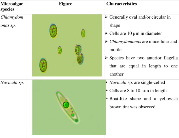
- Chlamydomonas sp
- Navicula sp
- Nannochloropsis sp
- Choricystis sp
- Chromochloris sp
- Tetraedron sp
- Chlorobotrys sp
- Coelastrella sp
- Kirchneriella sp
- Oscillatoria sp
Trichocysts, which are small organelles found beneath the cell membrane, burst under physical stress and emit slimy threads (Lebret et al., 2012). 7 small, non-motile spheres that cannot be separated with either light or electron microscopy (Andersen et al., 1998). It belongs to the family of common freshwater planktonic algae commonly found in tropical and temperate environments (Stoyneva et al., 2012).
They are distributed irregularly throughout the mucilage (Shubert and Gärtner, 2015). The size and shape of a cell can be used to identify between different species (Komárek and Fott, 1983).
Role of Microalgae
The steady, repetitive oscillation that gives it its name is thought to be caused by the secretion of mucus that pulls the filament away from the direction of secretion. This is achieved by supplementing the diet of farmed fish with natural pigments containing carotenoids, such as Astaxanthin produced by the microalgae Haematococcus (Perumal et al., 2012).
Isolation of Microalgae
Growth factors of microalgae
- Light
- Temperature
- Nutrient composition of media
- Mixing and aeration
The visual attraction of captive-bred finfish is one of the other applications of microalgae in aquaculture. The use of fluorescent light for indoor culture can promote better growth and cell division of microalgae (Laing, 1991). Changing the light intensity will affect the temperature, which indirectly affects the growth of microalgae (Huang et al., 2013).
Commercial media contain all kinds of micronutrients and macronutrients to aid in the growth of microalgae.
Microalgal growth
In order to ensure better contacts with cells and nutrients, it is also important to maintain homogeneous conditions with balanced ventilation.
Pigments of Microalgae .1 Chlorophyll
- Carotenoid
- Phycobiliproteins
Phycocyanin is a pigment used commercially in the pharmaceutical sector because it protects the photosystems against free radicals and can stop the oxidative damage caused by free radicals. According to Mathivanan et al. 2015), phycocyanin is a water-soluble, intensely fluorescent, blue-green light-harvesting pigment unique to some algae species and a member of the Rhodophyta and Cryptophyta families. Phycobiliproteins are widely used as nutraceuticals, natural colorants and in various biotechnological applications in the food, cosmetics, pharmaceutical and diagnostic industries (Becker, 2004).
Proximate composition of microalgae
Materials and Method
- Sample collection
- Determination of water quality parameters
- Determination of total ammonia nitrogen (TAN)
- Determination of nitrite-nitrogen (NO 2 -N)
- Determination of soluble reactive phosphorous (SRP)
- Collection of microalgae
- Centrifugation
- Determination of microalgal diversity
- Isolation of Microalgae
- Streak Plating Technique (Phang and Chu 1999) .1 Agar plate preparation
- Serial dilution
- Picking up method (Capillary method)
- Morphological identification of microalgae
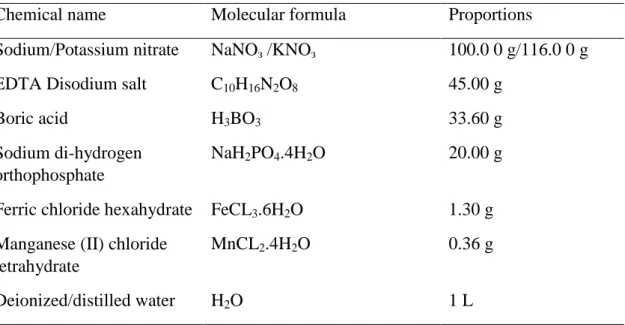
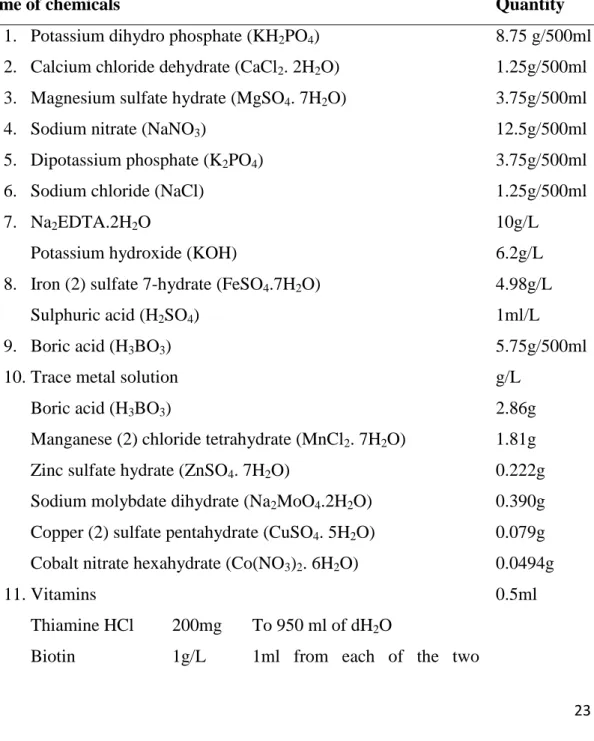
- Preparation of culture media for microalgal growth .1 Preparation of BBM (Bold basal medium)
- Preparation of conway Medium (Tompkins et al., 1995)
- Determination of growth curve
- Determination of cell density
- Determination of maximum absorbance (optical density)
- Experimental design for pigment and proximate composition determination In large sterile 2L borosilicate Erlenmeyer flasks, having 1.7L pure BBM and
- Determination of biomass
- Determination of productivity
- Volumetric productivity
- Areal productivity
- Lipid productivity analysis
- Specific growth rates (SGR)
- Determination of pigments
- Extraction of microalgae for chlorophyll determination
- Determination of chlorophyll
- Determination of phycobiliproteins
- Determination of proximate compositions .1 Protein determination
- Lipid determination
- Determination of amino acid
- Statistical analysis
After more than 2 min but less than 10 min, 1 ml of NED reagent (0.5 g of N-(1-napthyl)-ethylenediamine dihydrochloride dissolved in 500 ml of distilled water) was added and immediately mixed using vortex mixture. A sample score will be added to the following score once the target organism has been confirmed. A 1 mL aliquot of algal suspension from each culture was taken at their stationary phase and centrifuged at 1000 g for 5 min.
Finally, the absorbance of the separated hexane layer was determined using a spectrophotometer at a wavelength of 450 nm.
Chapter-4: Result
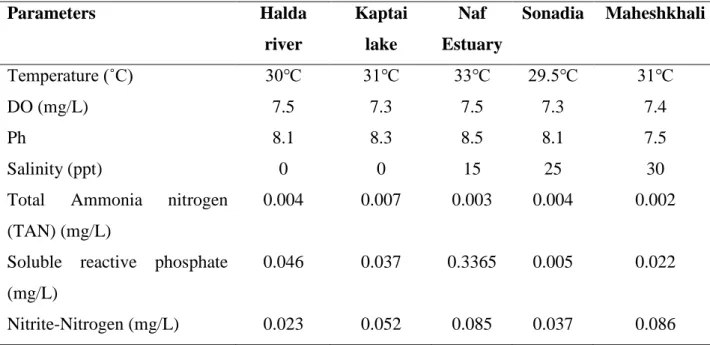
Water quality parameters of the sampling sites
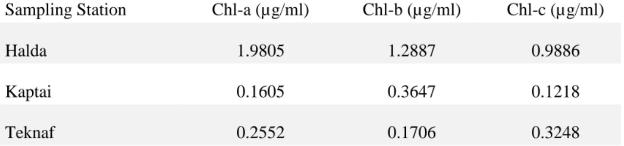
Chorophyll content of water collected from the sampling sites
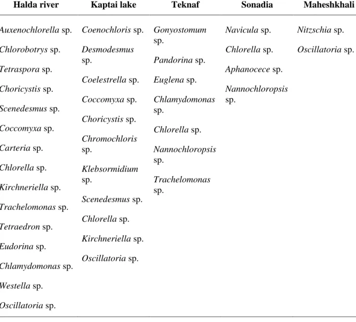
Isolated Microalgae from different sampling sites
According to John and Whitton (2002), isolated microalgae were Chlamydomonas sp., Navicula sp., Gonyostomum sp., Nannochloropsis sp., Choricystis sp., Chromochloris sp., Tetraedron sp., Chlorobotrys sp. the largest of which is Chlorobotrys sp. Among the fourteen microalgae, Chromochloris sp., Coelastrella sp., Kirchneriella and Oscillatoria sp3 were isolated from Kaptai Lake, where Choricystis sp., Tetraedron sp., and Oscillatoria sp4 from Halda River. Marine microalgae Chlamydomonas sp., Gonyostomum sp., Nannochloropsis sp., and Oscillatoria sp1 were isolated from Naf estuary where Navicula sp.
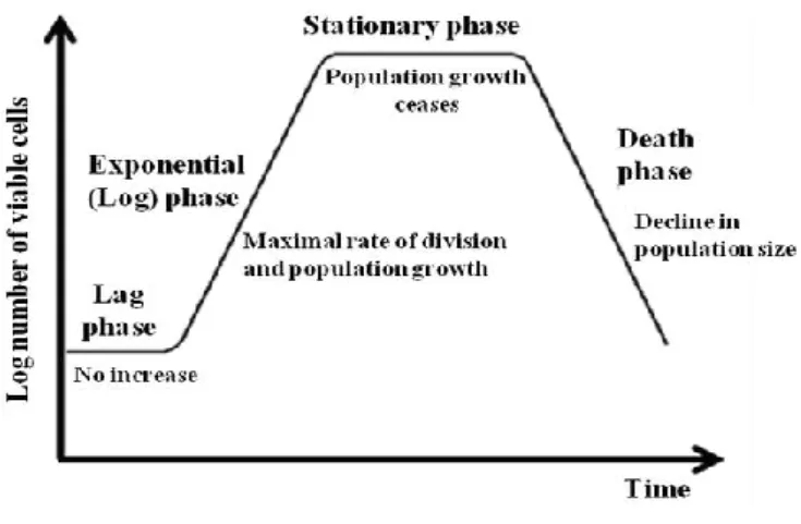
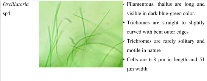
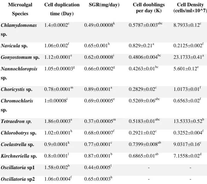
It contains filament-like clusters and the cells are cylindrical with tapers at the outer ends. The results of growth observation showed that the onset of the stationary phase (9–14 days) varied among the fourteen species. 41 exponential phase on days 2 to 10, stationary phase on days 9 to 10 and death phase from 10 days where Oscillatoria sp4 showed exponential phase on days 2 to 11, stationary phase on days 10 to 11 and death phase from 11 days.
Furthermore, in terms of cell density, no significant difference (p ˂ 0.05) was found between Navicula sp, Choricystis sp., Chromochloris sp.
Specific growth rate (SGR), cell duplication time, cell doublings per day and cell density on harvest of isolated freshwater and marine microalgae
46 Figure 4.2: Growth curve in terms of chlorophyll-a and optical density (absorption) of marine and freshwater microalgae Oscillatoria sp1 (K), Oscillatoria sp2 (L), Oscillatoria sp3 (M) and Oscillatoria sp4 (N). On the other hand, the significantly highest (p ≤ 0.05) amount of cell duplications was detected for Navicula sp. At the end of the exponential phase, the significantly highest and lowest (p ≤ 0.05) amount of cell density was detected from Gonyostomum sp.
In terms of cell density, no significant difference was detected from Navicula sp., Choricystis sp, Chromochloris sp. Values with different letters within each series indicate significant differences (p < 0.05) between species.
Volumetric, areal and lipid productivity of isolated freshwater microalgae In this study, different freshwater and marine tropical microalgae species were
Pigment content of isolated microalgae
- Chlorophyll
- Carotenoid
- Phycobiliproteins
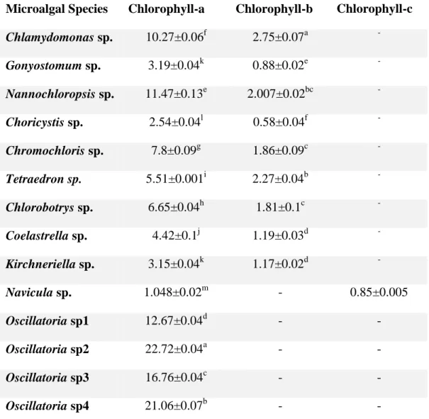
On day 13, the significant (p < 0.05) highest and lowest amount of carotenoid accumulations were detected from Chlamydomonas sp. Phycobiliprotein analysis showed that the content of phycocyanin, allophycocyanin and phycoerythrin was significantly (p < 0.05) highest in Oscillatoria sp4. A significant (p < 0.05) minimum and maximum (p < 0.05) amount of total phycobiliprotein content was achieved in Nannochloropsis sp.
Total lipid content of Chlamydomonas sp., Nannochloropsis sp., Chromochloris sp., Chlorobotrys sp., Coelastrella sp., and Kirchneriella sp. Chlamydomonas sp., Navicula sp., Gonyostomum sp., Nannochloropsis sp., Choricystis sp., Chromochloris sp., Tetrahedron sp., Chlorobotrys sp. Along with this, similar type of carbohydrate content was detected in Chlamydomonas sp., Navicula sp., Coelastrella sp and Oscillatoria sp4.
55 Figure 4.6: Lipid content (% dry weight) (mean ± SE) of isolated freshwater and marine microalgae grown in BBM and Conway Medium, respectively. In this study, Tables 4.6 and 4.7 showed the fatty acid content of marine and freshwater microalgae, respectively. On the other hand, the lowest amount of PUFA was found in Oscillatoria sp ppm) and the highest was recorded in Navicula sp. showed the highest amount of n-6 PUFA, with the lowest found in Choricystis sp.
In addition, Gonyostomum sp. showed the lowest and highest amount of n-3 PUFA, respectively, in this study. But no significant difference was found in the content of methyl icosapentaenoate and methyl docosahexanoate of microalgae species in this experiment.
Discussion
- Water quality parameters of the sampling sites
- Characterization of isolated microalgae
- Specific growth rate (SGR), cell duplication time, cell doublings per day and cell density on harvest of isolated freshwater microalgae
- Volumetric, areal and lipid productivity of isolated freshwater microalgae
- Pigments content of isolated freshwater and marine microalgae .1 Chlorophyll
- Carotenoid
- Phycobiliproteins
- Proximate composition of isolated microalgae
- Amino acid composition
Some global data are available regarding the SGR of Nannochloropsis sp. reported by Duong et al. 2015) showed similar value to the present study. Lipid productivity of Tetraedron sp. reported by Duong et al. 2015) was found lower than the present study. Total chlorophyll content (chlorophyll a and b) of Chlamydomonas sp. determined by Darwish et al. 2020) showed higher chlorophyll content than the present study.
In this study, Navicula sp. showed lower chlorophyll a content compared to other microalgae in this study. But this study of Nannochloropsis sp. showed a higher chlorophyll-a content than Fakhri et al. 68 chlorophyll a content in Coelastrella sp. was found higher than the previous work done by Iyer et al. But in the current study, Nannochloropsis sp. resulted in lower carotenoid content than Fakhri et al.
In this experiment, Nannochloropsis sp. demonstrated higher production of phycocyanin, allophycocyanin and phycoerythrin than the previous work of Islam et al. The protein content of Tetraedron sp. was found to be higher in the current study than the experiment of Duong et al. 2015). The result of the present study was supported by Thepsuthammarat et al. A previous study by Hasan et al. 2022) resulted in higher amount of SAFA and n-6 PUFA of Nannochloropsis sp. than the present study which is due to the varieties in growing conditions.
In the case of Choricystis sp., Menezes et al. 2015) reported a similar amount of SAFA as in the current study. Amino acid content of Nannochloropsis sp. in this study was found lower than the study by Hasan et al. 2022) and these changes may occur due to variations in growing conditions.
Conclusion
Recommendations and Future Prospects
Characterization of growth, chlorophyll content and lipid accumulation in a marine microalga Dunaliella tertiolecta under different nitrogen to phosphorus ratios. Effect of salinity on growth and chemical composition of the diatom Thalassiosira weissflogii at three cultivation phases. Notes on the taxonomy and nomenclature of the algal classes Eustigmatophyceae and Tribophyceae (synonym Xanthophyceae).
Molecular phylogenetics and biogeography of the eastern Asian-Eastern North American disjunct Mitchella and its close relative Damnacanthus (Rubiaceae, Mitchelleae). The freshwater algal flora of the British Isles: an identification guide to freshwater and terrestrial algae, Cambridge University Press, 2002. Effects of different salinity and pH on the growth and proximate composition of Nannochloropsis sp.
Influence of culture conditions on productivity and lutein content of the new species Scenedesmus almeriensis. Effect of light intensity and photoperiod on the growth and lipid content of the microalgae Nannochloropsis sp. Effect of nitrogen concentration on growth rate and biochemical composition of the microalga, Isochrysis galbana.
Appendices
Culture and maintenance of single isolated microalgae a b
- One way analysis of variance examining the difference in cell density on stationary phase of the isolated microalgae
- Appendix 3: One way analysis of variance examining the difference in carotenoid content of isolated microalgae
- Appendix 6: One way analysis of variance examining the difference in volumetric productivity of isolated microalgae
- Appendix 8: One way analysis of variance examining the difference in lipid productivity of isolated microalgae
- One way analysis of variance examining the difference in cell duplication time of isolated microalgae
- Appendix 11: One way analysis of variance examining the difference in SGR of isolated microalgae
- Appendix 12: One way analysis of variance examining the fatty acid content of isolated microalgae
Appendix N: Determination of Chlorophyll Content. a) Filtered sample on filter paper (b) Storing the filter paper in acetone (c) Homogenization of the sample (d) Determination of chlorophyll by optical density. Appendix O: Determination of carotenoid content. a) Centrifugation of the sample (b) Determination of absorbance with a spectrophotometer. a) Collection of microalgae (b) Collection from substrate (c) Dried biomass (d) Algae dust. Appendix Q: Determination of phycobiliprotein content (a) Soaking the sample in phosphate buffer (b) Mixing with a vortex mixer.
103 Appendix 4: One-way analysis of variance examining the difference in phycobiliprotein content of isolated microalgae. 104 Appendix 5: One-way analysis of variance examining the difference in the proximate composition (protein, lipids and carbohydrates) of isolated microalgae. 105 Appendix 7: One-way analysis of variance investigating the difference in surface productivity of isolated microalgae.