Production of Polyclonal Antisera of Vibrio cholerae O1 & O139 to Detect Pathogenic Vibrio cholerae
By
Rifah Tasnia ID-19336035
A thesis submitted to the Department of Mathematics and Natural Sciences in partial fulfilment of the requirements for the degree of
Bachelor of Science in Biotechnology
Department of Mathematics and Natural Sciences Brac University
January 2023
© 2022. BRAC University All rights reserved
Declaration
It is hereby declared that
1. The thesis submitted is my/our own original work while completing degree at Brac University.
2. The thesis does not contain material previously published or written by a third party, except where this is appropriately cited through full and accurate referencing.
3. The thesis does not contain material which has been accepted, or submitted, for any other degree or diploma at a university or other institution.
4. I/We have acknowledged all main sources of help.
Sincerely Yours,
………..
Rifah Tasnia ID-19336035
Enrolling Semester- Spring 2018
Department of Mathematics and Natural Sciences Brac University, Dhaka, Bangladesh
Approval
The thesis “Production of Polyclonal Antisera of Vibrio cholerae O1 & O139 to Detect Pathogenic Vibrio cholerae” submitted by Rifah Tasnia of Spring, 2018 has been accepted as satisfactory in partial fulfillment of the requirement for the degree of Bachelor of Science in Biotechnology on 31st January 2023.
Examining Committee:
Supervisor (Member)
_______________________________
Dr. Zahid Hayat Mahmud Scientist and Head,
Laboratory of Environmental Health, icddr,b
Supervisor (Internal) (Member)
Program Director
_______________________________
Dr. Iftekhar Bin Naser Associate Professor, Biotechnology
Department of Mathematics of Natural Sciences, Brac University
_______________________________
Dr. Munima Haque
Associate Professor and program director Biotechnology Department of Mathematics of Natural Sciences, Brac University
Departmental Head:
(Chair)
_______________________________
Prof. A.F.M. Yusuf Haider Chairperson, Department of
Mathematics and Natural Sciences, Brac University
Ethics Statement
I am Rifah Tasnia, student of B.Sc., Department of Mathematics and Natural Sciences, Brac University, do hereby declare that the thesis on “Production of Polyclonal Antisera of Vibrio cholerae O1 & O139 to Detect Pathogenic Vibrio cholerae” is an original and authentic record of my research work carried out by me for the degree of Bachelor of Science in Biotechnology, under the joint supervision and guidance of Dr. Zahid Hayat Mahmud, Scientist and Head, Laboratory of Environmental Health , Laboratory Sciences and Services Division (LSSD), International Centre for Diarrheal Disease Research, Bangladesh (icddr,b) and Dr. Iftekhar Bin Naser, Assistant Professor, Biotechnology, Department of Mathematics and Natural Sciences, Brac University, Dhaka, Bangladesh.
It has not been submitted by me for any other degree.
Sincerely Yours,
………..
Rifah Tasnia ID-19336035
Enrolling Semester- Spring 2018
Department of Mathematics and Natural Sciences Brac University, Dhaka, Bangladesh
Abstract
Cholera is endemic in Bangladesh due to serogroup O1, and people are still suffering from the disease seasonally in this geographic area. So, detecting the disease as soon as possible and making availability of materials to detect the disease is indispensable. Anti-sera are used to identify the serogroup of pathogenic Vibrio cholerae. Anti-sera are available commercially, but they are expensive, and getting the substance to the lab can take time. Thus, making antisera in the lab can be time and money efficient and can help local labs become self-sufficient. The purpose of this study and process was to show how to make the anti-sera on-site in the labs. The pathogenic strain of V. cholerae can be identified more quickly by utilizing V. cholerae antisera. The methods for recognizing V. cholerae serotype O1 and O139 are crucial as it is sensitive as well as repeatable because up to 60%-75% of all cholera cases are subclinical. During the study, the samples were taken from an area around ponds at a camp for Rohingya refugees where 42 suspected Vibrio were isolated using the common culture method. 26 of them had gelatinase activity and were oxidase positive. Based on colony morphology and gelatinase activity, isolates were obtained. On each isolate, common biochemical tests designed to identify Vibrio were conducted. 18 samples were determined to be Vibrio cholerae after conducting common biochemical tests. For molecular confirmation, multiplex PCR was then performed. ToxR genes were present in 18 of the isolates, but the lack of ctxA genes indicated that they were nonpathogenic O1/O139. To validate the findings and the prepared anti-sera the lab-prepared polyclonal antiserum was used where 3 samples agglutinated with the anti-sera confirming that they were nonpathogenic O139 Vibrio cholerae. The polyclonal antibodies were prepared by injecting dosage of dead cells of V. cholerae to New Zealand white rabbits and following all the procedures the polyclonal antisera were prepared which was further used to identify the pathogenic O1 and O139 serogroups of V. cholerae fast and cost effectively.
Keywords: Vibrio cholerae, Antisera, polyclonal, serotypes, environmental sample
This Thesis is Dedicated to my Family
Acknowledgement
Firstly, I would like to express my gratitude to Almighty Allah for providing me the strength to complete the task on schedule.
I would like to specially express my gratitude to Dr. Zahid Hayat Mahmud, PhD, Scientist and Head, Laboratory and Environmental Health, icddr,b for taking me under his mentorship and allowing me to avail his resources for my research. His support, inspiration and guidance during my thesis work, was utmost crucial in allowing me to undertake this project.
I would also like to thank Dr. Iftekhar Bin Naser, PhD, Assistant Professor Biotechnology Program, Brac University for guiding and monitoring my work progress throughout this thesis program and during the course of my undergraduate career. Without his support, this thesis project would not have come to fruition.
My special thanks goes to Dr. Shafiqul Islam, Assistant Scientist, Laboratory of Environmental Health, icddr,b for his inspiration and sincere guidance. Also I would like to thank Md. Rafiqul Islam, Research Investigator of LEH, icddr,b and Md. Moniruzzaman, Research Investigator of LEH, icddr,b for their kind support.
I would like to convey my indebtedness to Monir Hossain, Research Officer and Md. Sakib Hossain, Senior Research Assistant, Laboratory of Environmental Health, icddr,b. Without their support this thesis would not come to light. Their care and supervision helped me to successfully complete my dissertation without any barriers.
I would like to express my gratitude Atique Ul Alam, Research Officer of LEH, Raihana Habib Auroni & Rashedul Islam, Senior Research Assistant, icddr,b and Tanveer Hussain, Research Assistant for their help and support at different stages of my thesis work.
I am really thankful to laboratory technicians of icddr,b Moshiur Rahman, Md. Monnaf Ali and Shahjahan for their help and humble cooperation.
Professor A F M Yusuf Haider, Chairperson, MNS Department, has my sincere gratitude for upholding department regulations and delivering proper education to all students of the respective programs.
Last but not least, I am thankful to K.M Atiqul Hoque, my father, Shamshad Jahan, my mother, MD Tausif Uddin, my husband, and Tanzila Erash, my friend for their unparalleled love and support that helped me to get through turbulent times.
Sincerely, Rifah Tasnia ID: 19336035
Table of Contents
CHAPTER ONE: 13
INTRODUCTION 13
1. Introduction 14
1.1 Objectives of this study 15
CHAPTER TWO: 17
LITERATURE REVIEW 17
2.0 Literature review 18
2.1.1 Taxonomy 18
2.1.2 Historical Background of Vibrio cholerae 18
2.1.3 General Characteristics 22
2.1.4. Classification and antigenic type of V. Cholerae 29
2.1.6 Reservoir of infectious V. cholerae 39
2.1.7 Molecular method for detection of specific genes 40
2.1.8 Biofilms 42
2.1.9 Antibiotics against V. cholerae 44
2.1.10 Production of Antibodies 47
CHAPTER THREE: 50
MATERIALS 50
AND 50
METHODS 50
3.0 Materials and methods 51
3.1 Collection and revival of V. Cholerae strain: 52
3.2 Preparation of vaccines from the strains: 52
3.3 Sampled Animal- Housing and Feeding: 53
3.4 Blood sample test for pre-immunization: 54
3.5 Vaccine Doses and Intervals: 55
3.6 Absorption of serum: 57
3.6.1 Preparation of Cells: 57
3.6.2 Absorption in Live cells: 59
3.6.3 Absorption in dead cells: 60
3.6.4 Filtration of the serum: 61
3.7 Isolation and Validation of V. cholerae strain using prepared Antiserum 62
3.7.1 Media Preparation 62
3.7.2 Sample Processing and Isolation 63
3.8 Molecular Confirmation 64
3.8.1 Requirements for PCR 65
3.8.2 Boiled DNA template preparation for PCR 65
3.8.2 Preparation of Reaction Mixture for PCR 66
3.8.3 Multiplex PCR for detection of toxR and ctxA genes to identify pathogenic/non-pathogenic O1 and O139 66
3.8.4 Post-amplification detection: Agarose gel electrophoresis 67
3.9 Serological assay of the molecular confirmed V. cholerae 68
CHAPTER FOUR: 69
RESULTS 69
4.0 Results 70
4.1 Sample Processing isolation, identification, and determination of V. cholerae 70
4.2 Detection of toxR and ctxA genes by PCR 71
4.3 Serotyping of confirmed isolates 74
CHAPTER FIVE: 75
DISCUSSION 75
Discussion 76
CHAPTER SIX: 78
CONCLUSION 78
6.0 Conclusion 79
CHAPTER SEVEN: 80
REFERENCES 80
APPENDICES 91
Appendix I 92
Appendix II 94
Appendix III 95
List of figures
Figure number
Title of figure Page
no.
2.1 Cholera cases reported to WHO by year and by continent 1989-2012
……… 21
2.2 Basic structure of Vibrio cholerae……… 23
2.3 Monthly cholera outbreaks by region (Emch et al.2008) ………... 27
2.4 Classification stages of V. cholerae……… 31
2.5 The actions of cholera toxin……… 38
2.6 Involvement of VPS, RbmA, RbmC, and bap1 in the biofilm formation of Vibrio cholerae……….………... 44
3.1 Three New Zealand White rabbits chosen and marked…………... 54
3.2 Collecting blood sample to confirm no presence of immunity against cholera………... 54
3.3 Serology testing of V. cholerae with pre-immunized serum shows no clotting… 55 3.4 Rabbits are vaccinated intravenously and intramuscularly in Animal Resources Facilities, icddr, b………...…... 56
3.5 Enriched V. cholerae in BHI broth after overnight incubation………. 58
3.6 50ml falcon tubes in centrifuge machine……… 59
3.7 Eppendorf tubes in centrifuge machine to remove the pellet………. 60
3.8 Mixing live cells and distilled water in a vortex machine………. 61
3.9 Absorption of serum with filter membrane……… 62
3.10 Samples in Alkaline Peptone Water for enrichment………... 64
4.1 Medium sized yellow colonies on TCBS agar plate is indicative of V. cholerae ……….………... 70 4.2 V. cholerae on TCBS, CVA and GA plate. Here, the slot 13 is the positive control
and the slot 15, 23, 24, 28, 29 indicates putative V. cholerae due to their growth and distinctive color……….
71
4.3 toxR and ctxA genes showed after running agarose gel electrophoresis of multiplex PCR. Lane 1 shows 100 bp plus DNA ladder and lane 2 shows 779 bp and 564 bp amplicons of toxR and ctxA genes of V. cholerae O1, E.16434 (classical) control strain. Lane 3 represents no template control. Lane 4 to 19 show 779bp toxR genes for our samples. ……….
72
4.4 V. cholerae polyclonal anti-sera is taken in eight slots. The first one has positive control, and the fifth slot has negative control. 2nd, 3rd, 4th and 8th slots did not agglutinate with the anti-sera. 6th and 7th slots agglutinated with O139 antisera.
……….
74
List of Table Table
Number
Title of Table Page
no.
2.1 Cholera pandemics since 1817 (Adopted from hunter 1997) 19
2.2 Biotypes of V. Cholerae O1 33
2.3 3.1
Serotype Determination of V. Cholerae O1 Selected rabbits and their weight
35 53 3.2
3.3 3.4 3.5 3.6 4.1 4.2 4.3
Doses and Intervals of the Heat-killed V. Cholerae Vaccines Reagents Used in PCR
Components of PCR Reaction Mixture
Primers Used in Determination of the Presence of O1 and O139 Serotype PCR Conditions for toxR and ctxA Multiplex Reactions
Antibody Titration Checking Up to 400 Times Diluted Antiserum (After 1st Booster) Antibody Titration Checking Up to 400 Times Diluted Antiserum (After 2nd Booster) Total Count of Blood and Serum from the Rabbits
56 65 66 67 67 73 73 74
Abbreviations
WHO World Health Organization
CFR Case Fatality Rate
CFU Colony Forming Unit
FEDSD Field Epidemiology and Disease Surveillance Division
DNA Deoxyribonucleic acid
PCR Polymerase Chain Reaction
NIH National Institute of Health
ECDC European Center for Disease Control
TE Tris EDTA
TBE Tris Borate EDTA
TCBS Thiosulfate Citrate Bile Salt
cAMP Cyclic Adenosine Monophosphate
SEATO Southeast Asia Treaty Organization
APW Alkaline Peptone Water
TTGA Tellurite-Taurocholate-Gelatin Agar
GA Gelatine Agar
TSA Trypticase Soy Agar
IgG Immunoglobulin G
IgM Immunoglobulin M
TCP Toxin Coregulated Pilus
bp Base Pair
spp. Species (plural)
sp. Species (singular)
VBNC Viable but Non-Cultivable
ACE Accessory Cholera Enterotoxin
AC Adenylate Cyclase
CFTR Cystic Fibrosis Transmembrane Conductance Regulator
ZOT Zonula Occludens Toxin
dNTPs Deoxynucleoside Triphosphates
RNA Ribonucleic Acid
EPS Extracellular Polymeric Substances
SXT Self-Transmissible Transposon
pABs Polyclonal Antibodies
CVA CHROMagar Vibrio
icddr,b International Center For Diarrheal Disease Research, Bangladesh
CHAPTER ONE:
INTRODUCTION
1. Introduction
Millions of people have died from the terrible pandemic disease cholera, which is still a major health issue today. The word "cholera" comes from the Greek word "bilious," which means
"terrible sickness." The illness is known as "Olauta" in Bangladesh. Since Hippocrates and Lord Buddha, it has been known to happen (Barua, 1992; Pollitzer et al., 1959). In nations with weak socioeconomic situations, where everyone cannot be guaranteed access to safe water and proper sanitation, this issue is still quite problematic. With the exception of the most recent pandemic, which started in celeb instead of its endemic center in the Ganges and Brahmaputra delta, mankind has endured seven pandemics (International Centre for Diarrhoeal Diseases Research et al., 1993;
Pollitzer et al., 1959). With approximated cases which is around more than 5 million every year, the disease is endemic to the Indian subcontinent and continues to resurface elsewhere in Asia, Africa, and America (Glass & Black, 1992; Taylor et al., 1993). The condition is characterized by the loss of electrolytes and large amounts of fluid. The etiological agent of cholera is a toxigenic strain of the Gram-negative bacterium V. cholerae (Comstock et al., 1995). Typically, cholera is spread orally through feces, and a person only contracts the disease after ingesting the bacterium.
A normal infectious dose of the pathogenic V. cholerae serotype O1 is between 10^5 and 10^6 live cells. If given with food, an infectious dose can be initiated by a much lower number (Levine et al., 1981). Cholera's infectious dose is high in healthy adult males. Environmental aquatic reservoirs are linked to cholera transmission and yearly seasonal outbreaks. By consuming contaminated food and beverages, one contracts the primary infection. During an epidemic, transmission from person to person happens through contaminated food or drink (S. Holmberg et al., 1984). Consuming contaminated fish, oysters, crabs, or other shellfish during an epidemic time is another way that it can spread (Feachem, 1981).
Only 40% of the villages in Bangladesh, where 80% of the population lives, have hygienic latrines.
Common water bodies are contaminated by the typical methods of direct fecal discharge into ponds or rivers. People may come into touch with the cholera-causing agent when they bathe in, wash in, or drink from contaminated water. Primary transmission may occur when aquatic plants and animals from the marine environment are consumed as food (often without being fully cooked or through drinking water) (Miller et al., 1985). Cholera epidemics hit Bangladesh twice a year, with the first one peaking in the cooler months (September to December) and the second one being
water when the inter-epidemic span is ongoing, but it is possible to isolate it from patients and surface-water during the epidemic season (Comstock et al., 1995; Siddique et al., 1992).
There are two main serogroups present for epidemic Vibrio cholerae known as O1 and O139. The two primary serotypes are called Inaba and Ogawa which falls under the V. cholerae O1 serogroup.
They are known to replace each other throughout time, and the V. cholerae 0139 has recently developed (International Centre for Diarrhoeal Diseases Research et al., 1993). It is believed that the replacement of Ogawa and Inaba over time in an endemic area is a good indicator of the serotype's level of population immunity.
Vibrio cholerae, particularly the pathogenic strains of V. cholerae O1 and V. cholerae O139, cause the disease cholera which is a water-borne infectious disease, and V. cholerae non-O1 can also cause moderate-to-severe diarrhea that resembles cholera in Bangladesh as well as many different parts of world (Nair et al., 1994).
For the identification of the serotypes of V. cholerae O1 group-specific known as anti-A and type- specific marked as anti-B and anti-C antisera are used as they allow for the serological identification. Rabbits are immunized using cells of V. cholerae serotype Inaba and Ogawa that are heat-killed and are able to produce polyvalent antisera (Meeks et al., 2004). Antibodies are removed using group-specific antigen A against that antibody by cross-absorption which can create monospecific type sera (Mukerjee & Guha Roy, 1961). But the process can result in the concurrent deprivation of antibodies that are type-specific, which can make the production of such type antisera challenging. Sensitive as well as repeatable procedures are obtainable for the diagnosis of V. cholerae serotype O1, as up to 75% of the occurrence of cholera are subclinical (Merson, 1978). The objective of this work was to create an enzyme based on a highly specific polyclonal antibody for use in research laboratory and for epidemiological purposes.
1.1 Objectives of this study
The major purposes of this study are to prepare polyclonal antisera of V. cholerae O1 and O139 and validation of the prepared antisera by isolating V. cholerae from environmental samples.
• Preparing antisera with heat-killed vibrio strains in lab animals preferably New Zealand white rabbits.
• Isolation of V. cholerae from environmental samples
• Molecular detection of isolated samples of V. cholerae.
• Validation of prepared antisera by serotyping with the confirmed colonies.
CHAPTER TWO:
LITERATURE
REVIEW
2.0 Literature review
2.1 The Organism: Vibrio Cholerae
2.1.1 Taxonomy
Kingdom: Bacteria
Sub kingdom: Negibacteria Phylum: Proteobacteria
Class: Gamma proteobacteria Order: Vibrionales
Family: Vibrionaceae Genus: Vibrio
Species: Vibrio cholerae 2.1.2 Historical Background of Vibrio cholerae
Cholera became more common in the 19th century. In 1817, India recorded the first pandemic, which later spread to other regions of the world. This disease gradually attracted greater attention as it spread beyond India and started to cause major concern on a global scale.
There have been seven pandemics so far (Table 2.1). Six pandemics struck the world after the initial one in 1816–1817, starting in 1826, 1852, 1863, 1881, 1889, and 1961. There is still a seventh pandemic. By Pollitzer, Kamal, and Barua (Islam), the causes and effects from the previous and ongoing pandemics and how they extended to different parts of the universe were thoroughly examined. The location of this pandemic's inception is crucial information. The epicenter of all previous pandemics and epidemics happened to be formed at the Ganges delta of Bengal but for the last which is the present pandemic known as the seventh one happened to originate from Indonesia. The name of the place is Sulawesi which is an Indonesian island.
though it appeared that the disease had stopped spreading after the outbreaks, Bengal was always where it was still being reported. As a result, cholera has been an ongoing problem in Bengal since its inception. Bengal is regarded as the cholera's native land (International Centre for Diarrhoeal Diseases Research et al., 1993).
Cholera has almost certainly visited every nation on earth at some period, making it simpler to compile a list of those it didn't. The world's southernmost and northernmost regions normally did not become affected by this disease. In Asia, Chamchatka and Northern Siberia were unaffected.
The most northern portions of North America, such as Newfoundland and Greenland, as well as Western Europe, including Iceland, the Faroe Islands, Shetland, and the Orkney Islands, remained unaffected. The southernmost areas of Chile, Argentina, and the Falkland Islands in South America were cholera-free (International Centre for Diarrhoeal Diseases Research et al., 1993).
After the sixth pandemic, forty years of stillness ensued. The last pandemic after this was caused by the V. cholerae O1 El Tor strain, which started in 1961 in and around Indonesia and spread rapidly throughout most of Asia and Eastern Europe. In 1970, this El Tor biotype was also introduced to West Africa, where it expanded very quickly. After a century-long hiatus, it was once more introduced to Peru in 1991. Cholera epidemics in the South Asian subcontinent were known to be generated by V. cholerae O139 in 1992, and as a result, it had spread to eleven more Southeast Asian countries. Aside from a few isolated occurrences, this serogroup has not been documented outside of these nations (International Centre for Diarrhoeal Diseases Research et al., 1993).
Table 2.1: Cholera pandemics since 1817 (Adopted from hunter 1997) Pandemic Causative
Strain
Duration Origin Affected Regions
First O1 (Classical) 1817-1823 Bangladesh India, SE Asia, Middle East, East Africa
Second O1 (Classical) 1826-1851 Bangladesh India, SE Asia, Middle East, Africa, Europe, Americas
Third O1 (Classical) 1852-1859 Bangladesh India, SE Asia, Middle East, Africa, Europe, Americas Fourth O1 (Classical) 1863-1879 Bangladesh India, SE Asia, Middle East,
Africa, Europe, Americas Fifth O1 (Classical) 1881-1896 Bangladesh India, SE Asia, Middle East,
Africa, Europe, Americas Sixth O1 (Classical) 1899-1923 Bangladesh India, SE Asia, Middle East,
Africa, Europe, Americas Seventh O1 (El Tor) 1961-1975 Indonesia India, SE Asia, Middle East,
Africa, Europe, Americas
Eighth O139 1991- India India, SE Asia
2.1.2.1 Global Update of cholera
In its 2013 The World Health Organization (WHO) reported that the number of cholera cases started to fall in 2012 following several years of continuous growth beginning in 2007. With a combined total of 24,53,931 cases, a case-fatality ratio (CFR) of 1.2%, and a death toll of 3,034, this study found. This report indicates a 58% decline in the number of incidents since 2011. A total of 48 countries across all regions registered cholera occurrences to WHO during this time period, which is a 17% decrease over 2011. Cholera occurrences related to V. cholerae were registered throughout the world. Similar to 2011, 27 nations on the African continent reported instances. The number of countries reporting cases reduced from 15 in Asia during 2011 to 12 by 2012, and from 9 in the Americas during 2011 to 6 for 2012. In 30 nations, the WHO has received a report of cholera cases out from Americans, Asia, Europe, and Oceania. 23 of the 30 countries that reported cholera deaths occurred on the African continent, making up 67% of the total worldwide. On the American continent, the Dominican Republic and Haiti were responsible for 962 fatalities, or 31%
of the total. Throughout contrast, hundreds of thousands of cases of cholera were unrecorded in Asia due to the inadequate surveillance systems. More than two million episodes of severe watery diarrhea are reported annually in Bangladesh (WHO, 2011).
In 2010, during the month end of October a significant outbreak affecting the Dominican Republic and Haiti began. In 2012, 49 percent of all reported cases were associated with this outbreak.
Around 7367 cases were reported from Asia, which represents 3% of the global total and an 81%
drop from 2011 (38,298). Only important cases from Oceania were reported in Australia (Fig. 1.1).
In 2012, WHO confirmed few outbreaks in different countries, where Africa accounted for 29 cases where America only confirmed 4 and 5 cases were shown in Asia. WHO received reports of 1,19,995 Hispaniola cases, or 49% of the global total. A visual representation of cholera cases reported to WHO by year and continent till 2016 is given below (Lonappan et al., 2020).
Fig. 2.1: Report of Cholera cases by WHO by year and continent from 1989 to 2016
2.1.2.2 Cholera: Bangladesh Perspective
Bangladesh is endemic for cholera. Additionally, it is a major cause of illness and mortality.
Bangladesh, located within the delta of the Ganges, the place where all the cholera epidemic originated except the seventh one. According to studies, outbreaks of epidemics in Bangladesh typically happens two times a year, with the no. of cases being the highest between September and December, following the monsoon (Glass et al., 1982; Siddique et al., 1991, 1992). It was projected that the 1991 outbreak caused between 2.1 million and 2.3 million illnesses and over 8,000 deaths
(Siddique et al., 1992). From January to March 1993, a strain, that is new, of V. cholerae non-Ol, eventually named as V. cholerae 0139, which had never been known to trigger epidemics, started an outbreak of cholera in Bangladesh (International Centre for Diarrhoeal Diseases Research et al., 1993; Ramamurthy, 1993). This diarrheal illness killed more than 1,473 out of 1,07,297 patients in Bangladesh in 1993 (International Centre for Diarrhoeal Diseases Research et al., 1993).
2.1.2.3 Recent Outbreak Updates
Since the beginning of 2022, Pakistan and Bangladesh have witnessed an unprecedented increase in cholera, posing a growing health risk. As per the Field Epidemiology and Disease Surveillance Division (FEDSD) of National Institute of Health, Islamabad (NIH), the number of reported suspected cholera cases was 6,231 as of 17 April 2022. The statistical distribution of the cases varied throughout the nation, with Khyber Pakhtunkhwa, Sindh, Punjab, and Balochistan reporting, respectively, 2,596, 1,873, 956, and 697. As of 27 April 2022, the European Center for Disease Control (ECDC) reported 129 cases which has been confirmed by laboratory in Karachi, Pakistan. As per ECDC, the number of probable total cholera cases and deaths in Bangladesh is 495,433 and 29 respectively, inclusive of 33,832 cases from the Refugee Camps of Rohingyas in Cox's Bazar, Bangladesh. Till 13 March 2022, the number of laboratory-confirmed cases were 47.
In addition to that, the International Centre for Diarrhoeal Disease Research, Bangladesh (icddr,b) reports that, in Dhaka, daily hospital admissions due to diarrhoeal diseases exceeded 1000 for the first time in the last 60 years, with 1,057 patients getting admitted, alone, on 16th March. The count reached an all-time high of 1,334 admissions on 28th March. Current reports indicate that Dhaka has been primarily affected by a probable cholera outbreak since the start of the year.
However, it is also affecting other important cities, like Chittagong, Khulna, Barisal, Mymensingh, Rangpur, Sylhet, and Rajshahi. The government of Bangladesh, in collaboration with the World Health Organization, intends to provide oral vaccine of cholera to 2.3 million individuals (non- pregnant, older than 1 year) residing in diarrhea-prone districts.
2.1.3 General Characteristics
Vibrio cholerae, a member of the Vibrionaceae bacteria family, is a facultative anaerobe that moves by means of its flagellum. It's a Gram-negative bacterium that produces cytochrome C oxidase but does not create spores. Despite being facultative organisms, they develop considerably better in an aerobic environment. They thrive in alkaline conditions but are destroyed in conditions below pH 6. They are typically found in aquatic settings (freshwater, saltwater, wastewater, or brackish water) or in the intestine, vomit, and feces of human hosts (Harris, 2012). Its cells are bean-like rods measuring 1.3 m in length and 0.3 m in diameter. The optimal temperature for growth of Vibrio Cholerae is 37°C (98.6°F), but it can grow between 14°C and 40°C. Utilizing a single polar flagellum as a motility organelle, Vibrio cholerae is extremely mobile in liquid. Most Vibrio Cholerae can ferment glucose, maltose, sucrose, mannitol, dextrin, lactose, and starch;
however, V. cholerae species identification among fecal isolates in TCBS media is done by applying only sucrose fermentation. These isolates typically produce extracellular DNase, lipase, -galactosidase, and Ornithine Decarboxylase, and use acetate. They do not synthesize Phenylalanine deaminase, urease, and they cannot grow on media containing inositol and KCN.
This genus produces gas from glucose in a typical manner.
Figure 2.2: Basic structure of Vibrio Cholerae
V. cholerae has a vast array of strains and biotypes. Pathogenic along with nonpathogenic strains of V. cholerae differ in gene content related to their virulence. The O polysaccharides of V.
cholerae have 140 serogroups and based on its O antigen, the bacteria have more than 200 serotypes; nonetheless, the serotypes O1 in addition to O139 have been identified as the culprits responsible for the pandemic and epidemic cholera outbreaks (Baron, 1996). These can be distinguished by their feature to manufacture toxin of cholera, for which encoding is done by the ctx gene. The ctx gene was a means to detect choleragenic V. cholerae in environmental samples. Based on morphological and genetic traits (Baron, 1996). there can be two biotypes of Vibrio cholerae O1, classical and El Tor.
2.1.3.1 Physicochemical properties
Vibrio cholerae is an anaerobe which is facultative. It can reduce nitrates to nitrites. It produces gas and acid (mainly H2 and CO2) at the time of growth on glucose and sucrose fermentation. By traditional biochemical tests for indole production and string test, which are conducted in clinical laboratories, V. cholerae is positive. They are also citrate, oxidase and gelatin hydrolysis positive but urease and hydrogen sulfide and methyl red test negative. Most V. cholerae strains can grow over a wide range of temperatures (approximately 14–40°C). Maximum growth rate is observed in the narrow range of 37–40°C. V. cholerae can grow best at alkaline condition within a pH range:
6.5–9.0 (approximate) (Hollenbeck et al., 2014).
2.1.3.2 Role as normal flora
The small intestine is colonized by V. cholerae where they secrete the potent cholera enterotoxin.
In the bowel, the toxin is bound to the plasma membrane of intestinal epithelial cells and an enzymatically active subunit is released causing a rise in cyclic adenosine 51-monophosphate (cAMP) production. The high intracellular cAMP level is reason behind the cause of massive secretion of water and electrolytes into the intestinal lumen (Reidl & Klose, 2002).
2.1.3.3 Role as model organism
Research laboratories has been using V. cholerae for nearly a century because of the cholera pandemic of the Ganges Delta with an outbreak in Jessore, India, in 1817. It is studied because of its virulence factor, and fast-growth capacity. At 37°C this organism experiences ready growth on most of the laboratory media and upon overnight incubation, produces colonies.
2.1.3.4 Spectrum of Illness
The disease can be distinguished by a brief period of incubation (8 - 72 hours) followed by acute watery diarrhea, frequently along with vomiting, muscle cramps, in addition to consequences due to severe dehydration and metabolic acidosis. Rehydration is the primary treatment for cholera, although antibiotics have been demonstrated to be crucial and cost-effective adjuncts in severe patients and epidemic conditions. Antibiotics are not considered lifesaving under optimal treatment conditions, as patients can be treated with intravenous and oral rehydration fluids alone.
Antibiotics are regarded as the standard treatment for cholera since they lower by approximately 50 percent the length of disease, the volume of diarrhea, and the rehydration requirements. When treating a high number of cases, duration reduction and symptom alleviation are particularly significant, and antibiotic treatment minimizes the expense and effort required to combat an outbreak (Comstock et al., 1995)
2.1.3.5 Seasonality of V. cholerae
Bangladesh and other South Asian nations are regarded as the cholera endemic zone. A regular seasonal pattern is maintained by cholera, in Bangladesh (Glass et al., 1982; Islam et al., 1993), with the biggest peak happening during the post monsoon season (Sept. to Jan). The second peak occurring during the pre-monsoon season (Mar. to May). The lower Ganges Delta retained a distinct yearly pattern of outbreaks as the birthplace of cholera. For instance, Dhaka and Matlab are researched as the endemic center of cholera in Bangladesh. In 1961, The epidemiologic research of this illness was initiated in Bangladesh (then East Pakistan) after the creation of the Pakistan Southeast Asia Treaty Organization (SEATO) Cholera Research Laboratory. From 1964
to 1966, (Martin et al., 1969) noticed in Dhaka that cholera outbreaks peaked annually in November, December, or January. During the epidemic of 1965-1966, they detected another peak in April-May (McCormack et al., 1969), in a Matlab study, evaluated the seasonality of cholera.
The study was conducted between November of 1963 to June of 1966 and discovered that each year the cholera. outbreak was at its peak between November and January. Also, they detected a small secondary peak throughout April and June during the outbreak of 1966. (Merson et al., 1980) demonstrated in research conducted in Matlab, Bangladesh, between 1968 and 1977 that the occurrence of the classical biotype was highest between September and November. In March to April, a second, smaller peak was also detected. (Glass et al., 1982) analyzed the seasonal characteristics of both El Tor and conventional biotypes in Matlab, Bangladesh, where he accumulated 15 years of data. Similar seasonal patterns were identified for both El Tor and classical biotypes, as observed by (Merson et al., 1980). (Samadi et al., 1983) also investigated the seasonal characteristics of El Tor along with classic cholera. The forecasted peak of the El Tor outbreak was in October, while the classic pandemic peaked in December. The peak incidence of El Tor occurred before the onset of typical cholera, according to research. If the seasonality of cholera in Bangladesh (and the then East Pakistan) is analyzed based on available data within 1963 to 1980, it is clear that in endemic regions the incidence of cholera is typically seasonal (Figure 2.2).
During the interepidemic time, it is not possible to culture V. cholerae from water from the surface.
However, at the season of the epidemic, isolation of V. cholerae can be done from both the patient's body and water from the surface (Khan et al., 1984). When not causing human intestinal problems, V. cholerae can be discovered in a variety of aquatic settings, including estuaries, rivers, ponds, etc (Huq et al., 1995; Turner et al., 2009). It may survive in the aquatic environment either as free- living plankton type organism in the water column or in association with zooplankton and phytoplankton (Islam et al., 1990; Samadi et al., 1983). In natural settings, V. cholerae remains connected to Anabaena spp., while in an artificial aquatic environment, V. cholerae Ol persists in the mucilaginous sheath of a blue-green alga, A. variabilis (Islam et al., 1990). Some researchers investigated its survivability of the O1 variant in various aquatic conditions. In artificial sea water, in the presence of copepods and chitin, Pruzzo and his colleagues discovered that, V. cholerae O1 remained viable for 14 days, without losing its adherent characteristics (Stauder et al., 2010).
Feachem demonstrated that, at 4°C and 30°C, it can survive in clean water for up to one month and 2-14 days respectively (Feachem, 1981).
Fig 2.3: Monthly Cholera outbreaks by region (Emch et al. 2008)
Also, it is evident that, the epidemic in Bangladesh always reaches its peak during the winter season with a second small peak during the hot season.
2.1.3.6 Mode of Transmission of V. cholerae
Primarily, Vibrio cholerae transmission can happen by food and water that is contaminated with feces. For more than a century, the importance of water for transmitting cholera has been acknowledged. As was already established, this proof was shown in 1854, during the second cholera pandemic, by the London doctor and epidemiologist John Snow, who showed that illness was caused by consuming water from a system that pulled water from the Thames River below a sewage inflow. Several subsequent investigations have shown the importance of water in the dissemination of the disease (Glass & Black, 1992; Pollitzer et al., 1959). In a study conducted in Bangladesh it was portrayed that around 44% of shallow water which was sourced from communities with cholera, was positive in culture, for the organism, and not surprisingly, risk of infection associated with cooking, bathing, or washing with water from culture-positive sources
had a remarkable increase (Hughes et al., 1982). It has also been demonstrated that water contributed to the growth of diseases from South America. In a study conducted in Piura, Peru and Trujillo, it was determined that not boiling water was mainly linked with illness; contagion of urban water with feces was prevalent. In these and other research (Deb et al., 1986; Hughes et al., 1982; Ries et al., 1992; Taylor et al., 1993), the potential dangers provided by water storage containers, which are open, wide-mouthed and prevalent in houses in cholera-endemic regions, were also considered. Even though the supplied water to the family didn’t contain V. cholerae initially, the containers were contaminated easily, which raised the potential of disease transmission within the household. Adding to this, research conducted in Calcutta, India, revealed that the adoption of containers that had narrow-mouth and was used for storage of water (i.e. into which utensils, hands, etc. cannot be placed) significantly reduced disease transmission (Deb et al., 1986). Transmission of cholera is possible by consuming food also. Seafood can be contaminated via sources from the environment and may act as a vector for both epidemic and endemic sickness, especially if it is raw or only slightly cooked (Baine et al., 1974; Blake et al., 1980). Additionally, there is proof that irrigating vegetables with unrefined sewage water can harbor and spread V. cholerae O1 (Cohen et al., 1971).
Food within houses or other places can be affected (contaminated) by food-handlers (S. Holmberg et al., 1984; LOUIS et al., 1990), or organism may be contained in water used while preparing the food, if not boiled (Johnston et al., 1983). The source of a recent outbreak of cases in the US was tracked back to coconut milk that was frozen and was brought in from Thailand. Here, the floor on which the coconut meat was diced and later used to make coconut milk was washed with water from canals (Taylor et al., 1993). As previously said, food serves as an ideal medium for culture.
For example, cooked rice, which has been shown to promote rapid growth of V. cholerae (Kolvin
& Roberts, 1982), as well as neutral sauces like peanut sauce, serve this purpose. Additionally, it should be mentioned that acidic sauces with a pH of 5.0 or lower seem to protect against this bacterium (LOUIS et al., 1990). While food has repeatedly been blamed for spreading cholera to new areas (Glass & Black, 1992; Pollitzer et al., 1959), the organism's ability to thrive and the protection against gastric acidity suggest that food may have a bigger impact than previously thought on disease transmission in endemic areas. (Comstock et al., 1995).
Usually, cholera is transmitted by either contaminated food or water. In wealthy countries, the most common cause of death is seafood. In contrast, in underdeveloped nations, normal water is more prevalent. Most of the cholera cases in industrialized nations originate from contaminated food which occurs if the water is contaminated with sewage are used by people for harvesting oysters because V. cholerae accumulates in zooplankton and the oysters eat the zooplankton.
Cholera-infected individuals' rice-water stools contaminate water, and if the contaminated water is consumed by others, disease transmission can occur. Drinking contaminated water and consumption of food washed with contaminated water might cause infection. Cholera is infrequently transmitted directly from person to person.
2.1.3.7 Morphology of Vibrio Cholerae
Vibrio cholerae, a comma shaped bacillus which possesses a single polar flagellum. The name V.
cholerae is given because of their vibrating motility in fresh wet stool preparation. They are also called "comma bacilli” because of their unique comma shape. The bacteria are oxidase positive and do not form endospore or microcyst (Baumann, 1984b). The bacterium can grow in the media containing 0 to 6.5% salt but not in a media having 8% salt. V. cholerae grows at pH ranging 7.0 to 8.5 but a pH range of 7.5 to 8 is shown to be optimum and the ambient temperature is 37°C.
Most used broth for the enrichment of V. cholerae is alkaline peptone water (APW). For isolation of the bacterial colonies many selective and semisolid culture media have been developed and among them Tellurite-Taurocholate-Gelatin Agar (TTGA) which is also known as Monsur's media (Monsur, 1961) and Thiosulfate-Citrate-Bile salt-Sucrose (TCBS) agar have gained wide popularity around researchers. On TCBS agar V. cholerae produces typical yellow colonies; while on TTGA plate colonies are rather semi-transparent and grayish in color with dark spot in the center and also surrounded by a zone of opacity due to gelatinase activity. There are some non- selective media such as Gelatin Agar (GA), Trypticase Soy Agar (TSA) etc. which are also used for V. cholerae cultivation (Comstock et al., 1995).
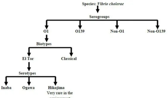
2.1.4. Classification and antigenic type of V. Cholerae
O antigen is the antigen used to characterize V. cholerae, which is primarily responsible for its pathogenicity. This antigen is stable at heated conditions and has a homopolymer containing the
amino-sugar D-perosamine (4-amino-4,6-dideoxy-D-mannose) in which the amino groups have been acetylated through 3-deoxy-L-glyco-tetronic acid. The variety of groups of this O antigen are known as serovars or serogroups. The rough (R) antigen of Vibrio cholerae is same across all species. It is difficult to differentiate between the R form and the S (smooth) form based on colony morphology alone, however identification of the R form can be done using the R antiserum. There is also a flagellar (H) antigen on the bacterium, although its utility for identification of species is restricted due to the existence of H epitopes shared by all species of Vibrio.
The O antigen of V. cholerae can be identified by a number of classification techniques. The most prevalent and frequently used system is Shimada and Sakazaki's (1973) typing technique, which employs antisera, that is raised against organisms which are heat-killed. This technique consists of about 200 distinct groups. Serogroups O1 and O139 of V. cholerae are toxigenic, while the remaining serogroups, designated non-O1/non-O139, are rarely toxigenic. V. cholerae Ol can be further characterized based on its morphological and antigenic features in two ways.
2.1.4.1 Serogroups of V. cholerae
Midway through the 1930s, a single antiserum was developed against the entire V. cholerae after discovery, where majority of vibrions isolated from cholera cases agglutinated with the raised antiserum. This test is characterized as an agglutination test because the bacteria in the elevated antiserum form clumps. The agglutination test became the primary criterion for detecting pathogenic V. cholerae over time. Based on O antigen, V. cholerae was split into six serologically identified groups (I to VI) (Gardner & Venkatraman, 1935). The majority of O antigens are thermostable polysaccharides. V. cholerae O1 or agglutinable V. cholerae is pathogenic V.
cholerae that may agglutinate with O antisera. Non-agglutinating vibrios (NAGs) or non-cholera vibrios are the colloquial terms for organisms that resemble vibrios morphologically but do not agglutinate in this serum (Group II to VI). According to current terminology, they are known as V. cholerae non-O1 or other Vibrio species. Non-O1 vibrios and other types of vibrios are prevalent in pond, lake, river, and ocean environments (Farmer et al., 1984).
From the action of cholera toxin, pathogenic V. cholerae O1 is the serogroup that causes severe watery diarrhea. In contrast, V. cholerae non-O1 is known to cause cholera to a considerably lesser
extent than V. cholerae O1 (Spira et al., 1981). A new serogroup of V. cholerae was discovered and designated as V. cholerae O139 in 1992, which is popularly known as Bengal (Islam et al., 1993; Ramamurthy et al., 1993). V. cholerae 0139 does not express O1 antigen due to the lack of at least two genes from the O1 biosynthetic gene cluster. On the basis of O antigen, more than 200 serogroups have been found to date (Colwell, 2002). V. cholerae Ol and non-Ol have a common heat-labile flagellar H antigen, although distinct species of vibrios possess a range of additional H antigens (Tassin et al., 1983). A common R antigen identification was done, and it was similar across all V. cholerae strains (Shimada & Sakazaki, 1973). The classification stages are shown below-
Fig 2.4 Classification stages of V. cholerae
2.1.4.2 Serovar and Biovar of V. cholerae Ol
All Vibrio cholerae strain has lipopolysaccharide in their structure. The lipopolysaccharides of V.
cholerae can produce powerful inflammatory reaction. The lipopolysaccharide consists of lipid A,
an endotoxin, core polysaccharide and O polysaccharide sidechain. The O polysaccharide can subdivide the V. cholerae into many serogroups. Cholera toxin (CT) is produced by V. cholerae O1 and it is linked to cholera pandemics and epidemics. There are further strains of V. cholerae O1 that don’t produce Cholera toxin because the gene for cholera toxin is absent (Comstock et al., 1995). The most important confirmation for identifying V. cholerae O1 is agglutination in polyvalent hyperimmune antisera produced against entire bacteria including all O1 antigenic components. Agglutination is really the cross-linking of many homogenous bacteria with immunoglobulin G (IgG) and immunoglobulin M (IgM) found in animal serum inoculated against that specific bacterial species (Baumann, 1984a).
2.1.4.2.1 Biotypes
Bio-typing is an important method of classification, epidemiologically for O1 strains of V.
cholerae. Classification of the two biotypes of serogroup O1 of V. Cholerae are done on the basis of various genotypic and phenotypic markers, and those are named as El Tor and classical. The serotypes of El Tor are then namely as Hikojima and Ogawa, Inaba (Comstock et al., 1995). The most dominant serotype is Ogawa, on the other hand Hikojima serotype is found to be very unstable and rare in the environment as this is the transitional state. It expresses both Inaba and Ogawa properties. There are some distinctness between the El Tor and classical biotypes of V.
cholerae O1. The differences are as follows:
• The adaptability of El Tor is finer than the classical biotype as it can subsist in the environment as well as in the host (human) as the intestinal epithelium can be colonized better by them (Finkelstein, 1996).
• However, in the comparison of toxigenicity, the V. cholerae O1 classical biotype is lead to belief to be more virulent and toxigenic (Koelle et al., 2005).
• Phenotypically the El Tor biotype is responsive to both Mukerjee El Tor phage 5 and 50 IU of polymycin B, on the other hand the classical biotype is responsive to Classical phage IV. Besides, agglutination of biotype, El Tor, can be seen through erythrocytes of chickens, it also gives a positive Voges-Proskauer test reaction. The classical biotype does not give a positive reaction for the same test (Mandal et al., 2011).
V. cholerae O1 classical biotype, as a causative agent, is thought to have caused the first six cholera pandemics, resulting in both asymptomatic and symptomatic cases. After the 6th pandemic it is assumed to be extinct and then the seventh and present cholera pandemic is thought to be associated with the El Tor biotype. The appearance of this pandemic was first noticed in 1905 in the village of El Tor, Sinai, Egypt and therefore it is named as El Tor. More asymptomatic infections were caused by it compared to any other biotype. With help of current advancement, it was possible to alter V. cholerae O1 El Tor isolates to produce cholera toxin of the classical biotype. The O139 serogroup, also known as Bengal, is contained on different strains which are genetically diverse and both non-toxigenic and toxigenic; genetically, El Tor V. cholerae is closer to this strain (Ansaruzzaman et al., 2004).
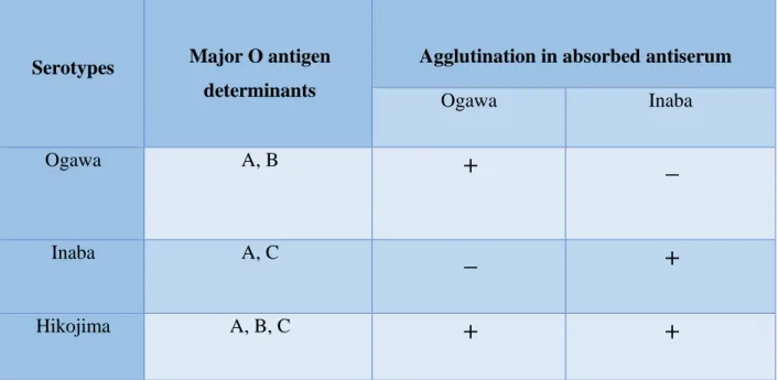
From O1 serogroup the three Hikojima Ogawa and Inaba serotypes are different, depending on the structures of their antigens. They are differentiated in terms of their somatic (O) antigen. The Ogawa serotype contains A and B antigens, the Inaba serotype contains A and C antigens, while Hikojima contains unstable antigenic types having all three antigens (A, B and C) which makes this serotype a transitional state. The Ogawa and Inaba serotypes are found to be positive for Ogawa and Inaba antisera, respectively. Hikojima serotype is found to be positive for both Ogawa and Inaba antisera (Shimada et al., 1994).
The two biovar or biotypes based on their phenotypic characterization is shown below. (Table 2.2) Table 2.2: Biotypes of V. cholerae O1
Biotypes Reactions
VP Test Resistance to polymyxin
B (50U)
Agglutination of chicken erythrocytes
Hemolysis of sheep red blood
cells
Lysis by Classical IV phage
Lysis by El Tor V phage
Classical
- - - - + -
El Tor
+ + + + - +
To examine biotypes for characterizing V. cholerae, the Voges-Proskaeur reaction, agglutination with chicken erythrocytes, and other tests are considered the most useful. Variations in the DNA sequences of the genes that encodes the toxin coregulated pilus (TCP) from the different type of strains are used in the second approach of bio typing (Keasler & Hall, 1993). In isolated colonies connected to the fifth and sixth pandemics, V. cholerae O1's characteristic biotype was discovered.
The El Tor biotype, however, predominated during the seventh pandemic and is now largely to blame for cholera outbreaks globally.
2.1.4.2.2 Serotypes
Inaba, Ogawa, and Hikojima serotypes are further subdivided under the serogroup O1 of V.
cholerae (Table 2.3). Hikojima is an uncommon condition, and this entire classification is founded on the three O antigen components A, B, and C, with A being either common or group specific. The B and C components are assumed to be unknown in nature, however the factor A is thought to be the homopolymer of D-perosamine. The serotype differences are mainly quantitative.
• The Ogawa- strain produces the antigens that are A and B. They also produce a small amount of C.
• The Inaba- strain produces only the A and C antigens. Specific Antisera from Inaba and Ogawa are made by absorbing with the other serotype.
• The Hikojima serotype contains all three factors (Sakazaki, 1992), and so, they react with both antisera of Inaba and Ogawa.
The subtype known as Hikojima, which is uncommon and unstable, is often referred to as a stage between Ogawa and Inaba. Some authority frequently fails to recognize this subtype and instead describe cultures as Inaba or Ogawa, and that depends on which serum elicits the strongest response (Mooi & Bik, 1997).
Table 2.3: Serotype determination of V. cholerae O1
Serotypes Major O antigen determinants
Agglutination in absorbed antiserum
Ogawa Inaba
Ogawa A, B
+ _
Inaba A, C
_ +
Hikojima A, B, C
+ +
2.1.4.2.3 Evolution of atypical El Tor strains
Classical strains have been steadily replaced by El Tor strains as the cholera source since the beginning of the seventh pandemic. The traditional biotype reportedly disappeared in Bangladesh in 1973 but reappeared in 1982. The classical biotype and El Tor biotype co-circulated for a decade (the latest isolation which was documented was in 1992) (Samadi et al., 1983; Siddique et al., 1991). Only in Bangladesh was the brief reappearance of Ol classical strains reported. Classical strains are assumed to be extinct; thus, the origin of the classical rst R and classical ctx alleles and their mechanism of transmission to El Tor strains in Bangladesh remain a mystery. Some classical strains may still exist in Bangladesh's aquatic ecosystems and may have served as gene donors.
Due to their low incidence or existence in a viable but non-cultivable (VBNC) condition, these strains may have evaded discovery by traditional culture procedures, but they still possess pathogenic potential. In the early 1990s, the existence of classical strains in Bangladesh's aquatic ecosystems was recorded, lending credence to the notion that they had not been totally eradicated in Bangladesh (Siddique et al., 1991).
Multiple microbial species and significant amounts of phage and free DNA are found in aquatic settings. It is known that natural cell lysis of V. cholerae and lytic phages play an essential role in the release of bacterial and phage DNA into the environment (Dziejman et al., 2005). The enhanced fitness of the El Tor strains may have contributed to the elimination of classical strains.
Consequently, a population of "homeless" free classical CTX prophage has evolved.
Typically, the free classical CTX prophages cannot produce functional virions. To secure their existence, they may infect additional hosts. By lateral gene transfer and recombination, the free classical CTX prophage may have entered the genetic background of a range of V. cholerae Ol and non-01/non-0139 serogroups or similar Vibrio species, such as V. mimicus (Bag et al., 2008).
Unknown is the selection pressure that causes aberrant El Tor strains to harbor classical CTX prophage. The classical biotype is associated with more severe diarrhea than El Tor strains, which may facilitate the formation of classical CTX prophage and, consequently, the capacity to create classical type CT in El Tor strains. This occurrence may have contributed to the spread of more toxic strains in the environment (Comstock et al., 1995).
2.1.5. Pathogenicity and clinical significance of V. Cholerae
The human small intestine becomes infected with the bacterium V. cholerae O1 and O139, which results in cholera (International Centre for Diarrhoeal Diseases Research et al., 1993). According to Chowdhury (1988), the following compounds are produced by cholera Vibrios:
Enterotoxin
Accessory Cholera Enterotoxin (Ace) RDE
Mucinade
Desquamating factor
Soluble haemolysin
The severity of infection and disease results from a series of interactions between the pathogenicity features of the pathogen and human host defensive mechanisms. The size of the inoculum, motility, chemotaxis, synthesis of key enzymes (including mucinase, protease, chitinase, and neuraminidase), presence of adhesions, and development of heat-labile cholera enterotoxin (CT) are determinants of the infection. CT generating strains are responsible for severe diarrhea, hypotension, and death within 12 hours if left untreated (Jesudason et al., 1993).
There are also some non O1 V. cholerae serotype can produce cholera toxin (CT), which is normally only produced by the epidemic type of V. cholerae O1 and O139. (Rahim & Aziz, 1992).
When sufficient organisms are swallowed and some cells survive the stomach's acidic environment, infection occurs. The organisms colonize the small intestine, grow rapidly, and produce cholera toxin in adequate quantities. In severe cases, there is significant diarrhea, and a considerable volume of rice-water stool that are clear fluids with mucus flecks and they pass without pain. One liter of fluid can be expelled per hour. Usually, there is vomiting and minimal appetite to preserve nutrients. If the patient is not treated, prostration will develop along with indicators of extreme dehydration, loss of skin elasticity, and no urine excretion. Extreme dehydration causes death to happen very quickly once symptoms appear (S. D. Holmberg &
Farmer III, 1984).
2.1.5.1 Cholera enterotoxin
Cholera enterotoxin, often known as CT or choleragen, is the cause of the severe dehydration diarrhea. This heat-labile protein could be generated by toxigenic strains of V. cholerae. Cholera enterotoxin V. cholerae generates several compounds that are extracellular and also toxic to eukaryotic cells. Although Koch (1884) suggested the existence of a chemical secreted by a comma-shaped bacillus in the human gut that causes cholera, the theory did not acquire traction until the 1970s. An Indian scientist, Sambhunath De, did not discover and define the Vibrio toxin until 1959, more than 140 years after the first epidemic began. Although not all Vibrio species produce toxins, the majority do. Twelve of the thirty identified species of this bacterium genus are pathogenic to humans (Choopun et al., 2002). Several scientists, including Finkelstein (1963), Richardson (1969), and Field (1979), took the effort to purify and determine the configuration of
the toxin of cholera. Nevertheless, vast majority of environmental isolates of V.cholerae ctx sequence (Comstock et al., 1995) are non-toxigenic V. cholerae. Strains or mutants of V. cholerae who are incapable of generating cholera toxin are also incapable of causing the disease, then again, presence of other toxins may induce a lesser type of diarrhea. Cholera toxin is the best researched biochemical and genetic virulence component of V. cholerae (Baudry et al., 1992).
2.1.5.1.1 The cholera toxin- Structure and action
An ADP-ribosylating toxin of the A-B type is cholera toxin. This toxin is a member of the enterotoxin family and is composed of two functional polypeptides, A and B. (Field, 1979). CT's structure is characteristic of the group of toxins with A-B subunits. One A subunit, five identical B subunits, make up the molecule of CT. In animal or intact cell systems, none of the components alone have substantial secret genic activity. While the A subunit performs a specific intracellular enzymatic function, the B subunit aids in the binding of holotoxin to the receptor of eukaryotic cell.
Fig 2.5: The actions of cholera toxin
After being released by bacteria in the infected gut, cholera toxin binds to enterocytes, which are intestinal cells (epithelial cell). Interaction between the B subunit of toxin that are pentameric and GM1 ganglioside which is a receptor of intestinal cell results in binding and endocytosis of the toxin. By separating the Al domain from the A2 domain, the active A1 enzyme is generated.
Through an ADP ribosylation process, the active A1 portion of A subunit that has toxin reaches the cytosol and initiates protein G. Continuously, G protein in its GTP-bound state stimulates to create cAMP from adenylate cyclase (AC). The elevated cAMP levels stimulate the transmembrane conductance regulator for cystic fibrosis (CFTR). When the CFTR is activated, infected enterocytes release a substantial number of ions and water, which causes diarrhea with a lot of fluid (Zhang, 2006).
2.1.5.1.2 Additional accessory V. cholerae toxins
Besides cholera toxin, V. cholerae yields other more toxins. These include zot (zonula occludens toxin), accessory cholera enterotoxin (ace), neuraminidase, disulfide isomerase, protease, and hemolysin-cytolysin toxin, among others (Kurazono et al., 1995).
2.1.5.1.3 Zonula occludens (zot) toxin
By altering the intracellular structure of the mucosa of the small intestine, the zot toxin produced by V. cholerae makes it more absorbent. Hydrostatic pressure may cause Zot to leak water and electrolytes into the lumen, causing diarrhea (Uzzau et al., 1999).
2.1.5.1.4 Accessory cholera enterotoxin (ace)
Trucksis and his colleagues found the gene encoding V. cholerae's accessory cholera enterotoxin (Trucksis et al., 1993). The gene, according to the researchers, results from an open reading frame that comes right before the zot gene.
2.1.6 Reservoir of infectious V. cholerae
The reservoir of any contagious agents is animal, any person, plant soil, arthropod, or blend of these, where this infective agent ordinarily exists and proliferates, where it hinges primarily on for
subsistence, and from which it can be transmitted to a susceptible host. Koch (1884) first hypothesized that aquatic environments may serve as reservoirs for vibrios when during the epidemic of cholera in 1883, he isolated bacillus that had comma like shape, from a reservoir in Calcutta. After Koch, numerous studies have been conducted to examine the possibility of aquatic environments as vibrio reservoirs.
(Hood & Winter, 1997) ensured that existence of V. cholerae in water is conditional on adjourned particles. They noticed that the survival duration of viable cells in filtered and centrifuged saltwater decreased in proportion to the degree of filtration and centrifugation. As the process of filtering as well as centrifugation diminish the impurity, the organism's feasibility may be influenced by the occurrence of particles. Also, the serotype V. cholerae 01 favors an epibiotic environment. These can be found from estuarial and marine water. Also, water that are particle-free and so they concluded that this V. cholerae 01 can exist in planktonic form for a period.
Although cholera is prevalent in many tropical regions, the marine environment serves as a reservoir for the vibrios, as the majority of cholera outbreaks began in coastal locations (Lowenhaupt et al., 1998).
2.1.7 Molecular method for detection of specific genes
The general evaluation criteria for describing systems include Reproducibility, typeability,