Urogenital Tract Associated Lymphoid Tissues in Native Chicken ( Gallus gallus domesticus ) of Bangladesh.
A clinical report submitted in partial fulfillment of the requirement for the degree of Doctor of Veterinary Medicine (DVM)
By:
Minara Begum Munni Roll No: 18/06 Reg No: 02063
Intern ID: 06 Session: 2017-18
Faculty of Veterinary Medicine
Chattogram Veterinary and Animal Sciences University
Khulshi, Chattogram-4225, Bangladesh
Urogenital Tract Associated Lymphoid Tissues in Native Chicken ( Gallus gallus domesticus ) of Bangladesh.
Supervised by:
(Dr. Mohammad Lutfur Rahman) Professor and Dean
Faculty of Veterinary Medicine
Chattogram Veterinary and Animal Sciences University
Khulshi, Chattogram-4225, Bangladesh
List of contents
List of tables ⅳ
List of figure ⅴ
List of abbreviation ⅵ
Abstract ⅶ
Chapter 1: Introduction 1
Chapter 2: Materials and methods 2
Chapter 3: Results and discussion 3-15
Conclusion 16
References 17-19
Acknowledgment 20
Biography 21
List of Table
Title Page
Table 1: Frequency of Intraepithelial lymphocytes and aggregated lymphoid 8 tissue per 5 microscopic fields (40X) in male urogenital system of native
chickens.
Table 2: Frequency of Intraepithelial lymphocytes and aggregated lymphoid tissue per 5 microscopic fields (40X) in female urogenital system of native chickens.
9-10
Table 3: Intraepithelial lymphocytes per 5 microscopic fields (40X) in the female urogenital tract of the native chicken.
10-11
Table 4: Aggregated Lymphoid tissue per 5 microscopic fields (40X) in the female urogenital tract of native chicken.
11-12
Table 5: Intraepithelial lymphocytes per 5 microscopic fields (40X) in the male urogenital tract of the native chicken.
12-13
Table 6: Aggregated Lymphoid tissue per 5 microscopic fields (40X) in the male urogenital tract of native chicken.
13-14
List of Figures
Title Page
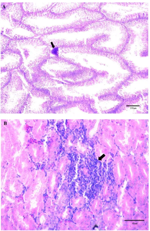
Fig 1: Hematoxylin and Eosin staining of Testes (A; male of 180 days) and 15 kidney (B; female of 90 days) of native chicken. The arrows in the images
indicate aggregated lymphoid tissue. Scales for low magnification:100µm, for high magnification:40µm.
Fig 2: Hematoxylin and Eosin staining of Kidney (C; male of 180 days) and 16 magnum (D; female of 90 days) of native chicken. The arrow in the images
indicated intraepithelial lymphoid tissue. Scales for high magnification:40µm.
List of abbreviations
Abbreviation Elaboration
MALT Mucosa-associated lymphoid tissue
ILT Intraepithelial lymphoid tissue
et al et alia (and others)
DVM Doctor of Veterinary Medicine
DLS Department of Livestock Services
N Number
C.T Connective tissue
Ig Immunoglobulin
µm Micrometer
> Greater than
≤ Equal or less than
H & E Hematoxylin and Eosin
DPX Dibutyl phthalate polystyrene xylene
Abstract
Considering the lack of research on local immunity of urogenital tract in native chickens than the high yielding chicken, the aim of this study was to determine the frequency of intraepithelial lymphocytes (ILT) and aggregated lymphoid tissue in that tract along with its morphological features. In addition, the study also aimed to establish the impact of bird’s age on the frequency of mucosal lymphoid tissue in uro-genital tract. For this purpose, different organs of the urogenital system from both male and female native chickens (age 1, 30, 90, and 180 days) were collected and stained with hematoxylin and eosin stain (H & E). From the present study, the lymphocytes can be characterized by spherical nucleus and clumps of chromatin near the nucleus membrane and scanty cytoplasm. The aggregated lymphoid tissue can be characterized by a cluster of a few cells to large aggregates of lymphocyte that lacks a limiting membrane, located in lamina propria usually below the basement membrane. The present study reveals both ILT and aggregated lymphoid tissue increased in the oviduct (at 90 days than 1 day);
infundibulum, isthmus and uterus at 180 days than at 90 days. Aggregated lymphoid tissue in the testes of males and kidneys of both males and females, ILT in male ureter was increased with age. However, there was no significant variation (p> 0.05) in the mean of both ILT and aggregated lymphoid tissue with age. The highest number of ILT was seen in Epididymis at 90 days cock and aggregated lymphoid tissue in the infundibulum at 180 days hen. The study suggests that apparently the lymphoid tissue in mucosa of the urogenital tract in native chicken increases with age but statistically there is no significant variation of the mean of lymphoid tissue with age.
Keywords: ILT, Aggregated lymphoid tissue, Urogenital tract.
Chapter 1: Introduction
Lymphocytes are immunocompetent cells that are capable of achieving a local defense mechanism, normally present in the mucosa of the urogenital tract because they are regarded as a ubiquitous migratory cells wandering wherever they can penetrate (Dym and Romrell, 1975). These lymphocytes in mucosa may be present in solitary or diffuse form, known as mucosa-associated lymphoid tissue (MALT). They form the first line of defense against pathogens that enter the body through the mucosal surfaces. Though there are many similarities between mammal and avian mucosal immune systems, there are unique features in the avian mucosal immune system. The avian mucosal immune system lacks of capsulated lymph nodes but the presence of diffuse lymphoid tissue (Nochi et al., 2018). Spleen, mucosa-associated lymphoid tissue (MALT), germinal centers and diffuse lymphoid tissue are the secondary avian lymphoid organs.
Bangladesh has a large and rapidly growing poultry sector in which chickens are considered the most preferred poultry species for egg and meat purposes. The current chicken population in Bangladesh is 319.689 million (DLS, 2022-23). One of the greatest threats to this growing industry is disease outbreaks. To combat this disease immunocomponent cells like lymphocytes play a vital role. For this purpose, sensitization of these cells is required. This sensitization occurs when they come in contact with pathogens. Due to the scavenging nature of native chickens, they come into closer contact with pathogens. Hence, they have more immune potential than high- yielding chicken breeds.
Despite such positive quality of native chicken, there is less information about tract- associated lymphoid tissue and their immunological contribution as most investigations were performed on high–yielding birds (Nagy and Olah, 2010). Therefore, this study was designed based on the following objectives:
1. To understand the distribution and frequencies of lymphoid tissue in urogenital tracts in native chickens along with morphological features.
2. To find out the relation of ages with the presence of lymphoid tissue in this tract.
Chapter 2: Materials and Methods
1. Study area and study population:
Hatazari upazila of Chittagong district was selected as the study area for this study and the study population was non-descriptive deshi chicken (Gallus gallus domesticus).
2. Collection of native chicken:
A total 24 chickens (both male and female) were collected from Hatazari of age 1 day, 30 days, 90 days, and 180 days (3 from each group).
3. Permanent slide preparation:
Samples from the urinary and genital tract from both native male and female chickens (age 1, 30, 90,180 days) were collected and fixed in 10% buffered formalin. Then the tissue was dehydrated in graded alcohol, cleared in the xylene, embedded in paraffin and finally sectionized in a rotatory microtome machine (Model HM 340E) at 4-6 m thicknesses. Then the tissue section was stained in Hematoxylin and Eosin (H & E) stain and mounted by Dibutylphthalate polystyrene xylene (DPX) for the studies of the distribution of urogenital tract-associated lymphoid tissue.
4. Microscopic study:
After slide preparation, a microscopic study of lymphoid tissues of the uro-genital tract at different aged chickens was performed and 5 photomicrographs were taken using a photomicroscope (Leica ICC50HD Microscope camera).
5. Data analysis:
After the microscopic study, all data was stored in an Excel sheet (Microsoft Excel- 2010). Then data were transferred to STATA -14.2 (STATA Corp., Texas, USA) to perform statistical analysis. An unpaired t-test was done to compare the means of different variables between the two groups (Value of day 1 chicks considered as baseline). A p-value of equal to or less than 0.05(p≤0.05) was considered significant for this test. Results were expressed as arithmetic mean± standard deviation (Mean±SD).
Chapter 3: Results
The urinary system of birds consists of paired kidneys and ureters. Unlike the mammal urinary system, the renal pelvis and urinary bladder are absent in the avian urinary system. Each kidney consists of three separate lobes. Each lobule is subdivided into a wide cortex and a small medulla cone. From histological studies, it reveals that the cortex consists of renal corpuscles (which is composed of Bowman’s capsule and glomerulus), proximal convoluted tubules, distal convoluted tubules and collecting ducts. The parietal layer of Bowman’s capsule is usually lined by a layer of simple squamous epithelium. The proximal convoluted tubules, distal convoluted tubules and collecting ducts were lined by simple cuboidal epithelium. The medulla consists of thick and thin segments of the loop of Henle. The thin segments of the loop of Henle are lined by simple squamous epithelium. The study also reveals the frequency of aggregated and intraepithelial lymphocytes (ILT) in chicken kidneys. The frequency of ILT in male kidney was 3 (1days),2 (30days),1 (90days),4 (180days) [Table 1] and in female kidney was 2 (1 days),4 (30days),5 (90days),5 (180 days) [Table 2]. The aggregated lymphoid tissue in the kidney at the age of 1, 30, 90, 180 days was 0,2,2,3 respectively in males [Table 1] and 2,0,3,2 respectively in females [Table 2]. There has been no significant variation of the mean of ILT and aggregated lymphoid tissue with age in any sex.
Ureter is consisting of tunica mucosa, sub mucosa, muscularis and serosa. The mucosal layer of the ureter formed simple longitudinal folds which gave the lumen a stellate appearance. The lining epithelium varied from simple cuboidal columnar to pseudostratified columnar at different regions of the ureter in tunica mucosa. The frequency of ILT in male ureters at 1, 30, 90, 180 days were 2,5,3,7 respectively [Table 1] and in females 1,1,4,2 respectively [Table 2]. The frequency of aggregated lymphoid tissue in the ureter at 1, 30, 90,180 days in males was 2,1,2,4 respectively [Table 1] and 1,1,2,0 respectively in females [Table 2]. There was no significant variation in the mean of both ILT and aggregated lymphoid tissue with age.
An avian female reproductive system is composed of two parts: ovary and oviduct. The histological study reveals the ovary is covered by a single layer of low cuboidal or
squamous cells called the germinal epithelium, which is continuous with the mesothelium of visceral epithelium. Underlying the germinal epithelium, there is a dense connective tissue layer called tunica albugenia. The ovary is composed of cortex with follicles, fibrocytes, collagen, reticular fiber, medulla with blood vessels, nerves and lymphatics. Numerous ovarian follicles are seen in stroma (connective tissue) of cortex.
Among them the largest follicle is a mature follicle consisting of a large antrum, a cumulus oophorus, corona radiate, granulosa cells, theca interna and theca externa. The ILT and aggregated lymphoid tissue in the ovary at 1, 30, 90, 180 days chicken was 0,3,1,3 and 0,1,2,1 respectively [Table 2]. There was no significant variation in the mean of ILT and aggregated lymphoid tissue with age.
The avian oviduct consists of five parts: infundibulum, magnum, isthmus, uterus and vagina. All of their parts were undifferentiated in day-old chick and 30-day-old chicken.
The frequency of ILT and aggregated lymphoid tissue in the oviduct was 0, 2 and 0, 1 respectively at the age of 1 and 30 days [Table 2]. All the five segments of the oviduct could easily differentiate at 90 and 180 days. Histological study reveals infundibulum wall consisted of folded tunica mucosa, tunica muscularis layer and externally tunica serosa. The epithelium of the mucosal fold was ciliated simple columnar epithelium and non-ciliated secretory cells. The frequency of ILT and aggregated lymphoid tissue at the age of 90 and 180 days were 3, 6 and 3, 11 respectively in the infundibulum [Table 2].
The magnum was composed of numerous mucosal folds. The mucosa was lined by pseudo-stratified columnar, ciliated columnar and secretory cells (goblet). The frequency of ILT and aggregated lymphoid tissue in magnum was 3, 5 and 3, 1 at the age of 90 and 180 days respectively [Table 2]. The fold of the isthmus was not as large as a magnum.
The epithelium lining of the isthmus was pseudo-stratified columnar. The frequency of ILT and aggregated lymphoid tissue in isthmus was 2, 4 and 1, 2 at the age of 90 and 180 days [Table 2]. The mucosal fold of the uterus of chicken was lined by ciliated and no-ciliated cells of pseudo-stratified epithelium. The number of ILT and aggregated lymphoid tissue in the uterus at the age of 90 and 180 days were 5, 7 and 4, 5
respectively [Table 2].
Testes are normally surrounded by a thin connective tissue (tunica albuginea) layer. The present study reveals the testes are composed of mainly two components: stroma and parenchyma. The stroma consist of capsule and interstitial connective tissue (C.T) and the parenchyma consists of seminiferous tubules and interstitial C.T. Seminiferous tubules contains sertoli cells and germ cells (spermatogonia).The frequency of ILT and aggregated lymphoid tissue in testes of native chicken was 2, 4, 3, 3 and 0, 1, 1, 3 at the age of 1, 30, 90, 180 days respectively [Table 1]. The lining epithelium of Vas deferens was pseudo-stratified columnar epithelium. The frequency of ILT and aggregated lymphoid tissue in vas deferens was 0, 1, 3, 2 and 1, 1, 2, 1 at the age of 1, 30, 90,180 days respectively [Table 1]. Ductus epididymis is lined by pseudo-stratified columnar epithelium with stereocilia. The frequency of ILT and aggregated lymphoid tissue in ductus epididymis was 1 and 0 on day 1; 3 and 3 on day 30; 9 and 1 on day 90; 2 and 1 on day 180 [Table 1].
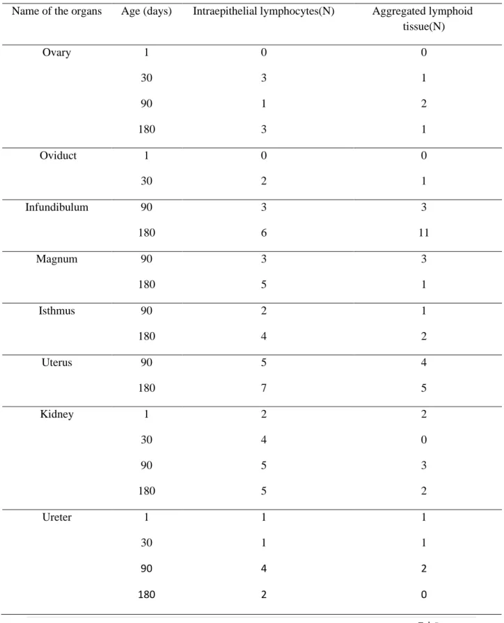
Table 1: Frequency of Intraepithelial lymphocytes and aggregated lymphoid tissue per 5 microscopic fields (40X) in the male urogenital system of native chickens.
Name of the organs Age (days)
Intraepithelial lymphocytes(N) Aggregated lymphoid tissue(N)
Testes 1 2 0
30 4 1
90 3 1
180 3 3
Vas deferens 1 0 1
30 1 1
90 3 2
180 2 1
Epididymis 1 1 0
30 3 3
90 9 1
180 2 1
Kidney 1 3 0
30 2 2
90 1 2
180 4 3
Ureter 1 2 2
30 5 1
90 3 2
180 7 4
Table 2: Frequency of Intraepithelial lymphocytes and aggregated lymphoid tissue per 5 microscopic fields (40X) in female urogenital system of native chickens.
Name of the organs Age (days) Intraepithelial lymphocytes(N) Aggregated lymphoid tissue(N)
Ovary 1 0 0
30 3 1
90 1 2
180 3 1
Oviduct 1 0 0
30 2 1
Infundibulum 90 3 3
180 6 11
Magnum 90 3 3
180 5 1
Isthmus 90 2 1
180 4 2
Uterus 90 5 4
180 7 5
Kidney 1 2 2
30 4 0
90 5 3
180 5 2
Ureter 1 1 1
30 1 1
90 4 2
180 2 0
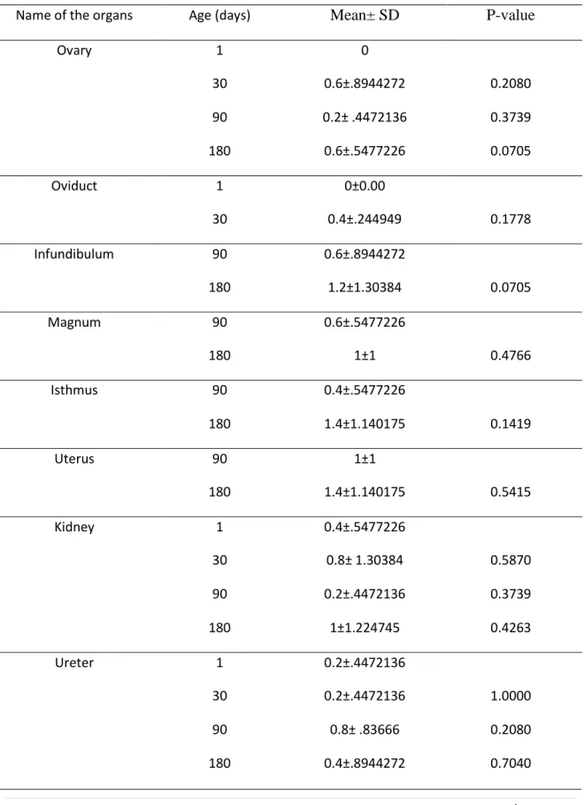
Table 3: Intraepithelial lymphocytes per 5 microscopic fields (40X) in the female urogenital tract of the native chicken.
Name of the organs Age (days) Mean± SD P-value
Ovary 1 0
30 0.6±.8944272 0.2080
90 0.2± .4472136 0.3739
180 0.6±.5477226 0.0705
Oviduct 1 0±0.00
30 0.4±.244949 0.1778
Infundibulum 90 0.6±.8944272
180 1.2±1.30384 0.0705
Magnum 90 0.6±.5477226
180 1±1 0.4766
Isthmus 90 0.4±.5477226
180 1.4±1.140175 0.1419
Uterus 90 1±1
180 1.4±1.140175 0.5415
Kidney 1 0.4±.5477226
30 0.8± 1.30384 0.5870
90 0.2±.4472136 0.3739
180 1±1.224745 0.4263
Ureter 1 0.2±.4472136
30 0.2±.4472136 1.0000
90 0.8± .83666 0.2080
180 0.4±.8944272 0.7040
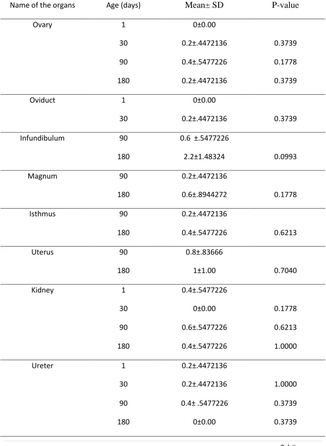
Table 4: Aggregated Lymphoid tissue per 5 microscopic fields (40X) in the female urogenital tract of native chicken.
Name of the organs Age (days) Mean± SD P-value
Ovary 1 0±0.00
30 0.2±.4472136 0.3739
90 0.4±.5477226 0.1778
180 0.2±.4472136 0.3739
Oviduct 1 0±0.00
30 0.2±.4472136 0.3739
Infundibulum 90 0.6 ±.5477226
180 2.2±1.48324 0.0993
Magnum 90 0.2±.4472136
180 0.6±.8944272 0.1778
Isthmus 90 0.2±.4472136
180 0.4±.5477226 0.6213
Uterus 90 0.8±.83666
180 1±1.00 0.7040
Kidney 1 0.4±.5477226
30 0±0.00 0.1778
90 0.6±.5477226 0.6213
180 0.4±.5477226 1.0000
Ureter 1 0.2±.4472136
30 0.2±.4472136 1.0000
90 0.4± .5477226 0.3739
180 0±0.00 0.3739
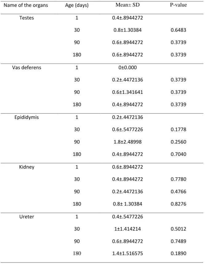
Table 5: Intraepithelial lymphocytes per 5 microscopic fields (40X) in the male urogenital tract of the native chicken.
Name of the organs Age (days) Mean± SD P-value
Testes 1 0.4±.8944272
30 0.8±1.30384 0.6483
90 0.6±.8944272 0.3739
180 0.6±.8944272 0.3739
Vas deferens 1 0±0.000
30 0.2±.4472136 0.3739
90 0.6±1.341641 0.3739
180 0.4±.8944272 0.3739
Epididymis 1 0.2±.4472136
30 0.6±.5477226 0.1778
90 1.8±2.48998 0.2560
180 0.4±.8944272 0.7040
Kidney 1 0.6±.8944272
30 0.4±.8944272 0.7780
90 0.2±.4472136 0.4766
180 0.8± 1.30384 0.8276
Ureter 1 0.4±.5477226
30 1±1.414214 0.5012
90 0.6±.8944272 0.7489
180 1.4±1.516575 0.1890
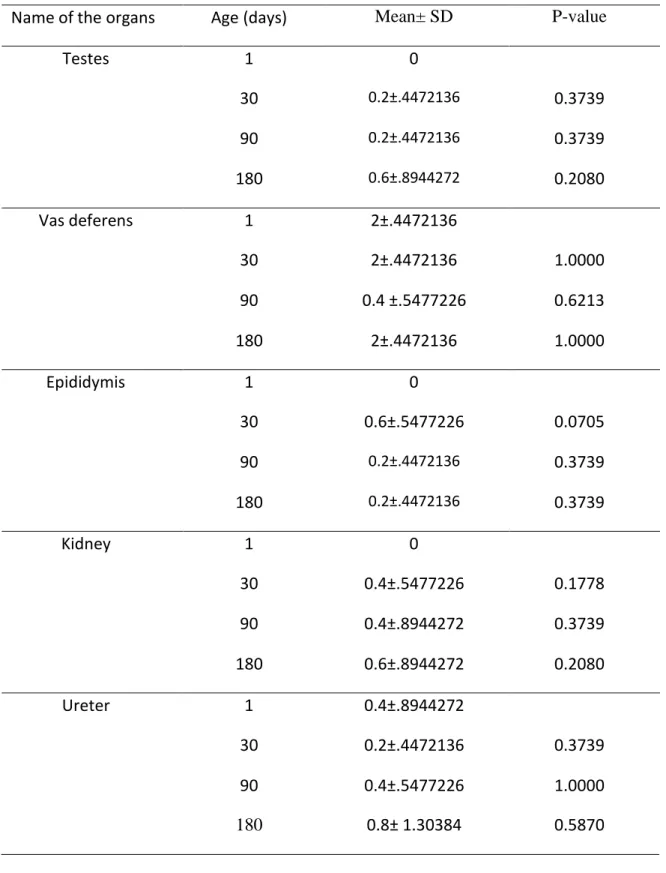
Table 6: Aggregated Lymphoid tissue per 5 microscopic fields (40X) in the male urogenital tract of native chicken.
Name of the organs Age (days) Mean± SD P-value
Testes 1 0
30 0.2±.4472136 0.3739
90 0.2±.4472136 0.3739
180 0.6±.8944272 0.2080
Vas deferens 1 2±.4472136
30 2±.4472136 1.0000
90 0.4 ±.5477226 0.6213
180 2±.4472136 1.0000
Epididymis 1 0
30 0.6±.5477226 0.0705
90 0.2±.4472136 0.3739
180 0.2±.4472136 0.3739
Kidney 1 0
30 0.4±.5477226 0.1778
90 0.4±.8944272 0.3739
180 0.6±.8944272 0.2080
Ureter 1 0.4±.8944272
30 0.2±.4472136 0.3739
90 0.4±.5477226 1.0000
180 0.8± 1.30384 0.5870
Fig 1: Hematoxylin and Eosin staining of Testes (A; male of 180 days) and kidney (B;
female of 90 days) of native chicken. The arrow in the images indicates aggregated lymphoid tissue. Scales for low magnification: 100µm, for high magnification: 40µm.
Fig 2: Hematoxylin and Eosin staining of Kidney (C; male of 180 days) and magnum (D; female of 90 days) of native chicken. The arrow in the images indicated intraepithelial lymphoid tissue. Scales for high magnification: 40µm.
Discussion
Observed structural differences between avian and mammal’s urinary organs are supported by Reshag et al., (2017) and Nabipour et al., (2009). The revealed histological features of the kidney were supported by Ghaji (2021) and Reshag et al., (2017). They reported that the avian kidney has two types of nephrons; reptilian and mammalian types. The mammalian type, which had big renal glomeruli, displayed a well-developed loop of Henle with thin and thick segments. The second kind was the reptilian type, which had small renal glomeruli and was distinguished by lacking the loop of Henle.
The presence of ILT and diffuse lymphoid tissue in the kidney is supported by Hadipour (2010) but shows dissimilarities in the case of the relation of age and increased frequencies of lymphocytes in the kidney. This may be due to a lack of exposure to disease, different environments and dissimilarity in breed selection. The histological findings except the presence of lymphoid tissue in the ureter are supported by Oliaii and Mobini, (2017). They reported the absence of lymphoid tissue in lamina propia in the ureter in Japanese quail. But the presence of lymphoid tissue in the avian ureter is supported by Islam et.al., (2001). The histological findings of the ovary are supported by Reza Dr. S. (20th edition). The normal histological structure was also supported by wani et al., (2017) and the additional finding of ovary and oviduct like ILT and aggregated lymphoid tissue were supported by Biswal, (1954) who stated the presence of lymphocyte in the stroma in the ovary and oviduct. He also stated that the variation of the presence of aggregated lymphocytes which lack limiting membrane varies from ovary to ovary. The presence of immunologically important cells (Lymphocytes) in ovary and oviduct was also supported by Withanage et al., (1997). He stated that the presence of lymphocyte in ovaries and oviduct are related to two aspects. Firstly, ovaries and oviduct are predilection sites for many pathogens that can transmit various infectious agents through eggs like avian encephalomyelitis, lymphoid leucosis, mycoplasmosis, pullorum dis., fowl typhoid etc. (Blaxland et al., 1982). Secondly, hen’s oviduct can be considered as a site for maternal Ig transfer to eggs. The presence of Ig eg. IgA and IgM in egg white and IgG in egg yolk (Yamamota et al., 1975; Rose and Orlans, 1981) may be related to the presence of lymphoid tissue and lymphocytes in the
chicken oviduct. The presence of lymphoid tissue and Ig-containing cells (B lymphocyte, plasma cell) in the oviduct was reported by (Biswal, 1954 and Lebacq- Verheyden et al., 1972). Trautmann and Fiebiger (1952) also described nodular aggregates of lymphocytes in the wall of the chicken oviduct. The current study reveals the increased frequency of ILT in chicken oviducts at 180 days rather than 90 days. This result shows a similarity with Kowalezyk et al, (2020) where T-lymphocyte population was lower in 32weeks birds than 38 weeks. Almost similar result was reported by Johnston et al., (2012) who stated that by 165 days of age, there is an increase in lymphocyte numbers from the levels at 130 days of age throughout the reproductive tract with the ovary. The presence of lymphocytes in the epithelial lining of the excurrent ducts of the testis in domestic fowl is supported by Aire and Malmquist, (1979) who stated that more lymphocytes were located closer to the basal lamina than anywhere in the epithelial layer of the excurrent ducts of the domestic fowl. It reported that the frequency of lymphocytes was higher in efferent ductules than in epididymis and rete testis. Dym and Romrella, (1975) also described the presence of lymphocytes in the tubuli recti, rete testis, ductuli efferentes, epididymis, and ducts deferens. The presence of aggregated lymphoid tissue is supported by King and McLelland, (1975). According to it, solitary, non-capsulated lymphatic nodules occur in virtually all the parenchymatous organs and their ducts in domestic fowl and wild birds.
Conclusion
In the present study, the result has been concluded that intraepithelial lymphocytes and aggregated lymphoid tissue are present in all the organs of the urogenital system.
Apparently lymphoid cells and tissues increase in frequency with age but statistically there is no significant variation in the mean of ILT and aggregated lymphoid tissue with age.
References
Aire, T. A., & Malmquist, M. (1979). Intraepithelial lymphocytes in the excurrent ducts of the testis of the domestic fowl (Gallus domesticus). Cells Tissues Organs, 103(2), 142-149.
Biswal, G. (1954). Additional histological findings in the chicken reproductive tract. Poultry science, 33(4), 843-851.
Blaxland, J. D., G. A. Cullen, R. F. Gordon, and F.T.W. Jordan, 1982. Diseases caused by bacteria, mycoplasmas and chlamydia. Pages 9–75 in: Poultry Diseases. 2nd ed. R. F. Gordon, and F.T.W. Jordan, ed. Bailliere Tindall, London, UK.
DLS (2022-23). Annual report of directorate of livestock services, farm gate, Dhaka, Bangladesh.
DYM, M., & Romrell, L. J. (1975). Intraepithelial lymphocytes in the male reproductive tract of rats and rhesus monkeys. Reproduction, 42(1), 1-7.
Ghaji, M. S. (2021). Morphmetrical and Histological Changes in Domestic Chicken Kidneys in Response to Salinated Water. Indian Journal of Forensic Medicine &
Toxicology, 15(3).
Hadipour, M. M. (2010). Histopathological study of A/chicken/Iran/772/99 (H9N2) influenza virus in commercial broiler chickens. Bulgarian Journal of Veterinary Medicine, 13(1), 38-44.
Islam, K. N., Khan, M. Z. I., Islam, M. N., Ahad, A., & Mazumder, M. S. (2001). Light microscopic structure of the ureters of Rhode Island Red (RIR) and White Leghorn Chicken (WLH) during their postnatal stages of growth and development. Journal of Biological Sciences 1(4), 272-4.
Johnston, C. E., Hartley, C., Salisbury, A. M., & Wigley, P. (2012). Immunological changes at point-of-lay increase susceptibility to Salmonella enterica Serovar enteritidis infection in vaccinated chickens. PloS one, 7(10), 48195.
King, A. S., & McLelland, J. (1975). Outlines of avian anatomy. Bailliere Tindall.
London, Pages 43-64.
Kowalczyk, J., Śmiałek, M., Tykałowski, B., Dziewulska, D., Stenzel, T., & Koncicki, A. (2020). Research Note: Effect of age on the distribution of lymphocytes in the oviduct in Turkey breeder hens. Poultry science, 99(6), 3009-3014.
Lebacq-Verheyden, A. M., Vaerman, J. P., & Heremans, J. F. (1972). Immunohistologic distribution of the chicken immunoglobulins. Journal of Immunology, 109(3), 652-654.
Nabipour, A., Alishahi, E., & Asadian, M. (2009). Some histological and physiological features of avian kidney. Journal of Applied Animal Research, 36(2), 195-198.
Nagy, N., & Oláh, I. (2010). Experimental evidence for the ectodermal origin of the epithelial anlage of the chicken bursa of Fabricius. Development, 137(18), 3019- 3023.
Nochi, T., Jansen, C. A., Toyomizu, M., & Eden, W. V. (2018). The well-developed mucosal immune systems of birds and mammals allow for similar approaches of mucosal vaccination in both types of animals. Frontiers in Nutrition, 5, 60.
Oliaii, A., & Mobini, B. (2017). The Histological Differences of the Ureter in Japanese quail (Coturnix japonica) Compared with Some Other Domestic Avian Species. International Journal of Morphology, 35(1).
Reshag, A. F., Abood, D. A., & Khayoon, E. S. (2017). Histological and histochemical characteristics of the kidneys in different avian species. Australian Journal of Basic and Applied Sciences, 11(16), 36-44.
Reza, Dr.S. (1989).The Essentials of Gross Anatomy Cell Biology &Histology (Part- 2).20th edition. Bangladesh.
Rose, M. E., & Orlans, E. (1981). Immunoglobulins in the egg, embryo and young chick. Developmental & Comparative Immunology, 5(1), 15-20.
Trautmann, A., & Fiebiger, J. (1952). Fundamentals of the histology of domestic animals. Fundamentals of the histology of domestic animals.
Wani, H., Darzi, M. M., Kamil, S. A., Wani, S. A., Munshi, Z. H., Shakoor, A., ... &
Shah, A. (2017). Histological and histochemical studies on the reproductive tract of Kashmir faverolla chicken. Journal of Entomology and Zoology Studies, 5(6), 2256-2262.
Withanage, G. S., Baba, E., Sasai, K., Fukata, T., Kuwamura, M., Miyamoto, T., &
Arakawa, A. (1997). Localization and enumeration of T and B lymphocytes in the reproductive tract of laying hens. Poultry science, 76(5), 671-676.
Yamamoto, H., Watanabe, H., Sato, G., & Mikami, T. (1975). Identification of immunoglobulins in chicken eggs and their antibody activity. Japanese journal of veterinary research, 23(4), 131-140.
Acknowledgment
I feel it is absolutely necessary for me to express my gratitude to "ALMIGHTY," for his incomprehensible grace and profound generosity in enabling me to successfully finish the task.
I would like to express my sincere gratitude and thanks to my respectful internship supervisor Prof. Dr. Mohammad Lutfur Rahman, Department of Anatomy and Histology and also Dean of Faculty of Veterinary Medicine, Chattogram Veterinary and Animal Sciences University for his academic direction, unwavering values, compassionate oversight, helpful counsel, ongoing inspiration, a warm feeling, and constructive criticism throughout the entire study.
I'm grateful to Prof. Dr. AKM Saifuddin, the director of external affairs, for arranging such a special internship program and making this kind of research work as a compulsory requirement of.
I also want to thank Dr. Rasel Prank (MS fellow, Department of Anatomy and Histology) for his kind guidance during my lab works.
I also want to thank all of the well-wishers, friends, and family members for their endless sympathy, considerate assistance, sacrifices, and prayers.
Biography
I am Minara Begum Munni, daughter of Mijanur Rahman and Jesmin Begum. I passed my Secondary School Certificate examination from Dwip Bandhu Mustafizur Rahman High School, Sandwip, Chattogram in 2015 (G.P.A-5.00) followed by the Higher Secondary Certificate examination from Ctg.Govt.Girls College, Chattogram in 2017 (G.P.A-5.00). Now I am an intern veterinarian under the Faculty of Veterinary Medicine at Chattogram Veterinary and Animal Sciences University, Bangladesh.