Shape and Vein Extraction on Plant Leaf Images
Using Fourier and B-Spline Modeling
Rahmadhani M. and Yeni Herdiyeni Email : [email protected]
Department of Computer Science, Faculty of Mathematics and Science, Bogor Agriculture University
ABSTRACT
Leaf features extraction on plant leaf image is still be a problem on automatic plant leaf identification. Shape and venation of leaf are a significant part of leaf for distinguishing a species of leaf from each other. Shape feature extraction with Hough transform and Fourier descriptor were implemented and their effectiveness on leaf shape recognition were compared. Effectiveness of both shape recognition methods were evaluated by recall-precision measurement. Recall-precision evaluation showed that leaf shape represented by Fourier descriptors is more effective than represented by Hough transform. Plant leaf image vein extraction using b– spline representation was implemented. An automatic initialitation of vein search parameter using Standard Hough Transform was proposed.
Keywords: leaf shape extraction, leaf vein extraction, fourier descriptor, b-spline.
Introduction
Automatic identification and classification of leaves is needed for finding biodiversity out (Hickey et al. 1999). But, quantitative data issue extracted from a leaf is still be a constraint of an automatic leaf identification development.
Some qualitative data had been extracted by Hickey et al. (1999) from leaf morphology. Based on leaf morphology too, Rasnovi (2001) got that derivative characters from leaf shape and vein are some effective characters for distinguishing leaf of tree from each others of some families in class Dicotyledoneae.
Shape feature extraction is often used on some indexing cases of content based image retrieval (CBIR). Hough transform (HT) and Fourier descriptor are often used for representation of shape of object in digital image processing. Leaf vein extraction was introduced by Kirchgeβner et al. (2002) using hierarchical b-spline modeling. But, this method need a starting point for vein structure searching interactively.
Experimental Methodology
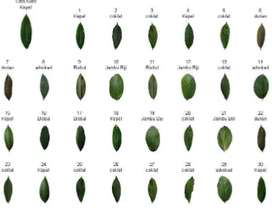
In this research, eleven kinds of leaves in campus Darmaga of Bogor Agriculture University were used. The images of leaves were amount 1100 in JPG format that were resized to 200x150 pixels. They were avocado (Persea americana), bisbul (Diospyros philippensis), cocoa (Theobroma cacao), durian (Durio zibethinus), jamblang (Syzygium cumini), guava (Psidium guajava), jambu bol (Syzygium malaccense), kepel (Stelechocarpus burahol), mangosteen (Garcinia mangostana), menteng (Baccaurea racemosa) dan jackfruit (Artocarpus heterophyllus).
First, an image was segmented. For shape feature extraction using Fourier descriptor, an image was segmented using thresholding. For vein extraction, an image was segmented using Canny edge detector.
Boundary of an object on image resulted by thresholding was got using 4-connected neighbouring. Point by point of boundary was traversed and represented as complex number,
. Sequence of complex number
was transformed using Fourier Transform, resulting . are called as Fourier descriptors.
Shape feature must be invariant to rotation, translation, and dilatation effect. For satisfying these, some adjustments on Fourier descriptors should be done. is ignored as Fourier descriptor due to translation by change the value
of , . Now, the sequence is remove the scaling factor .
On vein extraction, an automatic initialitation of structure searching parameter was proposed using Standard Hough Transform (SHT), instead of giving starting point interactively. SHT was used for line detecting on image resulted by Canny edge detector. One of endpoints of lines detected was used as structure searching starting point and the other as searching direction information (θ).
Vein points searching scans areas As, an 5x3 structure searching starting points and direction.
Veins are lines which are brighter than background (Figure 6). Therefore summing up the lines of the sampled area (Fig. 1 right) resulting in a vector of gray value sums along the lines of the area. The second derivative of each sum vector computed by the discrete Laplacian [1,-2,1] serves
as quality measure. It’s maximum indicates the
most significant vein direction (Kirchgeβner et al.
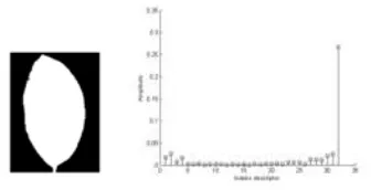
2002). Fourier descriptor magnitude are shown in Fig. 2.
Figure 2 Image resulted in segmentation and histogram of Fourier descriptor magnitude.
Dissimilarity between two histogram of Fourier descriptor magnitude was measured by mean of retrieved images are relevan images.
Evaluation of shape feature extraction based on CBIR involved recall-precision measure. Average precisions at each recall value are shown in Table 1
and visualize in Fig. 3. Comparasion with shape feature extraction with Hough Transform (HT) was done. HT method was taken from Wahyuningsih (2006).
Table 1 Average precision at each recall value
Recall
Precision 0.21846 0.31748
Figure 3 Recall-precision graphics of Fourier descriptor shape feature vs HT shape feature.
Fig. 3 show that graphic of Fourier descriptor shape feature is above graphic of HT shape feature at each recall value. It means leaf shape represented by Fourier descriptors is more effective than represented by Hough transform.
Images retrieved using HT shape feature (Fig. 4) are images that have silimar orientation, position, and size only with query image. In another hand, images retrieved using Fourier descriptors as shape feature (Fig. 5) are not images that have silimar orientation, position, and size only with query image. Fourier descriptor properties that invariant to rotation, translation and dilatation effect, increase average precision value.
Figure 4 Images retrieved using Hough transform shape feature.
2 Vein Extraction
An automatic initialitation of vein search parameter using Standard Hough Transform was proposed. SHT uses biner image of vein as input (Fig. 6 left) and detects existing lines. Detected lines (Fig. 6 center) were used for vein structure searching and the results were represented as b-spline curve (Fig. 6 right).
Figure 6 Vein biner image, detected lines and b-spline model of found vein structure.
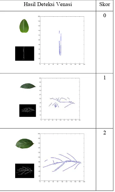
Visual evaluation of vein extraction results was done using 10 sampled images from each species. B-spline curve that contains main vein only was marked as 0. B-spline curve that contains main and side vein but not representative, was marked as 1. And the other one that contains main and side vein representatively was marked as 2.
Table 2 Extracted vein images (b-spline curve) and their marks
Hasil Deteksi Venasi Skor
0
1
2
On Table 2, it’s seen that image resulted on segmentation step determine result of vein extraction. Leaf image on first row physically has so unseen side vein that image contains main vein only. The second row image physically has main and side vein, but unclearly seen because of quality of photo. The leaf is wrinkle and the illumination of photo is poor. The third row image has so clearly seen main and side vein that produce representative result of vein extraction.
Table 3 shows visual evaluation result for all species used. Bisbul, durian, jamblang, and mangosteen are categorized as 0. They have unseen side vein. Kepel and menteng are categorized as 1. They have unclearly seen side vein. And the others have clearly seen main and side vein.
Table 3 Visual evaluation result for all species used
Species Total
Leaves with clearly seen main and side vein are reshown on Table 4 and 5, side of leaf invoked. Table 4 shows that most of the vein extraction result of leaves with up-side are not representative. Table 5 shows that more than fifty percent of the vein extraction result of leaves with bottom-side are representative.
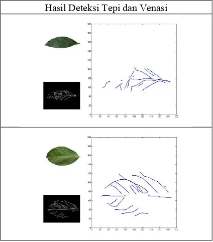
Table 5 Visual evaluation result for certain species categorized as 1. They physically has main and side vein, but unclearly seen because of quality of photo. The leaf are wrinkle or the illumination of photo is poor (Table 6). In another word, good quality of leaf photo is prerequirement for good vein extraction result.
Table 6 Result of vein extraction that marked as 1
Hasil Deteksi Tepi dan Venasi
Conclusion
Shape featue extraction using Fourier descriptors was successfully implemented. Leaf shape represented by Fourier descriptors is more effective than represented by Hough transform.
Vein extaction with automatic initialitation of structure searching parameter was successfully implemented. The results are determined by leaf condition and photo capturing technique. Bottom-side of leaf chosen and good quality of photo support representative of vein extraction result.
References
Hickey, LJ et al. 1999. Manual of Leaf Architecture - morphological description and categorization of dicotyledonous and net-veined monocotyledonous angiosperms by Leaf Architecture. Washington DC: Leaf Shape Description with Fourier Descriptors.
Rasnovi S. 2001. Kajian Pemakaian Morfologi Daun untuk Identifikasi Jenis pada Beberapa Famili Dikotiledon Berhabitus Pohon di Sumatera. Master Thesis. Program Pascasarjana Institut Pertanian Bogor.
Ta-Te L, Yud-Tse C, Wen-Chi L. 2002. Leaf Boundary Extraction and Geometric Modeling of Vegetable Seedlings. National Taiwan University.