Vol 9, No 2, Apil - June 2000 Pathology of melanoma
I 93
Rare
case
of melanoma studied
for
Its histopathological features
in
fndonesia
Achmad Tiarta', Mpu Kanokor, Masato Ueda2, Mochtar Hamzah3, Herman Cipto3, Masamitsu lchihashi2, Evert Poetiraya, Arman Muchhr4, Santoso Cornainl, Joedo ,rihartooot, Setyu*ati nudining.itti,
vorfiyotiô;J
-Abstrak
Banyak parameter hislopatologik unmk kepentingan pengelolaan yang optimal dan untuk menentul<an prognosis melanoma rnalignum kulit pimer stadium L Ketebalan nnror, level Clark dan bebasnya tepi sayatan adalah yang paling-peiting. Laporan pemeipsaan patologi yang baik ber+antunç pada cara penangaun jartngan biopsi elcsisi. Diantara iZO
t-*i
*an*er*itit
patla penelitian bersama Jepang - Indonesia didapatkan 9 kasus melanomayang terdii atas 6 perempuan dan 3 laki - laki. Hanya5 dâi 9 iasus yang dapat di laporkan dalam bentuk laporan histopatologi yang baik sedanglanr yang tain tidak absah, olei larena lcctèr.baiasan keterangan, pengelolaan jaringan el<sisi tidak benar; atau jaingan yang di peroleh merupakanjaingan
hasil biopsi insisi. Kebanyakan kasus berbkasi pada kaki, terutama twnit. Ketebalan rata - rata ttmor ialah 7.94 mm,Abstract
There are many hislopathological parameters for optimal m(Magement and determining prognosis of stage I pimary slcïn malignant melanoma patients. Tumor thickness, Clark level and twnorfree margin are the most impàrtant. Exceilent-pathologicàl report def,ends on the right procedure of handling and prepaing the excisional biopsy. Among 126 skin cancer cases anàlyzed
;i t"pà
- Indànesia joint study, there were 9 cases of malignant melanoma consisted of 6 femate and 3 male. Onty 5 from 9 caies can be-reportihg as an excellent diagnosis of pathology, while the others were not valid because of limited information. Wrong handlin7 à"dpr;p"*g
biopsies specimens or the specimen was an incisiona! biopsy tissue. The most frequent tuntor site was on foàt especialty solis. ivledian tumor thickness was 7.94 mm.Keyword
:
melanoma, pathological reportingThere are many histolopathological parameters which are important to predict the survival of stage I primary cutaneous malignant melanoma patients, among others
ulceration,
Clark level,
tumor's
thickness
in millimeters, prognostic index, risk group, post operativehistological
classification
(pTNM),
lymphocyric reaction, angioinvassion, microscopic satelitosis and associated with pigmented nevi. Further, immunologict
Dqrarluc,t! cf Atmtotrtic Patlology, Facultl, of Mc<!icittc, ^ Utrivcr.ritl' of lttlrncsia,:
[)qnrtmurt ofDcrrnato
ool of Mulicittc, Kobc 650-00
't I )r, 1,rr rr,,tr,,t t of I.) e rtt rtt t o
parameters and
DNA
aneuploidy also influence the prognosis of malignant melanoma patients.lAn
informative and accurate histophatological reportis
essential prerequisitefor
optimal
therapy for melanoma.Not
all
histological parametdrs have the same accuracyand
can
be
conductedin
routine examination.' However, tumor thickness, Clark level, dnd absolute removalof
margins of the lesion are the mostimportant.3
Histopathological evaluation on regionallymph
nodesis
importantfor
planning optimal treatment and determining the prognosis. Excellent pathological report of melanoma depends on theright
procedureof
handling and prep4ring rhe excisional biopsy specimen, especially in determining tumor thickness according to Breslow method.94
Tjarta et almalignant malanoma cases encountered
in
Japan -Indonesiajoint study of skin cancer.MATERIALS
AND METHODSTo
allow
better evaluationof
the histopathologicalfeatures of melanoma, both standard classification and
specific
guidelineswere
applied.They
includedhistologic types, depth
of
tumor cell invasion, tumorthickness, mitotic activity, prognostic index, melanotic
nevi
association, satellitosis, vascular invasion and ulceration. For clarity, they are respectively described briefly in the followings.Histologic types of malignant melanoma
Most
casesof
malignant melanoma are malignanttumors
of'
epidermal melanocytes." Basedon
the clinical characteristic and biological behaviour,it
isdivided into 4 types, i.e. lentigo malignant melanoma
(LMM),
superficial
spreading melanoma (SSM),nodular
melanoma(NM)
and
acral
lentigeneous melanoma (ALM).The characteristic of
LMM
and SSM are that tbey lasta long period and a slow horizontal growth, followed
by a
rapid radialgrowth.
This
changesof
growthwhich are characteristic for primary lesion is probably
due
to
oneor
more strainsof
cells which have an aggressive biologicalpotential.
The primary turnormay. last
for
a relatively long periodof
time duringwhich host
selectiveforces
act,
allowing
quite malignant strains of cells to grow. These cells grow to deeperpart.
Microscopically these cells looklike
a groupof
cells which do not contain melanin pigmentin
their cytoplasm. The groupof
tumor cells whichhas
the
characteristicsof
aggressiveness might develop at theinitial
evolutionof
the primary turnorand
will
manifest as NM; assuming that this is a tumor which is'very malignant from the beginning.3LMM
is a flat, brownish colored lesion which growsslowly and usually (rccurs in the elderly, Microscopically
it
can be observed that atypical melanocytesin
the basal epiderrnalregion
at
the
tumor margin
àneincreasing. Sometimes
it
forms melanocytes with manynuclei
or
nestsof
tumor cellsin
the disorganized epidermis.SSM
is a
lesionwhich
is
slightly elevatedat
thesurface
of
the skin and has a long lasting horizontalgrowth. Microscopically in the epidermis at the tumor
,
,"0
J Indonesmargin, tumor cells are seen scattered
in
the stratum"o-io*.
In
general the tumor cells are separated.Most of the tumor cells have the form
of
epithelioid cellswith
abundant cytoplasm, containing delicate melanin pigment granules, large nuclei and nucleoli.Tumor cells are usually monomorphus. Tumor cells progress
until
more than3
rete ridges outside the tumor.NM
is
a tumor whichis or
resembles an exophyticgrowth or
polypoid.
Microscopically no invasion of tumor cells are found in the epidermis at the margin of the tumor.Neurotropic melanoma showed infiltration
of
tumorcells along nerve sheath and angiotropic melanoma
showed infiltration of tumor cells along vascular vessels.
Acral lentigeneous melanoma occurs on the palms and soles and on the ungual and periu-ngual regions. The
soles being the most common site.'
In order to determine the histologic type of malignant melanoma
it
is
essentialto
takea
sectionfrom
themargin
of
the
tumor
on
routine
histopathologicalexamination.
The
depthof
tumor
cell
invasionin
cutaneousmalignant melanoma
Based on the depth of tumor cell invasion, Clark et ala
have divided malignant melanoma into 5 levels, i.e. I,
IL
Il,
IV andV.I-evel
I
lævel
II
I-evel
lll
tumor
cells
confinedto
epidermis (in situ)tumor
cells
have
penetrated basal membranes and invaded dermal papillae.tumor cells have already invaded dermal
papillae and have reached the boundaries
between
dermal
papillaean
reticulardermis.
tumor
cells
have
already
invadedreticular dermis.
tumor cells have already invaded sub-cutaneous tissue.
Iævel
IV
Iævel
V
It
is
relatively easyto
determine the levelof
tumorcells invasion
in
cutaneous malignant meianoma inVol 9, No 2, Apil - June 2000
irregular bundles. The border
of
dermal papillae andreticular dermis
can
be
easily
recognizedin
aspecimen stained with- Trichrome,
i.e.
Pico-Mallory,Masson or van Gieson.3
Thickness of the tumor
The
Clark's
level
V
malignant melanoma showsclinical
stagesI
(without
melastases)and
shows different 5-year survival rates.s'6 Besides,it is
oftendifficult
objectively to determine whetherit
is Clark's levelI
or
[I
malignant melanoma.In
this
relation, Breslowo suggested to take direct measurement of thedepth of tumor invasion by measuring the thickness of the tumor with an ocular millimeter.
The section is made from tumor tissues preserved in neutral formaline solution, by taking tangential section of the central part of the tumor, and stained with
H
&E. In cases of a large tumor several sections are taken.
Thç thickness of the tumor is measured with an ocular millimeter from the top
of
the granular layerof
theepidermis to the lowest point
of
tumor cells invasion. When the tumoris
ulcerated, measurementis
takenfrom
the baseof the ulcer
till
the deepest point oftumor cell invasion.
Measurement of tumor thickness is the best method to
determine
the
prognosisof
primary
cutaneous malignant melanoma comparedto
determining the depthof
invasion accordingto
Clark's method. Thepossibilities
of
recurrence
might
be
directlyproportional to the thickness
of
the tumor.oIt
means thatit is important to
determine the thicknessof
thetumor for further management. In patients with tumor
thickness not more than 1.50 mm, it is not necessary to dissect the regional lymph nodes. Different institutions
have
different
waysof
managingtumor and
theregional lymphnodes.T'E
For practical reasons measuring the thickness of tumor
is more reliable than to determine the level
of
tumorcells invasion according to Clark's method. However
in
routine
histopathologicalexamination
it
isrecommended always
to
mention the levelof
tumorcell invasion besides the thickness of tumor according
to Breslow's method.2
Mitotic Activity
The number of mitoses which was found on histologic
examination
of MM
was directly
conelated withPatlulogy of
melanoma
95prognosis. Ten-year survival
of
patientswith
nomitosis
or
one
mitosis noted
in a
high-powermicroscopic
field
was 87% comparedto
64Vo for lesionswith two
or more mitoses
per
high-power field.rPrognostic index of malignant metanoma
The prognostic index
of
malignant melanomais
the result of multiplication of the thickness of the tumor in millimeteron
measurement accordingto
Breslow's method with the mitotic rate per square millimeter on the standard of histologic"rray.t'to
In
stageI, a
more
accurate prognosiscan
be determinedby
prognosticindex
rather
than
thethickness
of
the tumor, particularly in patients with a tumor thicknessof
1.50 mm-
3.49mm. When
theprognostic index in this group was higher than 19, the death rate would be higher compared to a prognostic index less than 19.r0
It
is obvious that the prognostic index is important inthe selection of patients
with
malignant melanomawho have a poor prognosis and need further treatment,
for
instance dissectionof
lymphnodesor
immuno-therapy.e'lo
Prognostic
index can
be
determinedon
routine histopathological examination. The thickness of the tumor was measured accordingto
the
method of Breslow,i.e. the
distance betweenthe top
of
thegranular layer
of
the epidermisuntil
the lowest point of tumor cells invasion by using an ocular millimeter. The mitotic rate was counted at leastin.l.50
square milimeter(10
HPD
by using
40x
objectives lens,afterwards converse
into
I
squaremiliimeter.
Thearea
of
I
high power fieldis
evaluated through theformula 11 12.
Histologic association with melanocytic nevi
MM
appearsto
arise
histopathogicallyfrom
a melanocyic ic nevi in approximately ZOVoof
casesrr.ln
the remainderof
cases,MM
arise de novo that isfrom
melanocytesnot
associatedwith
melanocyticnevi.
Patientswith
MM
associatedwith
acquiredmelanocytic nevi had a better prognosis, i.e. five year
survival
of
havinglesions
that
ugh
no96
Tjarta et alMicroscopic satellitosis
These satellitosis were defined as discrete nest of tumor cells
at
least 0.05 mmin
diameter separatedfrom the main body
of
the tumor by reticular dermalcollagen
or
subcutaneousfat.
The presenceof
thesecalled "microscopic satellitosis" connotes
a
poorerorognosis.l Microscopic satellitosis also appear to
influence the
presenceof
tumorin
regional lymphnodes independent
of
the
tumor thickness and the increased tend"ncy to developed local recurrence.r2Vascular invasion
Vascular invasion protends
a
poorer prognosis. Theobservation
of
a thin liningof
endothelium is vital tobe certain that a neoplastic cells is
in
fact within thelumen of a vascular or lyrnphatic channel. Factor
VIII
immunoperoxidase staining can often be helpful in
defining
the
natureof
tumor cell within
vascular channel.l30
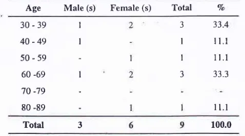
-39
r40-49
I50-59
60
-69
I70 -79
80 -89
33.4
ILl
1t.l
33.3
l
l.l
Total
3 100.0The age of patient ranged from 30 to 80 years, which peaked on 30 - 39 and 60 - 69 years.
d J lndones
Ulceration
The
clinical and
histopathological presenceof
ulceration in a malignant melanoma
(MM)
is a poorprognostic
sign.lr
Patients with ulcerated lesions hada
poorer
prognosis comparedto
those
withoutulceration. In general ulcerated lessions also tend to be
thicker than nonulcerated ones. However, multivariate
analysis, has shown
the
presenceof
ulceration assignificant factor even after the lesion thickness has
been taken into account.'
It
is
recomended that thebreadth
of
the ulcer be reported. Ulcer more than 3mm
in
breadth have been associatedwith
poorerprognosis.la
RESULTS
From 126 cases of skin cancer in this study there were
9
casesof
malignant melanoma, consistedof
6females and
3
males. The ratioof
the frequency infemale to male was 2
tol
(Tabel 1).Axilla
ll.l
Arm:Upper
I
-
I
ll.l
arm
Tumb
-
I
I
ll.1Table l. Age distribution of melanoma cases according to sex Table 2. Site distribution of melanoma cases according to sex.
Age
Male (s) Female (s) Total VoSite
Male(s)
Female(s) Total
Vo 3I
I
3
Foot
I
ISole
|
2Plantar
pedis
-
I2
22.23
33.3I
ll.1Total
6
9
100.0 [image:4.562.31.270.413.546.2]Vol 9, No 2, Apil
-
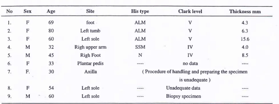
June 2000Table 3. Sex, age and site distribution by histological parameters
Pathology of melanoma 97
Sex age Site IIis type Clark level Thickness mm
l.
2.J.
4.
5.
6.
7. F F F M M F
F.
F M
69 80 60 32
45
33 30
foot lÆft tumb
læft sole
Righ upper arm
Righ Foot
Plantar pedis
Axilla
Left sole
Left sole
ALM ALM ALM
SSM
N
V V
v
Iv
IV
no data
4.3
6.3
15.6
4.0
8.5
54 60 8
9
( Procedure of handling and preparing the specimen is unadequate )
Unadequate data
Biopsy specimen
Only
5
caseswith
complete data. Other cases werelimited
data, or wrong procedureof
handling and preparingof
specimenand
biopsy specimens, orincisional biopsy specimen
only. Most
cases wereALM
type and Clark levelV
median tumor thickness was 7.944 mm.DISCUSSION
The data
in this
study showed that melanoma casesoccurred more often in females compared
to
males.The
previousfindings
showedthat
melanoma in Jakarta and also from outside Jakarta occurred moreoften in males as have been peported by usl5' 16' 17' l8 and other investigator.''
White
people exhibited awide
range
of
melanoma incidenceand
femalescommonly had a higher incidence than males.20'21
Non-white people exhibited in general a much lower incidence
of
melanoma.It
has been suggested thatskin
pigmentation protect âgainst melanoma.2rAll
groupsof
Africans have
a higher
incidence ofmelanoma than any group
of
Asians.
There was a predominance of either sex among these racial groups, althoughthe
combined ratefor
Asian
males wasnearly twice than of females.2l
In this study the tumors were mostly located at lower
on the soles and olantar nedis. r previous findingfs' Ie' tz'
tt
unoIt
appeared that melanoma were mostly sited at lower extremities, especially on the soles. This is similar tothe findings {Çported among Chinese of Hongkong,22 Puerto Rican,z3 Japanese2a and Philipines.2s Concerning
the
high
frequencyof
tumor site
at
the
lowerextremities many investigators considered its relation
to trauma and ultraviolet effect of sunshine.z'.
"
Suchfactors might also be taken into consideration for our
data, since
a large
proportionof
the
Indonesianpopulation, especially
in
rural areas were working inagricultural sectors. They
are
usedto
work
in traditional ways with naked foot which allows a longtime exposure
to
ultraviolet raysfrom
the sun and traumaon
their foot. Also the
more
frequent occurrenceof
melanoma on the feetof
males from outside Jakarta suggested the possibility of trauma as acontributing risk factor. [n contrast, the population of Jakarta was predominantly employed
in
industrialsectors, wearing shoes.l6' l8
In
Israel a higher incidenceof
melanoma was foundamonÊ agricultural workers in the Kibbuts than in the
city.'o
The role of trauma associatedwith
walking barefootedhad
been
emphasizedas
a
possible etioloqic factor,ls'le However, the Chineseof
HongKong," as shoe-wearing society, a similar result was
obtained, i.e. the majority
of
melanoma occurred onthe foot.
[t
was suggestedthat
many physical or chemical agents may be the factor causing changes in this areas. The tumors were most frequently found on the sole, indicating that weight-bearing as a rrauma is an important factor.z3'T
In Uganda
it
was observed that melanoma on the soleof
the feet
correspondedwith
that
of
discreteof
[image:5.553.29.518.98.281.2]98
Tjarta et al d J Indonesknowledge
of
the disease,or 2)
increased alertness aboutthe disease,
or 3)
improvementof
socio-economic status
of
the people, or4)
improvement ofhealth care facility. Total cases at present study were
too small to allow adequate statistical analysis. In this study
only
5
from
9
casesof
melanoma could bereported as an excellent pathological diagnosis. Four
cases could not be analyzed for the histological typing,
Clark level and tumor thickness because
of
limitedinformation
or
inadequate preparationof the
biopsyspeclmen.
Acknowledgement
We are grateful to the lnternational Cancer Research Grant system, Monbusho, Japan and the Dean, Faculty
of Medicine, University of Indonesia, Jakarta and we
would also thank
the
Director
of Dr.
Cipto Mangunkusumo National Central General Hospital fortechnical assistance.
We
appreciate
the
Dean's
approval
No. 8451P'102.H4.FK/B197of the
Japan-Indonesiacollaborative study. This work has been also supported
by
the
grant
no.
09042004,under
Ministry
ofEducation, Science, Sport and Culture, Government of
Japan and was
partly
supportedby the
IndonesianCancer Foundation, the Jakarta International Cancer Conference Fund
and
the
Terry
Fox
Foundation, Canada.REFERENCES
l.
Friedman RJ, Rigel DS, Kopf AW, Haris MH, Baker D. Cancer of the skin. Philadelphia, WB Saunders l99l; 117-98.2.
McGovern VJ, Mihm MG Jr, Baily JC. The classification of malignant melanoma and its histologic reporting. Cancer1973;32: lM6-57.
3. Australian Cancer Network. Guidelines for the management
of cutaneous melanoma. The Stone Press 20 Hermington
St. Epping 2121, Junç 1997
4.
Clark WH, FromL
SemadioEA,
Mihm MC. The histogenesis and biologic behaviourof
primary humanmalignant melanoma of the skin. Cancer Res 1969; 29: 705-14.
5. Lever WF, Lever
DS.
Histopathologyof
the skin.PhiladetphiaT'h ed. Lippincott, 1990: 780-96.
6.
AckermanAB.
Disagreements about classihcation of malignant melanoma. Am J Dermatopathol 1982: 4: 44'l-52.7.
Breslow A. Thickness, cross-sectional areas and depth of invasion in the prognosis of cutaneous melanoma. Ann Surg 1970;72:902-8.wooden smoke may be the factors causing melanoma
in this area.28
The present study indicated that the growth pattern (histological type) was mainly ALM type
(3
case), SSM andNM
typeis
I
case each. This pattem was different from the result of previous study in the same laboratory.ls NM type was the most frequent and onlya fe\À/ cases
of
LMM
andALM
type.No
SSM type had been found. However, the pattern was the same asthe melanoma among the Chinese of Hong Kong, the
tumor were mainly
ALM
type followed by SSM and NM type.22Among
the
Caucasians,SSM type was
the
most frequent, followed byNM, ALM
andLMM
type.2eNM
type have a bad prognosis compared toLMM
orSSM type.''u In
New
South Wales (Australia) and Alabama(USA)
the
incidenceof
NM
type
hasdecreased
during
25
years (1955-1980),while
the incidence of SSM typ" -hut increased significantly.3lEarly studies suggested that different histologic types
of
MM
correlatedwith
prognosis. However, later multivariate analysis did not show histologic type tobe
of
dominant prognostic significance when other values were taken into account.The difference in survival among these different types of MM is more probably related to difference in lesion
thickness and anatomic site.' The evaluation of the depth
of
tumor cells invasion (Clark level) revealedthat the majority
of
cases ware level V,.followed by levelfV.
This finding was similar to the result of theprevious study that most cases were level V, followed by level
[V
and level III.16'18 This finding seemed to differ from other studies.In
New South Wales andAlabama,
it
was reported that levelof
invasion havechanged. The incidence
of
melanomain
level[I
has increasedwhile
deeper
lesion (level
IV)
hasdecreasedsr. The death rate
of
levelII
was 8.3 Vo,level IIII was 35.2 Vo,level
fV
was 46.1 7o and level V was 52 Vo.aTumor thickness on 5 cases in this study ranged from
4.4 mm
to
15.6 mm with median thickness 7.94 mm. Previous studyin
this
laboratory,tumor
thickness rangedfrom
21
mm
to
20.25mm
with
medianVol 9, No 2, April - June 2000
8.
Breslow A. Prognostic factor in the treatment of cutaneousmalignant melanoma. J Cutan Pathol 1979; 6:208-12.
9.
Brestow A. Tumor thickness, level of invasion and nodedissection in stage I cutaneous melanoma. Ann Surg 1975:
182:5'12-5.
10. Schmoeckei
C,
Braun-Falco0.
Prcignostic index in malignant metanoma. Arch Dermatol 1978; I 14: 871 - 3. 11. Kopf AW, Gross DF, Roger GS, Rigei DS, Heliman Li.Levenstein
M,
et
al. Prognostic indexfor
malignant melanoma. Cancer 1987; 59: 1236-71.12. Friedman RJ, Riegel DS, Kopf AW. Favorable prognosis
for
malignant melanoma associatedwith
acquiredmelanocytic nevi. Arch Dermatol 1983 ; 119: 455-62.
13. Kelly JW, Sajabiel RW, Calderon W, Murillo L, Dubin RL, Blois
MS.
The frequencyof
local recurrence andmicrosatellites as
a
guideto
re-excision margins for cutaneous malignant melanoma. Ann Surg 1984; 200:759-63.
14. McGovem
VJ,
ShawHM,
MiltonGW.
Prognostic significanceof
the
histologic featureof
malignant melanoma. Histopathology 1979; 3: 385-93.15. Tjarta
A,
KanokoM,
MangunkusumoR.
Malignant Melanoma. Pathological aspectsof
primary cutaneousmaligant melanoma. In: Cancer in Asia Pacific. yayasan Kanker Indonesia, Jakarta, Indonesia
:
1988. Vol2:997-I 003.
16.ljarta
A.
The incidence and histopatological aspect ofprimary skin malignant melanoma in Jakarta, Indonesia. Presented on XIVth Intemational Pigment cell Conference,
Kobe October 3l-November 4, 1990.
17. 'ljarta A. The role of histophatological examination on the prognosis of cutaneous maligant melanoma. Indon. J. Oncol
l99l;
2:125-30.18. Tjarta
A.
Maligant melanomaof
the
skin. Some histopathological aspects and its prognostic value. Indon. J.Oncol 1992; 3: 1-6.
19. Pringgoutomo S. Skin Cancer In Indonesia. Natl Cancer
Inst Monogr 1963; l0: l9l-5.
Pathology of melanoma 99
20. Magnus K. Incidence of malignant melanoma of the skin in Norway, 1955 - 1970. Cancer 1973 32:1275-86.
21. Crombie IK. Racial differences in melanoma invidence. Br
J Cancer 1979; 40:185-93.
22. Colins JR. Melanoma
in
the Chineseof
Hongkong.Emphasis on volar and Subungual Sites. Cancer 1984; 54: 1482-8
23. Pantoja E, Llobet RE, Roswirz B. Melanoma of the lower Extremity among native Puerto Ricans. Cancer 1976; 38:
t420-3.
24. Seiji M, Takahishi M. Acral melanoma in Japan. Human Pathol 1982; 13 (7): 607-9.
25. Pantangco EE, Canlas M, Basa G, R. Observation on the
incidence, biology and pathology of skin câncer among Filipinos. Natl Cancer Inst Monogr 1963; l0: 109-25.
26. Anaise D, Steinitz R, Ben Hur N. Solar radiation a possible
etiological factor in malignant melanoma
in
Israel.Retrospective study (1960 - 1972). Cancer 1978; 42:299-304.
27. Hind MW. Anatomical distribution of malignant melanoma
of the skin among non- caucasians in Hawaii. Br J Cancer
1979;40: 497- 9.
28. Lewis MG, Kiryabwire JWM. Aspect
of
behavior andnatural history of malignant melanoma in Uganda. Cancer
1986;21:876-87.
29. Lonspari S, Mihnm MC Jr. Clinical and pathological corelation of malignant melanoma. J Cutan pathol 1979; 6:
I 80-94.
30. McGovern VJ. Melanoma. Histopathological diagnosis and
prognosis. New York: Raven Press, 1982; 158-81.
31. Balch CM, Soong S-J, Milton GW, Shaw HM, McGovern
VI,
Mc Carthy WH, et al. Changing trendsin
cutanemalignant melanoma over a quarter century in Alabama, USA and New South Wales, Australia. Cancer, 1983; 52: