Vol. 21, No. 2, May 2012 AFP effect on NF-κB translocation of dendritic cell 97
The effect of alpha fetoprotein on NF-κB translocation in lipopolysaccharide
induced monocyte-derived dendritic cell
Akterono D. Budiyati,1 Agus Setiyono,2 Elpita Tarigan,1 Heri Wibowo3
1 Division of Immunology, Mochtar Riady Institute for Nanotechnology, Tangerang, Indonesia
2 Division of Pathology, Department of Clinic, Reproduction and Pathology, Bogor Agricultural University, Bogor, Indonesia 3 Department of Immunology, Biomedical Graduate Program, Faculty of Medicine, Universitas Indonesia, Jakarta, Indonesia
Abstrak
Latar belakang: Alfa fetoprotein (AFP) merupakan antigen onkofetal yang berperan penting dalam perkembangan ontogenik dan onkogenik. Kadar AFP dalam darah pasien hepatocellular carcinoma (HCC) diketahui meningkat dibandingkan orang sehat. Publikasi terakhir menunjukkan bahwa AFP menyebabkan disfungsi sel dendritik derivat monosit sebagai antigen presenting cell (APC) yang dapat mengakibatkan respon antitumor menjadi tidak eisien. Penelitian pendahuluan ini bertujuan untuk mengetahui apakah efek AFP terhadap disfungsi sel dendritik derivat monosit sebagai APC adalah melalui jalur sinyal NF-κB dengan menggunakan lipopolisakarida (LPS) sebagai penginduksi aktifasi NF-κB.
Metode: Sel monosit dikultur dalam medium yang mengandung GM-CSF (800 ng/mL) dan IL-4 (1000 ng/mL) dengan atau tanpa penambahan AFP dan diinkubasi selama enam hari agar berdiferensiasi menjadi sel dendritik imatur. Sel dendritik matur kemudian diperoleh dengan menambahkan LPS ke kultur dan diinkubasi selama 30 menit. Deteksi translokasi NF-κB dilakukan menggunakan uji imunoluoresens (IFA).
Hasil: Pada kelompok kontrol, induksi LPS menyebabkan terjadinya translokasi NF-κB sedangkan kelompok AFP menunjukkan hasil yang berlawanan yaitu translokasi NF-κB tidak terjadi.
Kesimpulan: Penelitian ini menunjukkan bahwa AFP mencegah aktifasi dan translokasi NF-κB sehingga menyebabkan gangguan fungsi sel dendritik derivat monosit sebagai APC. Hasil yang diperoleh diharapkan memberikan pemahaman baru mengenai peran AFP dalam mekanisme supresi respon antitumor. (Med J Indones. 2012;21:97-101)
Abstract
Background: Alpha fetoprotein (AFP) is a tumor-associated Ag that has a function in both ontogenic and oncogenic growth and its serum level is elevated in patients with hepatocellular carcinoma (HCC). A recent study showed that the immunoregulatory effect of AFP was through impairment of dendritic cell function as antigen presenting cell (APC), a mechanism that is known to hamper eficient antitumor response. However, the underlying intracellular mechanism of action of AFP required elucidation. As an initial step to determine the signaling pathway of AFP, we analyzed whether LPS induced NF-κB translocation occured in AFP-treated monocyte-derived dendritic cell (MDDC), which was induced by lipopolysaccharide (LPS).
Methods: Monocytes were cultured in GM-CSF (800 ng/mL) and IL-4 (1000 ng/mL) containing medium and incubated for six days to generate immature MDDCs with or without the presence of AFP. Mature MDDC was generated by stimulation of the immature MDDC with LPS for another 30 minutes. The analysis of NF-κB translocation was measured by luorescent microscopy.
Results: Following activation of MDDC by LPS, the control group showed a marked nuclear staining of NF-κB. However, the AFP-treated group showed negative nuclear staining similar as observed in unactivated MDDC.
Conclusion: This study demonstrated that AFP prevented the activation and nuclear translocation of NF-κB and subsequently might cause the impairment of MDDC function as APC. This inding provides a new insight on the role of AFP in the suppression mechanism of anti tumor immune response. (Med J Indones. 2012;21:97-101)
Keywords: Alpha fetoprotein, dendritic cell, lipopolysaccharide, NF-κB translocation
Correspondence email to: [email protected]
have high level of AFP in their serum and tumor tissue,2 and increase in AFP might cause the dysfunction of DC. Dysfunction of DC is one of the critical mechanisms to
escape immune surveillance.4
Since our previous report proved that alpha fetoprotein
(AFP) inhibited synthesis of interleukin-12 (IL-12)3
andNF-κB is the transcription factor of IL-12,5 we assumed that nuclear factor kappa B (NF-κB) was involved in this mechanism. In order to deepen our
understanding on intracellular mechanism affected by
Alpha fetoprotein (AFP) is a 70 kDa oncofetal
protein and is detected both fetally and maternally during pregnancy and then its expression is turned off completely after birth.1 AFP is thought to play an intrinsic immunomodulator capacity to avoid rejection of the developing embryo by maternal immune system.1,2 Our previous study revealed this immunomodulatory properties of AFP might happen through the impairment of monocytes-derived dendritic
cell (MDDC) as antigen presenting cell (APC).3 A
AFP, we conducted a study to determine which element
in NF-κB signaling pathway that was directly inhibited by AFP. This preliminary report aimed to determine the effect of AFP on nuclear translocation of NF-κB using lipopolysaccharide (LPS) as activator for NF-κB
signaling pathway.5
METHODS
The experiment was designed as analytical laboratory experiment and was carried out at Mochtar Riady Institute for Nanotechnology (MRIN) laboratory. Informed consents were obtained from the donors, and
the ethical approval was granted from the Committee
on Health Research Ethics of MRIN.
Samples
A total of 30 mL blood was used in this study and was obtained by phlebotomy procedure from 3 healthy volunteers (n = 3) with the age of 25 to 35 years old.
We used vacutainer® with heparin (BD vacutainer) during the phlebotomy procedure. The peripheral blood mononuclear cells (PBMCs) were then prepared by
separating the phlebotomy product using Ficoll-hypaque
(GE Health Care, UK) density gradient separation.3 In brief, blood was suspended in equal volume of phosphate buffer saline (PBS) and then layered on icoll solution with ratio 4:3 (blood suspension/icoll volume). Centrifugation was then performed at 2500 rotation per minute (RPM) for 30 minutes. PBMCs
were then collected as a cloudy ring trapped between
icoll and plasma suspension. The PBMCs were then washed twice in PBS and spinned at 2000 RPM for 10
minutes.
Generation of monocyte-derived dendritic cells (MDDCs)
MDDCs were generated as described previously3 with some modiications. In brief, PBMCs from each volunteer were adhered to 12 well culture plates with a density of 1×10⁶/mL for 30 minutes at 37°C with 5% CO2. The non adherent cells were removed by gentle wash
and the adherent cells were then cultured in complete
medium RPMI 1640 (Sigma) supplemented with 10% heat-inactivated fetal bovine serum (Gibco), and rh-GM-CSF (800 U/mL, BD Bioscience Pharmingen) and rh-IL-4 (1000 U/mL, BD Bioscience Pharmingen] for another 6 days. Where indicated, puriied human cord blood AFP (FL-AFP, purity > 95%; Monobind method; Lee Biosolutions, Inc) was added at day 0. Based on our previous result, concentration of AFP 6.25 µg/mL gave a signiicance suppression effect to MDDCs, but still produced cells with viability > 90% compared to the non
AFP-treated cells (control).3 The concentration of 6.25
µg/mL was then used for all subsequent experiments.
Cell stimulation
NF-κB activation was performed as described
previously.6 In brief, immature MDDCs were activated by adding 1000 ng/mL LPS from Escherichia coli
(serotype 0111:B4; Sigma) and incubated for 30 minutes, one hour, four hours, or 24 hours respectively.
Detection of NF-κB translocation
From our previous study, we found that concentration of
6.25 µg/mL is the limit of signiicance of the suppressive effect of AFP on MDDCs.3 The same concentration was used to detect NF-κB translocation. In order to observe the nuclear translocation of NF-κB, the MDDCs
of each group were harvested and then pelleted by
centrifugation in microcentrifuge tubes for 3 minutes at 3500 RPM. The pellet was then washed twice with PBS
using similar centrifugation procedure. After washing,
the pellet was resuspended in PBS and smeared on poly
L-lysine glass slide. Fixation was performed on air
dried smeared cells using acetone for 10 minutes. The
cells were then incubated in phosphate buffered saline
(PBS) that contained bovine serum albumin (BSA) 1% for 30 minutes at room temperature (RT). After washing, polyclonal rabbit anti-p65 antibody (ReI A) 1 µg/mL, purchased from abcam (Ab6701), was used as primary antibody and incubated for 1 hour at RT. After washing, anti rabbit IgG antibody conjugated with Cy3 0.2 µg/mL (Sigma Aldrich C2306) was used as secondary antibody and incubated for 30 minutes in RT. After washing, 4’,6-diamidino-2-phenylindole (DAPI) was used as counter staining to mark the
nuclear location.
Visualization of NF-κB translocation
Following counter staining and washing step, observation of nuclear translocation was performed
under luorescence microscopy (Zeiss, axio40 FL). Under ultra violet excitation, NF-κB protein labelled by Cy3 have an emission peak at 570 nm (red emission) and DNA labelled by DAPI have an emission peak at 461 nm (blue emission). They were visualized using ilters that pass 605/70 and 445/50 emission respectively.
Data analysis
A positive microscopic examination of NF-κB translocation was deined as positive Cy3 staining in nuclear region on a per cell basis. The data were collected
Vol. 21, No. 2, May 2012 AFP effect on NF-κB translocation of dendritic cell 99
observed per ield of view. We used Mann-Whitney test
for statistical analysis to compare differences between AFP-treated group and untreated group.
RESULTS
LPS induces nuclear translocation of NF-κB
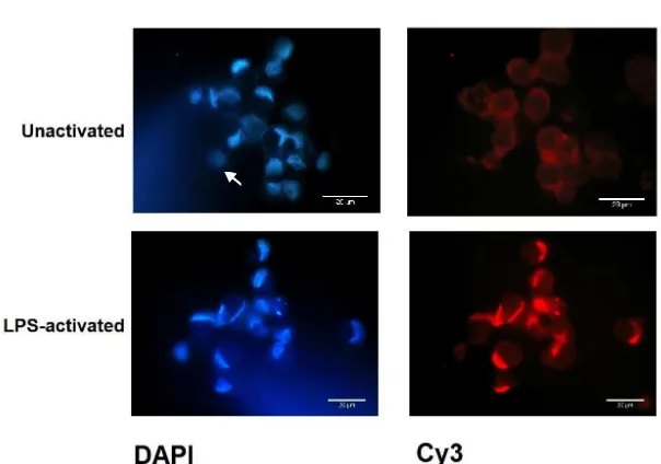
Figure 1 showed cytoplasmic staining without nuclear labeling was observed in unactivated MDDC of control group. Activation with LPS for 30 minutes caused rapid nuclear translocation of NF-κB. This was also observed
after 1 hour and 4 hours LPS activation, and then disappeared after 24 hours activation (data not shown). Based on this result, our experiment was then used 30
minutes as incubation period for LPS activation.
AFP inhibits the nuclear translocation of NF-κB
In contrast with the control group (Figure 2), the
AFP-treated group did not show any nuclear staining of
NF-κB following activation with LPS. Co-localization of
Cy3 and DAPI signal inside the nucleus was not found. The NF-κB signal was only observed in cytoplasmic
Figure 1. Nuclear translocation of NF-κB of the control group before (unactivated) and after LPS addition (LPS-activated) for 30 min -utes. Magniication 1000x, scale bar 20 µm relative to cell size. The arrow indicates positive DAPI staining and NF-κB label-ling within similar cell. DAPI= blue staining, NF-κB Cy3= red staining
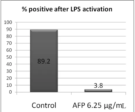
compartment. Based on percentage of positive nuclear
staining per ield of view, we got a signiicantly different
result between the control group compared to the
AFP-treated group (Figure 3, Mann Whitney, p < 0.001).
DISCUSSION
Alpha fetoprotein has long been known as an oncofetal immunoregulator that has an effect on immune cells. Our previous study revealed that the addition of AFP
on MDDC cultures at monocyte stage resulted in downregulation of MHC II expression, costimulatory molecules, maturation level and inhibition of IL-12 synthesis after LPS activation.3 Dendritic cell
dysfunction caused by the presence of tumor associated antigen such as AFP, can occur through several
mechanisms: 1) impairment of dendritic cell formation, 2) inhibition of maturation process, 3) induction of apoptosis, and 4) interference with the process of
antigen presentation.4
Several previous publications proved that NF-κB plays
an important role in the maturation process of dendritic
cells. NF-κB activation occurs in mature dendritic cells in response to stimuli such as LPS or TNFα and is characterized by nuclear translocation of NF-κB followed by increased expression of MHC II molecules, costimulatory molecules and proinlammatory cytokine, IL-12.5,6 If the activation of NF-κB is inhibited, either
pharmacologically or due to over expression of protein inhibitor, the maturation process will be inhibited.5,7
NF-κB is a heterodimer protein consisting of a 65 kDa DNA binding sub unit (p65 or ReIA) and an associated
50 kDa protein (p50 or NF-κB1). In most cell types, the p50/p65 heterodimer is located within the cytoplasm
as an unactivated complex form bound to its inhibitor,
namely inhibitor kappa B (IκB), which prevents entry
into the nucleus. Following cellular activation, i.e. LPS
induction, IκB is rapidly degraded allowing the p65 sub
unit to translocate to the nucleus.6 In order to measure NF-κB translocation, we used polyclonal rabbit anti-p65 antibody (ReI A) that recognized residues 500 to the terminus of human NF-κB p65 sub unit of both unactivated form of NF-κB located inside the cytoplasm
compartment and in activated form as p65 sub unit located inside the nucleus compartment.
In line with previous study, our result showed that LPS caused the activation of NF-κB.5 In contrast to this fact,
our AFP-treated group did not show nuclear positive
labeling after LPS activation. These results indicate that
the mechanism that is used by AFP in causing dendritic
cell dysfunction is via NF-κB signaling pathway. Several mechanisms have been proposed to explain these inhibition: 1) the over-expression of protein inhibitor,7 2) inhibition of proteosome activity that prevent IκB degradation,8 3) disruption of NF-κB-DNA binding activity.7 Our result showed that NF-κB was completely undetected in
nuclear area, suggesting that the inhibition process occurs
at the upstream level of this NF-κB signaling pathway.
Further research will be needed to determine whether this
inhibition caused by over expression of IκB or at earlier steps. There are many possibilities that can be explored to determine the speciic target that are affected by AFP, but
this experiment ensures that AFP plays role in the failure
of nuclear translocation of NF-κB.
In conclusion, this preliminary study demonstrated in-vitro that AFP prevented nuclear translocation of
NF-κB after LPS activation that lead to MDDC dysfunction as APC. This information can be used as a basic data
for further understanding of immune suppression mechanism in HCC, where AFP is increased.
Acknowledgment
We thank to all persons who kindly participate and contribute to this study, in particular the blood donors.
We also express our deepest gratitude to Dr. rer. nat. Ivet M. Suriapranata for critical reading and discussion. This research was supported by a grant from Mochtar Riady Institute for Nanotechnology.
REFERENCES
1. Mizejewski GJ. Biological roles of alpha-fetoprotein during pregnancy and perinatal development. Exp Biol Med. 2004;229:439–63.
Figure 3.Comparison of mean of percentage of positive cells having NF-κB translocation after LPS activation be -tween untreated group (control) and AFP-treated group (AFP 6.25 µg/mL). The data were generated from three independent experiments (*p < 0.001)
Vol. 21, No. 2, May 2012 AFP effect on NF-κB translocation of dendritic cell 101
2. Mizejewsk GJ. Alpha-fetoprotein (AFP)-derived peptides as epitopes for hepatoma immunotherapy: a commentary. Cancer Immunol Immunother. 2009;58:159-70.
3. Setiyono A, Budiyati AD, Purwantomo S, Anggelia MR, Fanany I, Wibowo GA, et al. Immunoregulatory effect of AFP on monocyte-derived dendritic cell. BMC Immunol. 2011;12:4.
4. Charry AP, Maxwell T, Lopez JA. Dendritic cell dysfunction in cancer: a mechanism for immunosuppression. Immunol Cell Biol. 2005;83:451-61.
5. Rescigno M, Martino M, Sutherland CL, Gold MR, Castagnolia PR. Dendritic cell survival and maturation are regulated by different signaling pathways. J Exp Med. 1998;188(11):2175-80.
6. Blaecke A, Delneste Y, Herbault N, Jeannin P, Bonnefoy JY, Beck A, et al. Measurement of nuclear factor-kappa beta translocation on lipopolysaccharide-activated human dendritic cells by confocal microscopy and low cytometry. Cytometry. 2002;48:71-9.
7. Yoshimura S, Bondenson J, Foxwell BM, Brennan FM, Feldmann M. Effective antigen presentation by dendritic cells is NF-κB dependent: coordinate regulation of MHC, co-stimulatory molecules and cytokine. Int Immunol. 2001;13:675-83.